Abstract
Context: Aikete injection is composed of acetylshikonin and β,β-dimethylacrylshikonin, which have been reported to have anti-tumor effects on a wide range of cancer cell lines. However, little is known about the effects of the combination of the two components on cancer cells.
Objective: To investigate the anti-proliferation activity of Aikete injection on human hepatocellular carcinoma (HCC) cells and its mechanism.
Materials and methods: 3-(4,5-Dimethylthiazol-2-yl)-2,5-diphenyl-tetrazolium bromide assay and growth curve assay were used to determine the inhibitory effect of Aikete injection on the proliferation of SMMC-7721 cells. Giemsa staining, Hoechst 33258 staining and flow cytometry were used to assess cell apoptosis. Expression of Bcl-2 and Bax was analyzed by reverse transcription-polymerase chain reaction and flow cytometry. H22 bearing mice were also used to determine the anti-tumor effect of Aikete injection in vivo.
Results: Aikete injection inhibited the proliferation of SMMC-7721 cells in both a dose- and time-dependent manner in vitro. The characteristics of apoptosis were observed in Aikete injection groups by Hoechst 33258 and Giemsa staining. In addition, Aikete injection induced cell cycle arrest at G2/M phase and downregulated the Bcl-2 expression and the ratio of Bcl-2/Bax in SMMC-7721 cells. The experiment in vivo showed that Aikete injection significantly inhibited the growth of H22 carcinoma, with an inhibitory rate of 34.37–57.99%.
Discussion and conclusion: The results demonstrated that Aikete injection suppressed the growth of HCC cells in vitro and in vivo by inducing cell apoptosis.
Introduction
Hepatocellular carcinoma (HCC) is the fifth most common cancer and the third most common cause of cancer-related death in the world. Although several trials have attempted to control the progression of liver cancer, little significant prolongation of survival and satisfactory efficacy were found, especially in China where HCC incidence rate was higher than Europe and America (CitationShariff et al., 2009). Furthermore, because of enhanced drug resistance and the side effects resulting from the application of chemotherapy, it is important to explore new medicine and new strategies for the treatment of HCC. Since traditional Chinese herbal plants can have unique efficacy and relatively small adverse reaction, this has led to more and more attention.
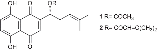
Shikonin, isolated from the root of traditional chinese herbal plants Lithospermum erythrorhizon Sieb. et Zucc.(Boraginaceae), is well known to contain various therapeutic components and has significant anti-tumor activity (CitationChen et al., 2002). Previous studies demonstrated that shikonin and its derivatives had potent anti-tumor activity which was likely mediated by inhibiting tumor cells proliferation in vitro and in vivo, such as human hepatoma cells SK-Hep-1 (CitationChen et al., 2003), human cervical epithelial cancer cells HeLa (CitationWu et al., 2005), human leukemia cells HL60 (CitationHashimoto et al., 1999), and S180 sarcoma mode (CitationKim et al., 2001). Aikete injection is composed of acetylshikonin and β,β-dimethylacrylshikonin (), the two main effective anti-tumor components of shikonin (CitationHu et al., 2006; CitationKundakovic et al., 2006; CitationZeng et al., 2009). Although the anti-tumor effect of acetylshikonin and β,β-dimethylacrylshikonin has been reported widely, little is known about the effects of the combination of the two components on cancer cells.
Figure 1. Chemical structure of acetylshikonin (1), and β,β-dimethylacrylshikonin (2).
Our present study focused on the effects of Aikete injection on the growth of human HCC cell line SMMC-7721 and H22 transplanted tumor, as well as the growth-inhibitory mechanism. Aikete injection exerted inhibition effects on HCC cells in vitro and in vivo by inducing cell apoptosis. Moreover, cell cycle arrest in Aikete injection-treated HCC cells was detected by flow cytometry, and the changes in apoptosis-related gene expression in HCC cells were observed by reverse transcription-polymerase chain reaction (RT-PCR). We further demonstrated that the possible inhibitory activity mechanisms of Aikete injection involved mitochondria pathways. The results suggested that Aikete injection is an attractive candidate drug that prevents tumor proliferation, not only by modulating the expression of genes associated with apoptosis but also by arresting the cell cycle.
Materials and methods
Reagents
Aikete injection was obtained from Huakang Pharmaceutical Co., Ltd. (Deyang, China). It was diluted with RPMI-1640 medium at different concentrations when needed. RPMI-1640, trypsin, 3-(4,5-dimethylthiazol-2-yl)-2,5-diphenyl-tetrazolium bromide (MTT) were purchased from the Gibco (Grand Island, NY); Hoechst 33258, diethyl pyrocarbonate were purchased from Sigma company (St. Louis, MO); Trizol was purchased from Invitrogen company (Carlsbad, CA); RevertAid™ First Strand cDNA Synthesis Kit was purchased from Fermentas company (Glen Burnie, MD). Mouse antibodies against Bcl-2, Bax, and β-actin were purchased from Santa Cruz Biotechnology (Santa Cruz, CA).
Cell cultures
Human hepatocarcinoma SMMC-7721 cell line and murine hepatocarcinoma H22 cell line were obtained from Nanjing Keygen Biotech (Nanjing, China). SMMC-7721 cells were routinely cultured in RPMI-1640 medium supplemented with 10% heat-inactivated calf serum, 100 U/mL penicillin, and 100 μg/mL streptomycin (Huabei Pharmaceuticals Ltd., China), in a humidified atmosphere of 5% CO2 at 37°C. All cells were harvested in their exponential growth phase.
MTT assay and growth curve assay
The inhibitory effect of Aikete injection on the growth of SMMC-7721 cells was measured by the MTT and growth curve assays.
For the MTT assay (CitationLuo et al., 2008), the cells were dispensed in 96-well plates at a density of 1 × 10⁵/well. After 24-h incubation, they were treated with Aikete injection at different final concentrations (0.1, 0.2, 0.4, 0.8, 1.6, 3.2, 6.4, 12.8, 25.6 µg/mL). The cells were incubated for 48 h, and 20 µL of MTT solution (5.0 mg/mL) was added to each well and incubated at 37°C for 4 h. The supernatant fluid was removed, and the MTT formazan was dissolved in 100 µL DMSO. Absorbance at 490 nm was measured with a microplate reader (Bio-Rad, Hercules, CA) using wells without cells as blanks. The 50% inhibitory concentration (IC50) at the 48 h was determined with the Bliss method.
For the cell growth curve assay (CitationVasilevskaya et al., 2004), SMMC-7721 cells in the logarithmic phase were cultured at a density of 1 × 10⁴ cells per well for 1–7 days. The wells were divided into three groups. Control negative group was cultured in drug-free medium; test group was exposed to various concentrations of Aikete injection; positive group was exposed to 0.05 µg/mL adriamycin. Every day three wells from each group were evaluated for cell growth using the Trypan blue dye assay.
Morphological changes
To observe the morphological changes of SMMC-7721 cells treated with Aikete injection, Giemsa staining and Hoechst 33258 nucleus staining were performed. SMMC-7721 cells suspension was adjusted to a density of 1 × 10⁵ cells/mL and cultured in cell culture plates. After a 24-h incubation, cells were exposed to various concentrations of Aikete injection for 48 h. After the medium was removed, cells were washed twice with PBS, fixed with 40 g/L formaldehyde, washed with PBS twice, and stained with Giemsa (0.75 mg/mL) for 15 min or Hoechst 33258 (5.0 mg/L) for 30 min. Stained cells were washed with PBS, and observed under a microscope or fluorescence microscope. Random fields were selected for photography. To observe the morphological changes of hepatocarcinoma H22 treated with Aikete injection, HE staining was performed.
Flow cytometry
The apoptosis rate, related gene expression and cell cycle of SMMC-7721 cells were determined by flow cytometry with DNA staining to reveal the total amount of DNA. SMMC-7721 cells were incubated with various concentrations of Aikete injection for 48 h. Cells were harvested from the treated and control cultures, washed twice with PBS and fixed in ice-cold 70% ethanol. The samples were concentrated by removing ethanol and treated with 1% (v/v) Triton X-100 and 0.01% RNase for 10 min at 37°C. Cellular DNA was stained with 0.05% propidium iodide (PI) for 20 min at 4°C in darkness. The cell cycle distribution and apoptotic cells were detected with FACScan (Becton-Dickinson, San Jose, CA).
RT-PCR
Expression of Bcl-2 and Bax mRNA was analyzed by RT-PCR. SMMC-7721 cells were plated at a density of 5 × 10⁵ cells/well into 6-well plates for 24 h. Then they were treated with various concentrations of Aikete injection for 48 h. Total RNA was isolated from the cells using Trizol reagent (Invitrogen) and reverse transcribed with the RevertAid™ First Strand cDNA Synthesis Kit (Fermentas) following the manufacturer’s instructions. Sequences of the primers are given in . β-Actin was used in each experiment as an internal control. PCR products were separated on 1% agarose gel stained with ethidium bromide and observed under ultraviolet light.
Table 1. Sequence of primers.
Animals
Kunming male mice, 5-week old, weighing 18–22 g, were obtained from the Experimental Animal Center at Sichuan University. The mice were housed in plastic cages with hardwood chip bedding in an air-conditioned room at 23 ± 2°C and 55 ± 5% humidity, and with a 12 h light/dark cycle on basal diet (animal center). The animal handling and experimental procedures were approved by the Animal Ethics Committee of Sichuan University.
In vivo anti-tumor activity
Five-week old male Kunming mice were inoculated with H22 cell suspension (1 × 10⁷/mL), 0.2 mL per mouse, through subcutaneous injection at right side axilla. Twenty-four hours after the tumor cell inoculation, the mice were randomized equally into six groups of ten mice each. Mice in three Aikete injection groups were given 0.5, 1.0, 2.0 mg/kg Aikete injection dissolved in castor oil through intraperitoneal injection (ip). The positive control group was injected with cyclophosphamide at 60 mg/kg; the negative control group was injected with an equal volume of normal saline only; the solvent control group was injected with an equal volume of castor oil simultaneously. Mice in the cyclophosphamide group received injections only on the first day, while mice in all other groups received injections once every other day (six times in total). From the forth day, tumor volume (V) was measured every other day, and calculated according to the formula: V (mm3) = ab2/2, where a is largest superficial diameter and b is smallest superficial diameter.
All mice were sacrificed 13 days after inoculation with H22 cells, and the transplanted tumors were exercised and weighed. To evaluate the anticancer activity of Aikete injection, tumor inhibitory rates were calculated as following formula: Tumor inhibitory rate (%) = 1-(tumor weight of treated group/tumor weight of control group) ×100%.
Statistical analysis
The data were expressed as mean ± standard deviation. The Statistical Package for Social Sciences version 13.0 (SPSS Inc., Chicago, IL) was used for standard statistical analysis including one-way analysis of variance and Student’s t-test. A value of P < 0.05 was considered statistically significant.
Results
Effects of Aikete injection on cell growth and proliferation by MTT assay and growth curve assay
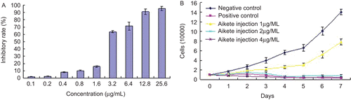
In order to assess the growth inhibition effects of Aikete injection on human liver cancer cells, the MTT assay and growth curve assay were performed. To measure the IC50 of the 48 h, SMMC-7721 cells were treated with Aikete injection at seven different concentrations (0.1, 0.2, 0.4, 0.8, 1.6, 3.2, 6.4, 12.8, 25.6 µg/mL). The results showed that the inhibition ratio increased significantly in a dose-dependent manner (), and the IC50 was 2.325 µg/mL. Time-dependent growth inhibition was observed at concentrations ranging from 1.0 to 4.0 mg/L by growth curve assay ().
Figure 2. Inhibitory effect of Aikete injection on SMMC-7721. (A) SMMC-7721 cells were treated with various concentrations of Aikete injection for 48 h, then tested with MTT; (B) the percentage of cell viability was determined by Trypan blue dye assay after 1, 2, 3, 4, 5, 6, and 7 days of treatment, respectively. Data were the mean ± SD of three independent experiments.
Effects of Aikete injection on morphological changes of SMMC-7721 cells
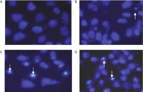
To observe the morphological changes of SMMC-7721 cells treated with Aikete injection, Giemsa staining and Hoechst 33258 nucleus staining were performed ( and ). The cells show marked morphological changes of apoptosis after treated with 2.0 µg/mL Aikete injection for 48 h, and the apoptotic cells with condensed and fragmented nuclei were seen in the Aikete injection-treated groups, but not in the negative control.
Figure 3. Morphological changes of SMMC-7721 cells detected by Giemsa staining (×200). SMMC-7721 cells incubated in the absence (A) or presence (B) of 2.0 µg/mL Aikete injection for 48 h were stained with Giemsa. Cells were viewed under microscope.
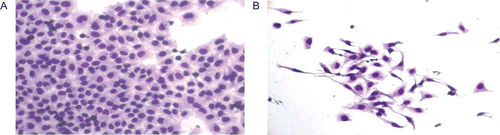
Figure 4. Morphological changes of SMMC-7721 cells detected by Hoechst 33258 staining (×400). After untreated (A) and treated with 1.0 µg/mL (B), 2.0 µg/mL (C), 4.0 µg/mL (D) Aikete injection for 48 h, cells were stained with Hoechst 33258 and observed under fluorescence microscope. Arrows indicate apoptotic cells.
Effects of Aikete injection on cell apoptosis and cell cycle arrest
DNA content in SMMC-7721 cells stained with PI was analysed by flow cytometry. After treatment with Aikete injection (1.0, 2.0, 4.0 µg/mL) for 48 h, cells were accumulated in the G2/M phase of the cell cycle. The proportion of cells at G2/M phase increased to 27.1% after treatment with 4.0 µg/mL Aikete injection. The apoptotic peak was detected, and the Aikete injection group had a much higher apoptotic peak than the control. The result showed that Aikete injection effectively induced cell cycle arrest and apoptosis in a dose-dependent manner ().
Table 2. Effect of Aikete injection on apoptosis rates and cell cycle of SMMC-7721 cells (n = 3, mean ± SD).
Effects of Aikete injection on the expression of apoptosis-related genes
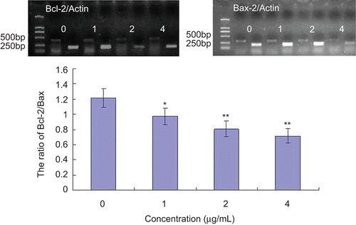
To assess the effect of Aikete injection on the expression of Bcl-2 and Bax, RT-PCR and flow cytometer analysis were performed in SMMC-7721 cells. Compared with the negative control group, flow cytometric analysis showed that the ratio of Bcl-2/Bax decreased in Aikete injection-treated groups in SMMC-7721 cells (). Besides, in Aikete injection groups the Bcl-2 expression and the ratio of Bcl-2/Bax were downregulated in SMMC-7721 cells, as measured by RT-PCR ().
Figure 5. Effect of Aikete injection on the expression of Bcl-2/Bax in SMMC-7721 cells. Different concentrations of Aikete injection were added to SMMC-7721 cell culture for 48 h and then cells were harvest to be processed. The expression of Bcl-2 and Bax were analyzed by flow cytometry. Data were the mean ± SD of three independent experiments. Compare with negative control: *P < 0.05 **P < 0.01
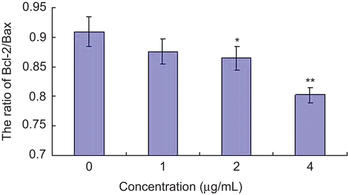
Figure 6. Effect of Aikete injection on Bcl-2 and Bax mRNA expression in SMMC-7721 cells. Cells were treated with various concentrations of Aikete injection for 48 h. The mRNA levels of Bcl-2 and Bax were analyzed by RT-PCR, and actin was used as a control. Data were the mean ± SD of three independent experiments. Compare with negative control: *P < 0.05 **P < 0.01.
Aikete injection inhibits the growth of mouse transplantable hepatoma
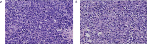
To determine whether Aikete injection inhibits tumor growth in vivo, H22 cells were injected into Kunming mice. Growth curve of hepatocarcinoma H22 showed that Aikete injection inhibited tumor growth significantly in a dose- and time-dependent manner from 0.5 to 2.0 mg/kg (). Treated with Aikete injection, the tumor inhibition rates ranged from 34.37 to 57.99% (). Compared with the saline group, Aikete injection groups showed no significant differences in thymus and spleen (data not shown). Besides, HE staining was performed to show the morphological changes of the Aikete injection-treated tumor. Compared with the negative control group, cell shrinkage, vacuoles in cytoplasm, pyknosis, and chromatin margination were observed in the 2.0 mg/kg Aikete injection group ().
Table 3. Effect of Aikete injection on H22 bearing mice (mean ± SD).
Figure 7. Anti-tumor effects of Aikete injection in vivo. (A) Growth curve of mouse transplantable hepatoma; (B) inhibitory effects of Aikete injection and cyclophosphamide on mouse tumor. Transplanted tumors from each group were shown: (B1) negative control; (B2) solvent control; (B3) cyclophosphamide 60 mg/kg; (B4) Aikete injection 0.5 mg/kg; (B5) Aikete injection 1.0 mg/kg; (B6) Aikete injection 2.0 mg/kg. Data were the mean ± SD of three independent experiments.
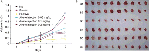
Figure 8. Morphological changes of H22 tumor cells treated with HE staining (×200). The transplanted tumors in the group of negative control (A) and Aikete injection 2.0 mg/kg (B) were exercised for HE staining and observed under microscope.
Discussion and conclusion
Apoptosis, the program of cellular suicide, is critical in tumorigenesis and tumor progression (CitationGreen and Evan, 2002). Inducing apoptosis appears to be a common mechanism of many anti-tumor agents used in chemotherapy. In recent studies, shikonin and its derivatives were demonstrated to induce apoptosis of tumor cells. It has been reported that shikonin could induce A375-S2 cells apoptosis and cell cycle arrest through caspase-9 dependent pathways (CitationWu et al., 2004a); acetylshikonin induced SGC-7901 cells apoptosis by downregulating Bcl-2 expression and upregulating Bax expression (CitationZeng et al., 2009); shikonin induced apoptosis in HeLa cells via caspase-3 activation, blockage of DNA synthesis, and cell cycle arrest (CitationWu et al., 2004b); shikonin could induce apoptosis in COLO 205 cells via modulating the Bcl-2 family, upregulating p27, p53, releasing cytochrome c, and sequentially activating caspases (CitationHsu et al., 2004).
In this study, we described the inhibitory effects of Aikete injection on the proliferation of cancer cell line SMMC-7721. We also demonstrated a preventive effect of Aikete injection on progression in mouse model. Aikete injection inhibited the growth of SMMC-7721 cells in a dose- and time-dependent manner as evaluated by MTT and cell growth curve assays. Several lines of evidence, including flow cytometry, Giemsa staining and Hoechst 33258 nucleus staining, consistently documented apoptosis in Aikete injection-treated SMMC-7721 cells. Treatment with Aikete injection resulted in marked morphological changes, cell cycle arrest in G2/M phase, and increased apoptosis rate (evidenced in elevated sub-G1 peak). We further demonstrated in vivo anti-tumor activity of Aikete injection in a mouse model. Aikete injection significantly inhibited the growth of tumor at the dose of 0.5–2.0 mg/kg. In treated mice, weight loss was not observed in all of the Aikete injection groups. We observed no severe side effects such as immune organ weight loss in either treated or N.S. control mice. Apoptosis was also found in H22 transplantable hepatoma by HE staining.
The mitochondrial-mediated signal transduction pathway plays a central role in the control of apoptosis. Bcl-2 and Bax are important components of this signal transduction pathway. The Bcl-2 family is a key regulator of the mitochondrial response to apoptotic signals in the intrinsic pathway. It comprises more than 20 different members that either positively or negatively regulate apoptosis (CitationCory and Adams, 2002; CitationLiou et al., 2003). It includes anti-apoptotic Bcl-2 members, such as Bcl-2, Bcl-XL, Bcl-W and Mcl-1, acting as potent suppressors of apoptosis by blocking the release of cytochrome c. Although pro-apoptotic members, such as Bax, Bak, Bad, Bcl-Xs, Bid, Bik, Bim, and Hrk, acting as promoters that have opposing functions and promote cell death (CitationMiyashita and Reed, 1993; CitationBurlacu, 2003; CitationKuwana and Newmeyer, 2003). The ratio of anti- to pro-apoptotic molecules determines the response to a death signal. Bcl-2 can promote cell survival by inhibiting apoptosis, while Bax can promote apoptosis by forming Bcl-2/Bax heterodimers (CitationWang et al., 2002; CitationBuchholz et al., 2003). Therefore, the ration of Bcl-2 to Bax is the key to determine cell survival or death. In order to explore whether apoptosis-related genes contribute to the inhibitory effect of Aikete injection on SMMC-7721 cells, we analyzed the relative Bcl-2 and Bax expression levels induced by Aikete injection in this study. Our results have suggested that Aikete injection may alter the expression of apoptosis-related genes. We found that Aikete injection downregulated the Bcl-2 expression and the ratio of Bcl-2/Bax in a dose-dependent manner. It is possible to postulate that Aikete injection may inhibits cell proliferation by inducing apoptosis via modulating Bcl-2 family members.
In conclusion, we demonstrated that Aikete injection can inhibit SMMC-7721 cell proliferation by inducing apoptosis via downregulating Bcl-2 expression and the ratio of Bcl-2/Bax, whereas it also has significant anti-tumor effects in vivo. These results suggested that Aikete injection was an attractive candidate for cancer therapy. Further investigation of Aikete injection as an anti-tumor compound is warranted.
Declaration of interest
The authors report no conflicts of interest. The authors alone are responsible for the content and writing of the paper.
References
- Buchholz TA, Davis DW, McConkey DJ, Symmans WF, Valero V, Jhingran A, Tucker SL, Pusztai L, Cristofanilli M, Esteva FJ, Hortobagyi GN, Sahin AA. (2003). Chemotherapy-induced apoptosis and Bcl-2 levels correlate with breast cancer response to chemotherapy. Cancer J, 9, 33–41.
- Burlacu A. (2003). Regulation of apoptosis by Bcl-2 family proteins. J Cell Mol Med, 7, 249–257.
- Chen CH, Chern CL, Lin CC, Lu FJ, Shih MK, Hsieh PY, Liu TZ. (2003). Involvement of reactive oxygen species, but not mitochondrial permeability transition in the apoptotic induction of human SK-Hep-1 hepatoma cells by shikonin. Planta Med, 69, 1119–1124.
- Chen X, Yang L, Oppenheim JJ, Howard MZ. (2002). Cellular pharmacology studies of shikonin derivatives. Phytother Res, 16, 199–209.
- Cory S, Adams JM. (2002). The Bcl2 family: Regulators of the cellular life-or-death switch. Nat Rev Cancer, 2, 647–656.
- Green DR, Evan GI. (2002). A matter of life and death. Cancer Cell, 1, 19–30.
- Hashimoto S, Xu M, Masuda Y, Aiuchi T, Nakajo S, Cao J, Miyakoshi M, Ida Y, Nakaya K. (1999). beta-Hydroxyisovalerylshikonin inhibits the cell growth of various cancer cell lines and induces apoptosis in leukemia HL-60 cells through a mechanism different from those of Fas and etoposide. J Biochem, 125, 17–23.
- Hsu PC, Huang YT, Tsai ML, Wang YJ, Lin JK, Pan MH. (2004). Induction of apoptosis by shikonin through coordinative modulation of the Bcl-2 family, p27, and p53, release of cytochrome c, and sequential activation of caspases in human colorectal carcinoma cells. J Agric Food Chem, 52, 6330–6337.
- Hu Y, Jiang Z, Leung KS, Zhao Z. (2006). Simultaneous determination of naphthoquinone derivatives in Boraginaceous herbs by high-performance liquid chromatography. Anal Chim Acta, 577, 26–31.
- Kim SH, Kang IC, Yoon TJ, Park YM, Kang KS, Song GY, Ahn BZ. (2001). Antitumor activities of a newly synthesized shikonin derivative, 2-hyim-DMNQ-S-33. Cancer Lett, 172, 171–175.
- Kundakovic T, Fokialakis N, Dobric S, Pratsinis H, Kletsas D, Kovacevic N, Chinou I. (2006). Evaluation of the anti-inflammatory and cytotoxic activities of naphthazarine derivatives from Onosma leptantha. Phytomedicine, 13, 290–294.
- Kuwana T, Newmeyer DD. (2003). Bcl-2-family proteins and the role of mitochondria in apoptosis. Curr Opin Cell Biol, 15, 691–699.
- Liou AK, Clark RS, Henshall DC, Yin XM, Chen J. (2003). To die or not to die for neurons in ischemia, traumatic brain injury and epilepsy: a review on the stress-activated signaling pathways and apoptotic pathways. Prog Neurobiol, 69, 103–142.
- Luo G, Guan X, Zhou L. (2008). Apoptotic effect of citrus fruit extract nobiletin on lung cancer cell line A549 in vitro and in vivo. Cancer Biol Ther, 7, 966–973.
- Miyashita T, Reed JC. (1993). Bcl-2 oncoprotein blocks chemotherapy-induced apoptosis in a human leukemia cell line. Blood, 81, 151–157.
- Shariff MI, Cox IJ, Gomaa AI, Khan SA, Gedroyc W, Taylor-Robinson SD. (2009). Hepatocellular carcinoma: current trends in worldwide epidemiology, risk factors, diagnosis and therapeutics. Expert Rev Gastroenterol Hepatol, 3, 353–367.
- Vasilevskaya IA, Rakitina TV, O’Dwyer PJ. (2004). Quantitative effects on c-Jun N-terminal protein kinase signaling determine synergistic interaction of cisplatin and 17-allylamino-17-demethoxygeldanamycin in colon cancer cell lines. Mol Pharmacol, 65, 235–243.
- Wang QF, Chen JC, Hsieh SJ, Cheng CC, Hsu SL. (2002). Regulation of Bcl-2 family molecules and activation of caspase cascade involved in gypenosides-induced apoptosis in human hepatoma cells. Cancer Lett, 183, 169–178.
- Wu Z, Wu L, Li L, Tashiro S, Onodera S, Ikejima T. (2004a). p53-mediated cell cycle arrest and apoptosis induced by shikonin via a caspase-9-dependent mechanism in human malignant melanoma A375-S2 cells. J Pharmacol Sci, 94, 166–176.
- Wu Z, Wu LJ, Li LH, Tashiro S, Onodera S, Ikejima T. (2004b). Shikonin regulates HeLa cell death via caspase-3 activation and blockage of DNA synthesis. J Asian Nat Prod Res, 6, 155–166.
- Wu Z, Wu LJ, Tashiro S, Onodera S, Ikejima T. (2005). Phosphorylated extracellular signal-regulated kinase up-regulated p53 expression in shikonin-induced HeLa cell apoptosis. Chin Med J, 118, 671–677.
- Zeng Y, Liu G, Zhou LM. (2009). Inhibitory effect of acetylshikonin on human gastric carcinoma cell line SGC-7901 in vitro and in vivo. World J Gastroenterol, 15, 1816–1820.