Abstract
Objectives: To investigate transdermal absorption enhancement of gel containing elastic niosomes loaded with gallic acid in the semipurified fraction isolated from Terminalia chebula Retz. (Combretaceae) galls.
Materials and methods: Nonelastic and elastic niosomes loaded with gallic acid in pure form or in the semipurified fraction were developed. Rat skin permeation by vertical Franz diffusion cells of gallic acid from various gel formulations containing elastic niosomes loaded with gallic acid or the semipurified fraction was performed.
Results: Elastic and nonelastic niosomes loaded with gallic acid or the semipurified fraction exhibited the mixture of unilamellar and multilamellar structures with negative zeta potential values and in the size range of 200–400 nm. Both loaded elastic and nonelastic niosomes showed good physical and chemical stability for 3 months. The percentages remaining of gallic acid in nonelastic were slightly higher than in elastic niosomes. Both elastic and nonelastic niosomes retarded rat skin permeation of the loaded pure gallic acid, while enhanced the loaded gallic acid in the semipurified fraction. However, elastic niosomes exhibited higher percentages of gallic acid through rat skin than the nonelastic niosomes.
Discussion and conclusion: This study has demonstrated the potential of niosomes, especially elastic niosomes, for the enhancement of chemical stability and rat skin transdermal absorption of gallic acid in the semipurified fraction from T. chebula galls, which will be beneficial for topical antiaging application.
Introduction
As known, many polyphenolic compounds have shown many interesting biological activities, such as antimutagenicity, anticarcinogenicity, and antioxidative activity. Gallic acid is a naturally occurring plant phenol. When in emulsion or lipid system, it has been found to have strong antioxidant activity (CitationNakatani, 1992; CitationMadsen & Bertelsen, 1995) and exhibit antimutagenicity. It is generally used in processed foods, cosmetics, and food packing materials to prevent rancidity induced by lipid peroxidation and spoilage. It is almost as effective as the tocopherol analogue (Trolox) and even more effective than several water soluble antioxidants, such as ascorbic acid (CitationCholbi et al., 1991).
Terminalia chebula Retz. (Combretaceae) is a native plant in India and Southeast Asia. In Thailand, this plant is commonly known as Samor Thai. All parts of this plant contain numerous phytochemicals. Kot Phung Pla (in Thai), which is the gall of T. chebula, is produced by the plant against insect bite with or without being accompanied by microorganism infection. It has been used in Thai folklore medicines as an astringent and purgative. It has been reported to retard the effects of aging and impart longevity as well as the boosting of immune systems (The Pharmacopoeia Commission of PRC, 1997; CitationZhu, 1998; CitationLemmens & Bunyapraphatsara, 2003). Moreover, the extracts from T. chebula galls exhibited high DPPH radical scavenging and potent matrix metalloproteinase-2 (MMP-2) inhibition indicating the potential for antiaging application (CitationManosroi et al., 2010a). This gall is rich in tannin, which is a hydrolyzable pyrogallol. Six phenolic compounds of the hydrolyzable tannins including gallic acid, punicalagin, isoterchebulin, 1,3,6-tri-O-galloyl-β-d-glucopyranose, chebulagic acid, and chebulinic acid have been found in this gall (CitationManosroi et al., 2010b). However, phenolic compounds are generally not chemically stable and when exposed to light and oxidation (CitationCheynier et al., 1990; CitationLiazid et al., 2007) they give skin irritation (CitationFrosch, 1992; Roberts et al., 1997) and low skin permeation owing to their large molecular structures. Hence, these limitations are obstacles of these phenolic compounds from T. chebula galls for an efficient topical use in cosmetic and pharmaceutical applications.
Recently, nanotechnology has become the center of interest especially nanovesicles including liposomes and niosomes for cosmetic sciences because of the ease of encapsulation of various bioactive compounds in these vesicles, an increase in physicochemical stability, the reduction in serious side effects and skin irritation and the enhancement of transdermal absorption and the accumulation of the loaded substances at the site of administration. These nanovesicles are bilayer spheroid vesicles composed of cholesterol mixed with phospholipids (liposomes) or nonionic surfactants (niosomes), which can entrap both water and oil soluble substances. In comparing to liposomes, niosomes offer higher chemical stability, lower cost, and greater availability of the surfactant composition than liposomes (CitationHandjani-Vila et al., 1979; CitationUchegbu & Vyas, 1998). CitationManosroi et al. (2008) have recently developed a novel elastic niosome for diclofenac diethylammonium. This elastic niosomes is superior for topical use because of its flexibility and deformability of being able to squeeze itself through the pore, which is much smaller than its diameter. From our previous study, the aqueous crude extract as well as its phenolic compounds from T. chebula galls demonstrated high potential in vitro antiaging activities (CitationManosroi et al., 2010a,b). In this study, transdermal absorption of gallic acid containing in the semipurified fraction isolated from T. chebula galls and the pure gallic acid loaded in this developed elastic niosome was evaluated for topical antiaging applications.
Materials and methods
Materials
Standard gallic acid, Tween 61 (polyoxyethylene sorbitan monostearate), cholesterol, and Sephadex-G-50 were purchased from Sigma Chemicals (St. Louis, MO, USA). Diaion HP-20 (particle size 200–600 µm) and octadecyl silica (ODS) gel (Chromatorex-ODS, 100–200 mesh) were purchased from Mitsubishi Chemical Co. (Tokyo, Japan) and Fuji Silysia Chemical, Ltd. (Aichi, Japan), respectively. Carbopol® 980 was purchased from Fluka (Buchs, Switzerland). All other analytical grade chemicals and reagents were purchased from Wako Pure Chemical Industries, Ltd. (Osaka, Japan).
Plant materials
T. chebula galls were collected from Chiang Mai Province in Thailand, during January to February in 2008. The specimen was authenticated by a botanist at Natural Products Research and Development Center (NPRDC), Science and Technology Research Institute (STRI), and deposited in the medicinal plant herbarium, Faculty of Pharmacy, Chiang Mai University in Thailand for authenticated specimen (voucher specimen no. 012102).
Preparation of semipurified fraction containing gallic acid from T. chebula galls crude extract
The semipurified fractions containing gallic acid were isolated and purified as previously described (CitationManosroi et al., 2010b). Briefly, 100 g of the dried powder was macerated and extracted with 400 ml of distilled water and sonicated in a bath sonicator for 1 h at room temperature (27 ± 2°C). The extract was filtered and concentrated under vacuum by a rotary evaporator (R-124 Buchi, Switzerland) and lyophilized (CHRIST, Martin Christ, Germany). The yields of the crude extracts were 49.85% w/w of the dried plant material. Approximately 50 g of the crude extract was applied on a Diaion HP-20 column (8 × 90 cm) eluted with 50% methanol (MeOH) in water to give 17.38 g. A portion of this fraction (8 g) was subjected to an ODS column (4 × 85 cm) eluted with MeOH-H2O-acetic acid gradient to give three fractions (250 ml of each fraction). The eluted solvent and the yield of each fraction were as followed: fr.1: MeOH/H2O/AcOH (2:8:0.1) (0.54 g), fr.2: MeOH/H2O/AcOH (4:6:0.1) (0.74 g) and fr.3: MeOH/H2O/AcOH (6:4:0.1) (0.50 g).
Identification of the phenolic components in the selected semipurified fractions
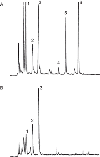
The total phenolic contents of the fractions from the ODS column were determined by a modified Folin-Ciocalteu method (CitationYeh & Yen, 2005), calibrating against gallic acid as the reference standard. Briefly, the fraction (500 µl) at the concentration of 10 mg/ml was mixed with an equal volume of 1N Folin-Ciocalteu reagent and 1 ml of 20% sodium carbonate (Na2CO3) and incubated for 25 min at room temperature (27 ± 2°C). The reaction mixture was then centrifuged at 5000 rpm for 10 min. The absorbance of the supernatant was measured at 730 nm using a spectrophotometer. The experiments were done in triplicate. The total phenolic contents were calculated on the basis of the calibration curve of the standard gallic acid. The semipurified fraction which gave the highest total phenolic content was selected to load in niosomes. The phenolic compounds in the selected semipurified fraction were analyzed using the gradient HPLC system with the evaporative light scattering detector (ELSD, SEDERE Sa., France). The equipment consisted of a binary pump (Senshu Scientific Co., Ltd., Tokyo, Japan) with a gradient controller (Senshu Scientific Co., Ltd.) and a degasser (Senshu Scientific Co., Ltd., Tokyo). The column used was a Capcellpak AQ 5 µm, 250 × 4.6 mm (Shiseido Co., Ltd., Tokyo, Japan). The injection volume of the sample was 20 ml and eluted by acetonitrile (solvent-A)/0.1% formic acid (solvent-B) at 0.5 ml/min. The gradient system started with 10% solvent-A at 5 min and then linearly changed to the 60% solvent-A in 75 min. The sample was dissolved by 10% solvent-A. The compounds which were isolated from the cold water crude extract of T. chebula galls including gallic acid, 1,3,6-trigalloylglucopyranoside, punicalagin, isoterchebulin, chebulagic acid, and chebulinic acid were eluted at 9.2, 13.7, 16.5, 30.1, 34.5, 41.9 min, respectively (CitationManosroi et al., 2010b). The chromatograms of the cold water crude extract of T. chebula galls and the selected semipurified fraction (fraction no. 1) indicating the peaks of the phenolic compounds were shown in .
Figure 1. The gradient high-performance liquid chromatography (HPLC) chromatogram of (A) cold water crude extract of Terminalia chebula galls containing phenolic compounds including gallic acid (1), 1,3,6-tri-O-galloyl-ß-d-glucopyranose (2), punicalagin (3), isoterchebulin (4), chebulagic acid (5), and chebulinic acid (6) and (B) the semipurified fraction.
Entrapment of the semipurified fraction and gallic acid in elastic niosomes
Elastic and nonelastic niosomal formulations were prepared by the modified chloroform film method previously described (CitationManosroi et al., 2008). Tween 61 mixed with cholesterol (at 1:1 molar ratios) of 20 mM was placed in a clean and dry round-bottomed flask. The mixture was dissolved in chloroform. The organic solvent was removed by a rotary evaporator under vacuum (R-124 Buchi, Flawil, Switzerland). The resulting film was dried overnight under vacuum at room temperature (27 ± 2°C). The film was rehydrated with 5 mM phosphate buffer (PB), pH 7.0 for nonelastic niosomes or 25% ethanol in PB for elastic niosomes. The niosomal dispersion was put in an ice bath (4°C) while sonicating by a microtip probe sonicator (Vibra Cell TM, Sonics & Materials, Inc., Newtown, CT, USA) at pulse on 3.0 and pulse off 1.0, 33% amplitude for 10 min. The dispersions were kept in vials tightly covered with aluminum caps. For the loading of gallic acid or the semipurified fraction, gallic acid or the semipurified fraction dissolved in PB was used to hydrate the film to obtain niosomal dispersion.
Physicochemical characteristics of the loaded elastic niosomes
Morphology, vesicle size, and zeta potential determination
A drop of niosomal dispersion was applied on a 300-mesh formvar copper grid on paraffin and allowed the sample to adhere on the formvar for 10 min. The remaining dispersion was removed and a drop of 2% aqueous solution of ammonium molybdate was applied for 5 min. The remaining solution was then removed. The sample was air-dried and examined with a transmission electron microscope (TEM 1200S JEOL; JEOL Ltd., Tokyo, Japan). The morphology and lamellarity of niosomes were observed. The diameter of the loaded and unloaded (empty) niosomes was determined using dynamic light scattering (DLS) by a Zetasizer 300HSA (Malvern Instruments, Malvern, Worcestershire WR14 1XZ, UK) based on photon correlation spectroscopy. The charges on the niosomal surface were determined using the phase analysis light scattering (PALS) (Malvern Instruments, Malvern, UK). Analysis time was kept for 60 s. The average zeta potential values and charges were determined. The time-dependent correlation function on the scattered light intensity was measured at a scattering angle of 90°. All samples were diluted to 30 times with freshly filtrated Millipore water for particle size and zeta potential measurement (n = 3).
Measurement of elasticity value
Elasticity of the loaded or unloaded elastic and nonelastic niosomes was carried out by the extrusion method previously described (CitationCevc, 1996; CitationJain et al., 2003). Briefly, the dispersions were extruded through a polycarbonate membrane filter with a pore size of 50 nm (Millipore, Billerica, MA, USA) at constant pressure (2.5 bar). The elasticity of the niosomes was expressed in terms of deformability index (DI) according to the following equation: Deformability index (DI) = j × [(rv/rp)2], where j is the weight of the dispersion, which was extruded in 10 min through a polycarbonate filter of 50 nm pore size, rv was the size of niosomes after extrusion, and rp was the pore size of the filter membrane.
Entrapment efficiency determination
The entrapment efficiencies of the pure gallic acid or gallic acid containing in the semipurified fraction loaded in elastic and nonelastic niosomes were determined by gel-filtration using Sephadex® G-50 as a packing material and PB (pH 7.0) as an eluent. Eluates were collected in tubes using a fraction collector (Foxy JR, Isco Inc., Lincoln, NE, USA) at the flow rate of 6 ml/min. The fractions containing niosomes detected at 470 nm (CitationCastanho et al., 1997) were pooled, collected, and dried with a freeze-dryer (CHRIST, Martin Christ, Osterode, Germany). The residues were dissolved in absolute ethanol and assayed for gallic acid contents by HPLC. The percentages of entrapment efficiencies in terms of gallic acid were calculated according to the following equation:
Physicochemical stability
The elastic and nonelastic niosomes loaded with the pure gallic acid or gallic acid in the semipurified fraction were put in vials covered with aluminum cap and stored at room temperature (27 ± 2), 4 ± 2 and 45 ± 2°C for 3 months. The physical characteristics (color, sedimentation, morphology, and particle size by DLS) as well as the remaining gallic acid contents determined by HPLC at 0, 0.5, 1, 1.5, 2, and 3 months were investigated.
Determination of gallic acid contents by HPLC
Amounts of gallic acid were determined by HPLC, LC1200 UV/VIS detector, and LC1100HPLC pump (AS 1000, Thermo Finigan, USA) using Luna® C18, 10 µm i.d., 250 × 4.0 nm, Phenomenex USA column, and a mobile phase containing MeCN/H2O/AcOH (10:90:1, v/v) at the flow rate of 1 ml/min. The samples were filtered through a 0.45-µm membrane filter, prior to the injection onto the HPLC column. An amount of 20 µl of the samples was injected into the column and monitored at 254 nm UV detector. The retention time of gallic acid was 3.8 min. Gallic acid contents were determined from the standard curve of the standard gallic acid, which demonstrated linear with high correlation (r2 = 0.9993). The following regression equation was obtained: y = (9 × 10⁷)x + 510586, where y was the peak area and x was the quantity of gallic acid (µg). The experiment was done in triplicates.
Preparation of gel containing the semipurified fraction loaded in elastic niosomes
The elastic and nonelastic niosomes loaded with the pure gallic acid or gallic acid in the semipurified fraction were incorporated into the gel base containing Carbopol® 980. Briefly, the 0.6% Carbopol® 980 gel was dispersed in the niosomal dispersion with gentle stirring, resulting in the gel containing 0.5% w/w of pure gallic acid, and the gel containing 0.08% w/w of gallic acid in the semipurified fraction.
In vitro rat skin permeation by Franz diffusion cells
The rat skin used in this study was taken from the male Sprague-Dawley rats weighing between 150 and 200 g obtained from the National Laboratory Animal Center, Mahidol University in Nakhon Pathom, Thailand. The in vitro rat skin permeation study protocol has already been reviewed and approved by the ethical committee of Faculty of Medicine, Chiang Mai University in Thailand since 27 April 2010. Rat skin permeation of phenolic compounds using gallic acid as a marker from various gel formulations was performed using vertical Franz diffusion cells having the contact area between the donor and the receiver chamber of 2.46 cm2 and the volume of the receptor compartment of 13 ml. The receptor compartment contained phosphate-buffered saline (PBS, pH 6.5), which was constantly stirred at 100 rpm with a magnetic bar and the controlled temperature at 32 ± 2°C throughout the experiment. The skin was prepared from the abdominal skin of the rats. After the abdominal skin of the rats was shaved and carefully separated, subcutaneous fat was carefully removed using a scalpel. The freshly skin was mounted on the receptor compartment with the subcutaneous side facing upward into the donor compartment, while the dermal side was in contact with the receiver medium. The gel (0.5 g) was placed into the donor compartment and covered with paraffin film. Cells were stopped at 1, 3, 6, 9, and 12 h. For the calculation method of gallic acid concentration in the SC, the rat skin was removed and swung twice in 100 ml of distilled water. The skin was stripped with 10 pieces of an adhesive tape using 3M Scotch Magic™ tape (1 cm × 1 cm) described by CitationPlessis et al. (1992). Each tape was charged with a weight of 300 g for 10s and then removed rapidly (Citationvan der Molen et al., 1997; Fresno-Contreras et al., 2005; CitationFoldvari et al., 2006). In each skin samples, the first tape strip was discarded (CitationPadula et al., 2010). The 2nd to 10th tape strips were used to analyze for gallic acid amounts in SC. For the calculation method of gallic acid concentration in the VED, the skin (VED) after stripping was cut into small pieces and used to assay for gallic acid contents in VED. The nine tape strips or the cut skin (VED) were placed in each vial with 5 ml of ethanol and then sonicated for 10 min in an ice bath and filtered. The filtrate was assayed for gallic acid by HPLC. The experiments were done in triplicates.
For the direct comparisons of skin permeation of gallic acid among gel formulations containing different gallic acid concentrations, the percentages of gallic acid amounts (%) in SC, VED, and receiving solution following transdermal absorption across excised rat skin from various gel formulations by Franz diffusion cells after 12 h were normalized and presented in . For the normalization, the percentages of gallic acid amount (%) were calculated by the following equation: percentages of gallic acid amount (%) = (gallic acid amounts found in SC, VED or receiving solution/the total gallic acid in 0.5 g of each gel formulation) × 100, where GS, GE, and GN gels containing 0.5% w/w of gallic acid, while SS, SE, and SN gels containing the semipurified fraction with 0.08% w/w of gallic acid
Statistical analysis
The results were presented as the mean ± SD of three independent experiments and analyzed by ANOVA at the significant level of P-value < 0.05.
Results and discussion
Total phenolic content and identification of the phenolic compounds in the selected semipurified fraction
After fractionation of the cold aqueous crude extract of T. chebula galls by Dianion column and ODS column chromatography, the semipurifed MeOH/H2O/AcOH (2:8:0.1) fraction, which gave the highest total phenolic content of 21.43 ± 1.31 mg GAE/g (), was selected to load in niosomes. A gradient HPLC chromatogram of this selected semipurified fraction is shown in . The peaks were identified by comparing the retention times with the isolated phenolic compounds (CitationManosroi et al., 2010b). The contents of gallic acid, 1,3,6-trigalloylglucopyranoside, and punicalagin in the semipurified fraction calculated using area under the peak, were found at 15.3, 17.8, and 38.4%, respectively.
Table 1. Percentage yields and total phenolic contents of fractions by column chromatography from the cold aqueous Terminalia chebula gall crude extract.
Characteristics of niosomes
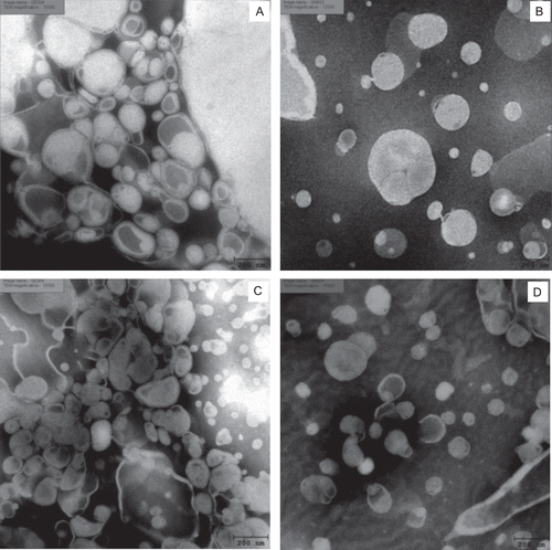
The mean size of niosomes, entrapment efficiency, zeta potential, and deformability index were listed in . A decrease in vesicular size was observed when 25% ethanol was incorporated. It has been reported that the higher ethanol concentration in the elastic vesicles, the lesser membrane thickness was observed owing to the formation of a phase with interpenetrating hydrocarbon chains (CitationDubey et al., 2007; CitationBarry & Cullis, 1995). Ethanol may also modify the net negative charge of the system leading to a decrease in the mean particle size of elastic niosomes (CitationLasic et al., 1998). Both elastic and nonelastic niosomes loaded with gallic acid or semipurified fraction gave larger mean sizes than the unloaded (blank) niosomes. All loaded niosomal formulations exhibited negative zeta potential value, larger size, and higher negative zeta potential value than the unloaded (blank) vesicles. In the buffer system, the vesicles such as niosomes are surrounded by the counter ions, which are opposite charges to the surface charges of niosomes (CitationMcLaughlin et al., 1971). The partial positive charge distribution of cholesterol molecules in the niosomal composition may be neutralized and dominated by the phosphate ions of the buffer system resulting in the negative zeta potential values. Visualized by negative staining of TEM, both nonelastic and elastic niosomes loaded with gallic acid or the semipurified fraction was in the mixture of unilamellar and multilamellar structures (). The entrapment efficiencies of pure gallic acid and gallic acid in the semipurified fraction in elastic niosomes (55.18 ± 3.87 and 24.34 ± 1.53 %) were higher than in nonelastic niosomes (29.72 ± 1.68 and 20.10 ± 3.91%) of about 1.86 and 1.21 times, respectively. The solubility of gallic acid in ethanol is higher than in water of about seven times (CitationAli et al., 2008). Thus, the 25% of ethanol composed in elastic niosomes may increase the solubility of gallic acid resulting in its higher percentages of entrapment efficiency in the bilayers of the elastic niosomes than in the nonelastic niosomes, which contained no ethanol.
Table 2. Size, entrapment efficiency, zeta potential, and deformability index (DI) of nonelastic and elastic niosomes loaded with gallic acid or the semipurified fraction containing gallic acid.
Figure 2. Negative-staining TEM images of elastic and nonelastic niosomes (Tween 61 mixed with cholesterol at 1:1 molar ratio, 20 mM) loaded with gallic acid and the semipurified fraction containing gallic acid: (A) elastic niosomes loaded with gallic acid (GE) (×15,000); (B) nonelastic niosomes loaded with gallic acid (GN) (12,000×); (C) elastic niosomes loaded with the semipurified fraction (SE) (15,000×); and (D) nonelastic niosomes loaded with the semipurified fraction (SN) (15,000×).
For vesicular deformability, the elastic niosomes loaded with pure gallic acid (10.98 ± 2.75) or semipurified fraction (10.75 ± 3.47) showed similar DI to the unloaded (blank) elastic niosomes (11.36 ± 1.55), while higher than the nonelastic niosomes loaded with gallic acid (2.50 ± 0.78) and the semipurified fraction (2.04 ± 1.13) of about 4.39 and 5.27 times, respectively (). Ethanol in the elastic niosomes may interact with the polar head group region of the surfactant molecules (Tween 61) resulting in the reduction of the melting point, thereby increasing the fluidity of the vesicles (CitationDayan & Touitou 2000; CitationTouitou et al., 2000). However, nonelastic niosomes also showed some deformability. In fact, a proper amount of surfactant molecule within the lipid bilayer can provoke a disruption and fluidization of the bilayers (CitationTouitou et al., 2000). Both elastic and nonelastic niosomes, indicated slight different of DI when loaded with gallic acid or the semipurified fraction (), indicating that the loaded compounds did not have any effects on the vesicular membrane elasticity, because the polar hydroxyl groups of gallic acid and other phenolic compounds in the semipurified fraction were located in the aqueous inner core, but not in the niosomal membranes.
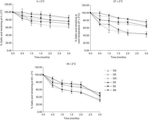
shows the percentage remaining of gallic acid in various formulations at different storage temperatures for 3 months. Stored at room temperature (27 ± 2°C) and 45 ± 2°C for 3 months, both pure gallic acid (GS) and gallic acid in the semipurified fraction (SS) in buffer solution were not only physical unstable with color change to brownish, but also the percentages of gallic acid remaining were less than 45 and 30% at room temperature (27 ± 2) and 45 ± 2°C, respectively. However, at 4 ± 2°C, the percentages of gallic acid remaining after 3 months of GS and SS were more than 60%. The increased temperature showed the less amounts of the remained gallic acid. These may be due to the decarboxylation of gallic acid to pyrogallol at high temperature (Jennifer et al., 1998). When gallic acid or the semipurified fraction containing gallic acid was loaded in elastic (GE and SE) and nonelastic niosomes (GN and SN), they showed no sedimentation, no layer separation, and no color change with higher percentages of gallic acid remaining at all temperatures for 3 months. The percentages of gallic acid remaining after 3 months of GE, SE, GN, and SN were 62.87, 64.55, 72.09, and 75.99; 74.32, 75.99, 83.00, and 85.29; 40.14, 43.54, 48.89, and 49.98% at 27 ± 2, 4 ± 2, and 45 ± 2°C, respectively. The percentages of gallic acid remaining in nonelastic niosomes were higher than elastic niosomes of about 1.12–1.21 times. This may be due to the evaporation of ethanol at elevated temperature during storage, which may fluidize the vesicular membrane and facilitate the leakage of gallic acid from the elastic vesicles. In all systems, the percentages remaining of gallic acid in the semipurified fraction, which contained the mixture of phenolic compounds including gallic acid, were slightly higher than the pure gallic acid. The polyphynol pro-oxidant activity of other phytochemicals in the semipurified fraction, such as punicalagin may have some degradation protection for gallic acid (CitationRice-Evans et al., 1996; CitationHalliwell, 2008).
Figure 3. The percentages of gallic acid remaining in various formulations at different storage temperatures (27 ± 2, 4 ± 2, and 45 ± 2°C) for 3 months. GS = gallic acid in phosphate buffer solution; GE = elastic niosomes loaded with gallic acid; GN = nonelastic niosomes loaded with gallic acid; SS = the semipurified fraction in phosphate buffer solution; SE = elastic niosomes loaded with the semipurified fraction; SN = nonelastic niosomes loaded with the semipurified fraction.
In vitro transdermal absorption of gallic acid through excised rat skin from various gel formulations
The cumulative amounts and percentages of gallic acid per area in different skin strata from various gel formulations after 12 h investigated by Franz diffusion cells were presented ( and ). Each 0.5 g of GS, GE, and GN gel containing 0.5% w/w of gallic acid, and of SS, SE, and SN gel containing the semipurified fraction with 0.08% w/w of gallic acid was placed onto the donor compartment. The cumulative amounts (µg/cm2, n = 3) through skin of gallic acid in all gel formulations increased with times. The gel containing pure gallic acid (GS) exhibited the highest cumulative amount after 12 h at 898.98 ± 18.96, 21.87 ± 1.00 and 15.89 ± 0.75 µg/cm2, which were 35.96, 0.87, and 0.64% in SC, VED, and the receiving solution, respectively (). The gel containing the semipurified fraction (SS) showed the lowest cumulative amount after 12 h of gallic acid at 1.12 ± 0.86 and 0.55 ± 0.17 µg/cm2, which were 0.19 and 0.09% in VED and the receiving solution, respectively. For SC, the semipurified fraction in gel (SS) showed the percentages of gallic acid at 41.01% higher than the fraction loaded in elastic (SE) (32.92%) and nonelastic niosomes (SN) (33.99%) gel. Gallic acid in the pure form appeared to penetrate into the skin more efficiently, while gallic acid in the semipurified fraction exhibited lower penetration with some were remained on the upper layer of the skin (SC). This can be explained with two reasons. First, the pure gallic acid which was a small molecule (MW = 170.12) and had higher amount (2500 µg of gallic acid) at initial in the donor chamber of about six times of gallic acid in the semipurified fraction (400 µg of gallic acid) may result in higher penetration. Second, gallic acid in the semipurified fraction may be highly polymerized or associated with other polyphenolic compounds by hydrogen bonding (CitationHaslam, 1996), thereby retarding the skin permeation from its large complex molecular structure.
Figure 4. Cumulative amounts (µg/cm2) of gallic acid from various gel formulations versus time (hours) in stratum corneum (SC) (A), viable epidermis and dermis (VED) (B) and receiving solution (C) following transdermal absorption across excised rat skin by vertical Franz diffusion cells. ![]()
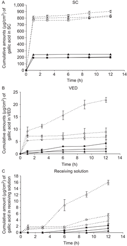
Table 3. The percentages of gallic acid amounts (%) in stratum corneum (SC), viable epidermis and dermis (VED), and receiving solution following transdermal absorption across excised rat skin from various gel formulations by Franz diffusion cells after 12 h.
Gel containing pure gallic acid loaded in elastic (GE) and nonelastic niosomes (GN) showed lower cumulative amounts at 12 h of gallic acid in SC and VED of 821.44 ± 42.70 and 837 ± 31.35; 8.84 ± 0.49 and 6.67 ± 1.63 µg/cm2, respectively, than the unloaded gallic acid in gel (GS) (898.98 ± 18.96 and 21.87 ± 1.00 µg/cm2 in SC and VED, respectively). However, gel containing the semipurified fraction loaded in elastic and nonelastic loaded niosomes (SE, SN) showed higher cumulative amounts at 12 h of gallic acid in VED and the receiving solution of 4.21 ± 1.11 and 1.99 ± 1.42; 2.22 ± 0.31 and 1.32 ± 0.52 µg/cm2, respectively, than the unloaded semipurified fraction in gel (SS) (1.12 ± 0.86 and 0.55 ± 0.07 µg/cm2 in VED and the receiving solution, respectively) (). Elastic niosomes loaded with pure gallic acid (GE) and the semipurified fraction (SE) showed higher percentages of gallic acid amount at 12 h of 0.35 and 0.70; 0.21 and 0.22% in VED and receiving solution, respectively, than their nonelastic niosomes loaded with pure gallic acid (GN) and the semipurified fraction (SN) (0.27 and 0.33; 0.14 and 0.22% in VED and receiving solution, respectively) (). Therefore, both elastic and nonelastic niosomes demonstrated the skin permeation retardation of the loaded gallic acid, whereas exhibited the skin permeation enhancement of gallic acid in the semipurified fraction. Hence, elastic and nonelastic niosomes appeared to reduce the systemic effect of the loaded pure gallic acid since about less than 3.0 and 4.5 times of the loaded in elastic and nonelastic niosomes than the unloaded pure gallic acid was found in the receiving solution. For gallic acid in the semipurified fraction loaded in elastic niosomes, the niosomes did not only enhance transdermal permeation of gallic acid to VED, but also reduced the remaining gallic acid on the upper layer of the skin (SC). This may be not only due to the small vesicular size and high deformability of elastic niosomes () that ethanol may interact with the polar head group region of the Tween 61 molecules and provide the reduction of vesicular melting point. This resulted in the soft and flexible characteristics of the elastic vesicles that enhanced the penetration of gallic acid in the semipurified fraction into the deeper layers of the skin. Ethanol may also fluidize the bilayer structure of SC (CitationKirjavainen et al., 1999) leading to the enhancement of gallic acid penetration.
Generally, the local effects of many compounds such as antioxidative effect and side effects, for example, skin irritation are related to the compounds concentrations in the skin. In this study, gallic acid contained in the gel was higher (2500 µg of gallic acid in gallic acid gel formulation) than gallic acid containing in the semipurified fraction (400 µg of gallic acid in semipurified gel formulation) of about six times. Also, the pure gallic acid gel demonstrated higher cumulative amounts in SC, VED, and receiving solution in vitro rat skin permeation by Franz diffusion cells of 3.67, 19.52, and 28.89 times than that in the semipurified fraction in gel. So, the local effects of this gallic acid gel are expected to have more than that in the semipurified fraction gel. In fact, our further study (CitationManosroi et al., 2010c) has found that gallic acid gel cause skin irritation on rabbits’ skin by the closed patch test while the semipurified fraction gel did not give this effect. In addition, when the pure gallic acid loaded in nonelastic or elastic niosomes, no rabbit skin irritation was observed. This may be due to the reduction of the direct contact between gallic acid and the skin when gallic acid was loaded in niosomes (CitationManosroi et al., 2010c).
Conclusion
This study has found the three phenolic compounds including gallic acid, 1,3,6-trigalloylglucopyranoside, and punicalagin in the selected semipurified fraction (F1, MeOH/H2O/AcOH = 2:8:0.1) isolated from the T. chebula gall cold aqueous crude extract. The elastic and nonelastic Tween 61 niosomes loaded with pure gallic acid or the semipurified fraction containing gallic acid exhibited the mixture of unilamellar and multilamellar structures with negative zeta potential values and in the particle size range of 200–400 nm. Both elastic and nonelastic niosomes loaded with gallic acid or the semipurified fraction containing gallic acid showed physical stability with no layer separation and chemical stability for 3 months of more than 70, 60, and 40%, while only more than 60, 40, and 30% of the remained gallic acid in the unloaded gallic acid at 4, 27, and 45°C, respectively. For rat skin transdermal absorption by Franz diffusion cells, these niosomes retarded the permeation of the loaded pure gallic acid indicating of no risk of systemic effect, which will be beneficial for topical application. However, they enhanced the permeation of the loaded gallic acid in the semipurified fraction. Although elastic niosomes did not significantly enhance physicochemical stability of gallic acid in comparing to nonelastic niosomes, they have demonstrated the potential of transdermal absorption through rat skin of gallic acid containing in the semipurified fraction from T. chebula galls. This result can be applied for the further development of semipurified fraction from T. chebula galls for antiaging by loading in elastic niosomes.
Declaration of interest
This work was supported by the Thailand Research Fund (TRF) under the RGJ-PhD program, Natural Products Research and Development Center (NPRDC), Institute for Science and Technology Research and Development (STRI), Nanoscience and Nanotechnology Research Center Project, Faculty of Science, Chiang Mai University, Thailand.
References
- Ali D, Hassan SG, Nasrolah H. (2008). Solubility of gallic acid in methanol, ethanol, water and ethyl acetate. J Chem Eng Data, 53, 776–778.
- Barry AL, Cullis PR. (1995). Direct NMR evidence for ethanol binding to the lipid-water interface of phospholipid bilayers. Biochemistry, 33, 8082–8088.
- Castanho MARB, Santos NC, Loura LMS (1997). Seperating the turbidity spectra of vesicles from the absorption spectra of membrane probes and other chromophores. Eur Biophys J, 26, 253–259.
- Cevc G. (1996). Transfersomes, liposomes and other lipid suspensions on the skin: Permeation enhancement, vesicle penetration, and transdermal drug delivery. Crit Rev Ther Drug Carrier Syst, 13, 257–388.
- Cheynier V, Rigaud J, Souquet JM, Duprat F, Moutounet M. (1990). Must browning in relation to the behavior of phenolic compounds during oxidation. Am J Enol Vitic, 41, 346–349.
- Cholbi MR, Paya M, Alcaraz MJ. (1991).Inhibitory effects of phenolic compounds on CCl4-induced microsomal lipid peroxidation. Experientia, 47, 195–199.
- Dayan N, Touitou E. (2000).Carriers for skin delivery of trihexyphenidyl HCl: ethosomes vs. liposomes. Biomaterials, 21, 1879–1885.
- Dubey V, Mishra D, Jain NK. (2007).Melatonin loaded ethanolic liposomes: Physicochemical characterization and enhanced transdermal delivery. Eur J Pharm Biopharm, 67, 398–405.
- Foldvari M, Kumar P, King M, Batta R, Michel D, Badea I, Wloch M. (2006).Gene delivery into human skin in vitro using biphasic lipid vesicles. Curr Drug Deliv, 3, 89–93.
- Fresno Contreras MJ, Jiménez Soriano MM, Ramírez Diéguez A. (2005).In vitro percutaneous absorption of all-trans retinoic acid applied in free form or encapsulated in stratum corneum lipid liposomes. Int J Pharm, 297, 134–145.
- Frosch PJ. (1992). Cutaneous irritation. In: Rycroft RJG, editor. Text Book of Contact Dermatitis. Berlin: Springer-Verlag, p. 54.
- Halliwell B. (2008).Are polyphenols antioxidants or pro-oxidants? What do we learn from cell culture and in vivo studies? Arch Biochem Biophys, 476, 107–112.
- Handjani-Vila RM, Ribier A, Rondot B, Vanlerberghie G. (1979).Dispersions of lamellar phases of non-ionic lipids in cosmetic products. Int J Cosmet Sci, 1, 303–314.
- Haslam E. (1996).Natural polyphenols (vegetable tannins) as drugs: Possible modes of action. J Nat Prod, 59, 205–215.
- Jain S, Jain P, Umamaheshwari RB, Jain NK. (2003). Transfersomes: A novel vesicular carrier for enhanced transdermal delivery: Development, characterization and performance evaluation. Drug Dev Ind Pharm, 29, 1013–1026.
- Jennifer SB, David AC, Grady G, Tonalee CK. (1988). Aqueous thermal degradation of gallic acid. Geochimica et Cosmochimica Acta, 52, 341–344.
- Kirjavainen M, Urtti A, Valjakka-Koskela R, Kiesvaara J, Mönkkönen J. (1999).Liposome-skin interactions and their effects on the skin permeation of drugs. Eur J Pharm Sci, 7, 279–286.
- Lasic D, Weiner N, Riaz M, Martin F. (1998). Liposomes. In: Lieberman A, Rieger M, Banker G, editors. Pharmaceutical Dosage Forms: Disperse Systems. New York: Marcel Dekker, p. 8–43.
- Lemmens RHMJ, Bunyapraphatsara N (2003). Plant Resources of South-East Asia: Medicinal and Poisonous Plants. Leiden: Backhuys Publishers, p. 434.
- Liazid A, Palma M, Brigui J, Barroso CG. (2007). Investigation on phenolic compounds stability during microwave-assisted extraction. J Chromatogr A, 1140, 29–34.
- Madsen HL, Bertelsen G. (1995). Spices as antioxidants. Trends Food Sci Tech, 6, 271–277.
- Manosroi A, Jantrawut P, Manosroi J. (2008).Anti-inflammatory activity of gel containing novel elastic niosomes entrapped with diclofenac diethylammonium. Int A Pharm, 360, 156–163.
- Manosroi A, Jantrawut P, Akihisa T, Manosroi W, Manosroi J. (2010).In vitro anti-aging activities of Terminalia chebula gall extract. Pharm Biol, 48, 469–481.
- Manosroi A, Jantrawut P, Akazawa HManosroi A, Jantrawut P, Akazawa H, Akihisa T, Manosroi J. (2010b). Biological activities of phenolic compounds isolated from galls of Terminalia chebula Retz. (Combretaceae). Nat Prod Res, 24: doi 10.1080/14786419.2010.488631.
- Manosroi A, Jantrawut P, Akihisa T, Manosroi J. (2010c). In vitro and in vivo skin anti-aging evaluation of gel containing niosomes loaded with phenolic compounds from Terminalia chebula galls. Skin Res Technol. In Press.
- McLaughlin SG, Szabo G, Eisenman G. (1971).Divalent ions and the surface potential of charged phospholipid membranes. J Gen Physiol, 58, 667–687.
- Miguel ARBC, Nuno C, Santos Luís MSL. (1997). Separating the turbidity spectra of vesicles from the absorption spectra of membrane probes and other chromophores. Eur Biophys J, 26, 253–259.
- Nakatani N. (1992). Natural antioxidants from spices. In: Huang MT, Ho CT, Lee CY, editors. Phenolic Compounds in Food and Their Effects on Health II: Antioxidants and Cancer Prevention. Washington, DC: American Chemical Society p. 72–86.
- Padula C, Fulgoni A, Santi P. (2010).In vivo stratum corneum distribution of lidocaine, assessed by tape stripping, from a new bioadhesive film. Skin Res Technol, 16, 125–130.
- Plessis J, Egbaria K, Weiner N (1992). Influence of formulation factors on the deposition of liposomal components into the different strata of the skin. J Soc Cosmet Chem, 43, 93–100.
- Rice-Evans CA, Miller NJ, Paganga G. (1996).Structure-antioxidant activity relationships of flavonoids and phenolic acids. Free Radic Biol Med, 20, 933–956.
- Roberts MS, Anderson RA, Swarbrick J. (1977). Permeability of human epidermis to phenolic compounds. J Pharm Pharmacol, 29, 677–683.
- The Pharmacopoeia Commission of PRC. (1997). Pharmacopoeia of the People’s Republic of China (I). Beijing: Chemical Industry Press, p. 61.
- Touitou E, Dayan N, Bergelson L, Godin B, Eliaz M. (2000). Ethosomes - novel vesicular carriers for enhanced delivery: characterization and skin penetration properties. J Control Release, 65, 403–418.
- Uchegbu IF, Vyas SP. (1998). Non-ionic surfactant based vesicles (niosomes) in drug delivery. Int J Pharm, 172, 33–70.
- van der Molen RG, Spies F, van‘t Noordende JM, Boelsma E, Mommaas AM, Koerten HK. (1997). Tape stripping of human stratum corneum yields cell layers that originate from various depths because of furrows in the skin. Arch Dermatol Res, 289, 514–518.
- Yeh CT, Yen GC. (2005). Effect of vegetables on human phenolsulfotransferases in relation to their antioxidant activity and total phenolics. Free Radic Res, 39, 893–904.
- Zhu PY. (1998). Chinese Materia Medica. Amsterdam, Netherlands: Harwood Academic Publishers, pp. 663–666.