Abstract
Context: Vernonia cinerea (L.) Less [Compositae (Asteraceae)] is used traditionally for several medical purposes such as inflammation, pain, fever, and cancer.
Objectives: The present study identified the bioactive constituents in the methanol extract of Vernonia cinerea leaf and evaluated its antioxidant activity and acute toxicity.
Methods: The identification of phytochemicals was accomplished by GC-MS and the major antioxidant phenolic compounds in the extract were quantified by HPTLC analysis. To quantify the essential elements, atomic absorption spectrophotometeric analysis was carried out. Total phenol and flavonoid content was measured by Folin-Ciocalteau reagent and 2% aluminium chloride, respectively.
Results: GC-MS analysis identified the presence of 27 phytoconstituents. The predominant phenolic compound in the extract as quantified by HPTLC was gallic acid (1.92 mg/g) followed by rutin (0.705 mg/g), quercetin (0.173 mg/g), caffeic acid (0.082 mg/g) and ferulic acid (0.033 mg/g). The following elements were quantified: Fe (0.050 ppm), Mn (0.022 ppm), Co (0.0180 ppm), Pb (0.029 ppm), Hg (3.885 ppm) and Se (4.5240 ppm). The antioxidant activity of the extract increased with increasing concentration and the correlation (r2) for all in vitro assays were satisfactory.
Conclusions: V. cinerea extract has significant (p < 0.05) antiradical activity. Hence, V. cinerea may have potential medicinal value and can be used in the formulation of pharmacological products for degenerative diseases.
Introduction
Oxidative stress has been implicated in the pathology of many diseases such as inflammatory conditions, cancer, diabetes, and ageing (CitationMarx, 1987). Free radicals induced by peroxidation, have gained much importance because of their involvement in several pathological conditions such as atherosclerosis, ischemia, liver disorder, metal toxicity and pesticide toxicity (CitationPanday et al., 1994). The use of plant and natural products is beneficial in protecting against the oxidative stress induced damage, as they are less toxic, or practically nontoxic, relative to synthetic compounds at their optimum protective doses (CitationJagetia & Baliga, 2002). Synthetic antioxidants like butylated hydroxyl anisole (BHA) and butylated hydroxyl toluene (BHT) have harmful effects on liver (CitationHettiarachchy et al., 2010). Therefore, screening of natural products presents a major avenue for the discovery of new pharmacological products.
Vernonia cinerea (L.) Less [Compositae (Asteraceae)] (CitationHenry et al., 1987) is an herbaceous plant commonly called as Purple Fleabane and as Sahadevi in Sanskrit. The stem is slender, grooved and ribbed. The leaves are variable in shape, broadly elliptic or lanceolate, membranous or rather coriaceous. The flowers are pinkish and purple, in minute heads in rounded or flat-topped corymbs. It has many therapeutic uses in different traditional medicine of the world. Different parts of the plant are of different therapeutic values. The juice of the plant is given to children with urinary incontinence. The leaves are eaten as a potherb. A decoction of it is also given in diarrhea, stomachache, cough and bronchitis. No adverse effect was reported on usage of this plant as a drug (CitationLatha et al., 2010). The whole plant has several pharmacological properties; it is used for malarial fever, worms, pain, infections, diuresis, cancer, abortion, and various gastro-intestinal disorders (CitationBero et al., 2009; CitationPratheeshkumar & Kuttan, 2011a; CitationJain & Puri, 1984). Phytochemical analysis of V. cinerea showed the presence of steroids, triterpenoids, sesquiterpenes, flavonoids, and tannins (CitationHall et al., 1979; CitationJakupovic et al., 1986; CitationMisra et al., 1993). The structure of triterpene 24-hydroxytaraxer-14-ene was identified and elucidated. The other triterpenes namely, β-amyrin acetate, β-amyrin benzoate, lupeol and its acetate, β-sitosterol, sigmasterol and α-spinasterol were also isolated. Vernolides are the major sesquiterpenoids and are reported as the active principle in this plant (CitationKuo et al., 2003). Sesquiterpenes are a class of terpenes that consist of three isoprene units and have the molecular formula C15H24, which are found naturally in plants and invertebrates. Biochemical modifications such as oxidation or rearrangement produce the related sesquiterpenoids. Studies showed that V. cinerea has good antioxidant and anti-inflammatory properties and the administration of its extract can inhibit the elevated proinflammatory cytokine level in carrageenan-induced paw edema mice model (PratheeshCitationKumar & Kuttan, 2009). It also has protective role against radiation-induced oxidative stress, DNA damage and inflammatory conditions (CitationPratheeshkumar & Kuttan, 2011b). Hence identification, quantification and isolation of phytoconstituents responsible for the several pharmacological properties of the herb are necessary.
The present study was designed to identify and quantify the bioactive constituents in the leaf extract by GC-MS and HPTLC analyses respectively. Atomic absorption spectrophotometric analysis was done to quantify the essential elements in the extract. The free radical scavenging and antioxidant properties of V. cinerea leaf extract were carried by several standard in vitro methods. The leaf extract was prepared with 90% methanol and subjected to several assays like DPPH radical scavenging, ABTS radical cation-scavenging, hydroxyl radical scavenging, nitric oxide radical scavenging, PMS-NADH system superoxide-radical scavenging, in vitro anti-lipid peroxidation, oxidative hemolysis inhibition activity. The antioxidant property of the extract was evaluated with a range of concentration (10–500 µg/ml). The linear regression equation analysis (r2) was calculated to study the correlation of the antioxidant property for each assay. The half maximal inhibitory concentration (IC50) values were also calculated for each assay. Finally we tested the in vivo acute toxicity in rats.
Materials and methods
Chemicals
Thiobarbituric acid (TBA), 1,1-diphenyl-2-picrylhydrazyl (DPPH), 2,2'-azino-bis(3-ethylbenzothiozoline-6-sulphonic acid) diamonium salt (ABTS), nitroblue tetrazolium (NBT), phenazine methosulphate (PMS), nicotinamide adenine dinucleotide-reduced (NADH), α-tocopherol, 2,2-azobis (2-amidinopropane) dihydrochloride (APPH) were purchased from Sigma Aldrich. All other chemicals and reagents were of analytical grade and purchased from Merck and Himedia Chemicals.
Collection and preparation of plant extract
Plant material (wild Vernonia cinerea leaves) was collected between October and November 2008 from around the villages of Panruti, Postal code-607805, Tamil Nadu, India and authenticated by Prof. Dr. P. Jayaraman, Director, PARC, National Institute of Herbal Science, Chennai, India (Specimen number - PARC/2008/562). About 500 g of the shade-dried, powdered leaves of V. cinerea were exhaustively extracted with 90% methanol using Soxhlet apparatus. The residue was filtered and concentrated under reduced pressure and the extract was used for analyzing the phytochemicals and antioxidant activity.
GC-MS analysis
The V. cinerea extract was analyzed by GC-MS (GC Clarus 500 Perkin Elmer) equipped with Elite-1 column (100% dimethyl poly siloxane), 30 × 0.25 mm × 1 μm (df) and mass detector: Turbomass gold Perkin Elmer. The temperature program was set as follows, 110°C for 2 min hold rose at 10°C/min up to 200°C and rose at 5°C/min up to 280°C with 9 min hold. Injector temperature was set at 250°C. The mass range was scanned from 45 to 450 (m/z). The control of the GC-MS system and data peak processing was controlled by means of Turbomass software version 5.2. Compound identification was verified based on the relative retention time and mass fragmentation pattern spectra with those of standards and the NIST Version (2005) LIB database of the GC-MS system.
HPTLC fingerprint analysis
For HPTLC fingerprint profile, stock solution (10 mg/ml) of the extract was prepared in methanol. Sample (10 µl) was spotted on a pre-coated Silica gel 60 F 254 HPTLC plate (Merck) and the plate was developed in Toluene: Ethyl acetate: Acetic acid (8:1:1) solvent system (CitationRavishankara et al., 2002). The plate was scanned using CAMAG TLC scanner 3 at 280 nm. The Rf values, spectra, λmax and peak areas of the resolved bands were recorded. Relative percentage area of each band was calculated from the peak areas. The presence of flavonoids was confirmed by spraying the plates with 5% aluminum chloride in ethanol.
Elemental analysis
About 250 mg of the V. cinerea leaf sample was weighed and 5 to 10 ml of concentrated H2SO4 was added to it. The acid digestion was further initiated by heating up to 440°C using a digesdahl apparatus. The samples were made free from organic matter and the resulting solution was made colorless by adding 5 to 10 ml of H2O2. The digested material was made up to 100 ml. The elemental analysis of the digested samples was determined by an Atomic Absorption Spectrophotometer (AAS), Perkin Elmer. Hg and Se were estimated using a hydride generator attached to AAS. The calibration was performed using a blank solution to zero the instrument. Results were calibrated using standard linear calibrations and the mean of triplicate values were calculated.
Evaluation of radical scavenging and antioxidant activity
The amount of total phenolics and total flavonoids in the extract was measured spectrophotometrically using Folin-Ciocalteu’s reagent and 2% aluminum chloride respectively (CitationDjeridane et al., 2006). The antioxidant activity of methanol V. cinerea extract was assessed by several in vitro assays based on their ability to scavenge or donate hydrogen/electrons against the stable free radicals. Demonstration of antiradical activity was performed with a wide range of concentration (10–500 µg/ml). The antiradical activity of extract was done by DPPH radical scavenging assay (CitationBraca et al., 2001). ABTS radical cation-scavenging activity of the extract was done by an improved method (CitationBaltrusaityte et al., 2007). ABTS.+ radical cation was produced by mixing 7 mM 2,2'- azino-bis (3-ethylbenzothiozoline-6-sulphonic acid) diamonium salt (ABTS) and 2.45 mM potassium persulfate (K2S2O8), incubated at room temperature in dark. The hydroxyl radical scavenging activity of the extract was measured with Fenton reaction (CitationWenli et al., 2004). Nitric oxide radical scavenging activity was determined by generating nitric oxide from sodium nitroprusside and measured by Griess’ reaction (CitationGreen et al., 1982). Superoxide-radical scavenging assay was done using PMS-NADH system (CitationAo et al., 2008). The effect of V. cinerea extract on lipid peroxidation in liver homogenate was determined by the method of CitationOhkawa et al. (1979) with minor modifications (CitationKumar et al., 2000). Freshly excised goat liver was processed to get 10% homogenate in cold phosphate buffered saline (pH 7.4) using a glass Teflon homogenizer and filtered to get a clear homogenate. The degree of lipid peroxidation was assayed by estimating the thiobarbituric acid reactive substances (TBARS). Oxidative hemolysis inhibition assay of the extract was done by using APPH, a peroxy radical initiator which causes oxidation of lipids and proteins in cell membrane resulting in lysis of RBC (CitationHe et al., 2000). All the experiments were done in triplicates and measured using Shimadzu UV-Vis spectrophotometer (UV-1700). The results were expressed as % inhibition which is calculated by the following equation:
ODcontrol is the absorbance without the extract; ODsample is the absorbance with the extract. Ascorbic acid and α-tocopherol were used as standard antioxidant compounds for comparing the antioxidant activity of the V. cinerea extract.
Animals
Twelve healthy young adult Wistar albino rats of weight variation not exceeding ± 20% of the mean weight (130–160 g) were selected for the present study. All the animals were acclimatized to laboratory condition for a week before commencement of experiment. The animals were housed in poly carbonate cages (3 in each cage) at controlled room temperature of 28–30°C and a relative humidity between 30–70% and a constant light-dark schedule (12 h light/dark cycle). The rats had free access to water and dry rat pellets. The experimental protocol was approved by institutional animal ethics committee (CPCSEA/CRIS/PHARMA/54/2008).
Animals were divided in to two groups of six animals each (comprising 3 males and 3 females). Group I animals served as control and received only vehicle. Group II animals treated with plant extract (2000 mg/kg p.o single dose of V. cinerea extract). The dose was determined based on the previous studies where whole plant extract was used to study the anti-inflamatory effect (CitationMazumder et al., 2003). The animals were kept under observation for 24 h for any gross behavioral changes and mortality. The animals were also observed for pre-terminal morbidity, mortality and toxic reactions, if any for 14 days post exposure to the plant extract. Animals were sacrificed and blood and tissue samples were collected on 15th day. Biochemical and histological parameters were measured in all groups. Liver and kidneys were removed from the sacrificed animals and preserved in 10% buffered formalin for histological examination.
Biochemical assay
Glucose, urea, uric acid, creatinine, serum alanine transaminase, serum aspartate transaminase, alkaline phosphatase, lactate dehydrogenase, and α-glutamyl transaminase were analyzed using in BAYER–RA-50 semi auto analyzer
Statistical analysis
Data were presented as mean ± SD. Results were evaluated by one-way ANOVA followed by Duncan multiple range tests. Difference was considered significant when p value ≤ 0.05 for test sample, ≤0.01 for standard. Statistical Software Graph pad prism version 5.01 was used for analysis.
Results and discussion
Identification and quantification of bioactive constituents
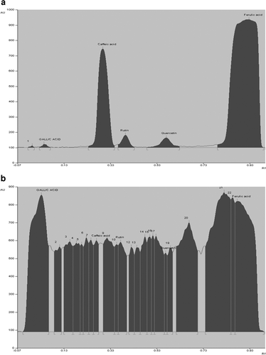
Medicinal plants have a long history of use in the treatment of several health disorders including cancer and degenerative diseases (CitationHartwell, 1982). Herbal preparation in particular has potential uses in the field of pharmacology and medicine (CitationHsu, 1967). The studies on biological and chemical constituents of the genus Vernonia had resulted in the isolation of a number of highly oxygenated sesquiterpene lactones with a 7,11-double bond and an oxygen function at C-13, some of which exhibited significant cytotoxic activities (CitationJakupovic et al., 1986). Several sesquiterpenoids are being isolated and studied for their biological activity like antiplasmodial, immunomodulatory, cytotoxicity, chemoprotective and antioxidant effects (PratheeshCitationKumar & Kuttan, 2009, 2010, 2011c). Some of the previously elucidated sesquiterpenoids by spectral analysis are vernocinolide-A, vernolide-A, vernolide-B. Biological evaluation showed that vernolide-A has potent cytotoxicity against human KB, DLD-1, NCI-661, and HeLa tumor cell lines. Vernolide B had marginal cytotoxicity against KB, NCI-661, and HeLa tumor cell lines (CitationKuo et al., 2003). In the present study, GC-MS analysis showed that V. cinerea leaf extract contained a variety of bioactive phytocompounds (). By comparing the MS spectral data with that of standards and MS library, 27 phytochemicals were identified (). Among the identified compounds, there were five sesquiterpenes namely, caryophyllene, α-guaiene, α-humulene, valencene and α-bulnesene. Sesquiterpenes are the major compounds identified in the leaf extract which is in agreement with previous report (CitationKuo et al., 2003). Phytol a diterpene and squalene a triterpene were also identified. Sesquiterpenes, diterpene contain the isoprene units linked in a head to tail fashion whereas triterpenes and carotenoids (tetraterpenes) contain two C15 and C20 units, respectively, linked head to head. The other major group of compounds identified in the leaf extract belongs to fatty acid and fatty acid ester (). HPTLC analysis quantified the major phenolic compounds in the extract and they were found to be gallic acid (1.92 mg/g) followed by rutin (0.705 mg/g), quercetin (0.173 mg/g), caffeic acid (0.082 mg/g) and ferulic acid (0.033 mg/g). Gallic acid was predominantly found in the extract. The chromatographic profile of the methanol extract of V. cinerea leaves is shown in and .
Table 1. Phytocomponents identified in the V. cinerea leaf extract by GC-MS analysis.
Figure 1. GC-MS analysis of the phytoconstituents in the methanol extract of V. cinerea leaf extract showing the retention time (min) of the compounds in X axis and percentage (%) of peak area in the Y axis.
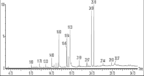
Figure 2. HPTLC analysis of the phytochemicals present in the V. cinera leaf extract showing Rf values. Detection was at 280 nm. ) Chromatogram of standard phenolic compounds. ) Chromatogram of V. cinera leaf extract. The numbers indicate the unidentified compounds.
Elemental analysis
Plants are the excellent source of minerals and many of these minerals have been classed as essential elements, necessary for utilization by the body to ensure good health. Macro minerals are needed by the body in relatively large amounts (e.g. sodium, potassium, chlorine, calcium, phosphorus, magnesium). Micro/trace minerals are needed in small amounts (e.g. selenium, iron, zinc, copper, manganese, molybdenum, cobalt). In this study essential trace element quantified by AAS is shown in . These trace elements (Fe, Mn, Co and Se) are essential to human health through organic source, because they have essentially been processed. Plants take up minerals from the ground, digest them, making them ionic so that when consumed by humans, assimilation into the body occurs much more easily and toxicity by accumulation does not occur. However, micro minerals from inorganic sources, such as heavy metals, cannot be used by the body as they tend to build up in the tissues. Manganese is component of the following important enzymes, Mn-superoxide dismutase, important in preventing damage to tissues caused by lipid oxidation, pyruvate carboxylase, and arginase. Selenium is required as an essential cofactor of antioxidant enzymes such as glutathione peroxidase. Dietary Se supplements in renal transplant recipients indicated an increase in activity of glutathione peroxidase and reductase, relieving oxidative stress, decreasing plasma and LDL lipid peroxidation, and producing an antiatherogenic effect, suggesting a protective role of Se against hyperlipidaemia, hypertension and other cardio vascular disease (CitationHussein et al., 1997). It also has chemopreventive effects on N-nitrosodiethylamine induced hepatoma in rats (CitationThirunavukkarasu et al., 2001). Cancer, cardiovascular disease, diabetes and arthritis have been extensively shown to be associated with Se deficiency in a well controlled intervention trial (CitationFleet & Mayer, 1997). Cobalt is part of vitamin B12 and iron is required for many proteins and enzymes, notably hemoglobin to prevent anemia. Hence the presence of trace elements in the leaf extract can be used for the production of pharmacological products against degenerative diseases and cancer.
Table 2. Elemental present in V. cinerea leaf extract.
Evaluation of radical scavenging and antioxidant activity
Phytoconstituents were reported to have antioxidant property in suppressing the initiation or propagation of chain reactions. Phenolic compounds have one or more aromatic ring bearing hydroxyl groups that act as reducing agents. Total phenol content of V. cinerea leaf extract was determined in comparison with standard gallic acid and found to be 167.48 ± 0.57 gallic acid equivalents mg/g dry sample. Flavonoids are one of the most diverse and wide spread groups of natural compounds and are probably the important natural phenols. These compounds possess a broad spectrum of chemical and biological activities including radical scavenging properties (CitationKhatiwora et al., 2010). Total flavonoid content of V. cinerea extract was determined in comparison with standard quercetin and found to be 5.72 ± 0.04 quercetin equivalents mg/g dry sample.
The free radical scavenging activity of the methanol extract and the possible mechanism involved based on the response to five different in vitro methods covering the major radicals, viz., DPPH, ABTS, hydroxyl, nitric oxide, and superoxide was demonstrated. The antioxidant property of extract was further supported by inhibiting the in vitro lipid peroxidation and oxidative hemolysis. V. cinerea leaf extract exhibited a strong antioxidant activity against the free radicals in a dose-dependent manner as shown in . It showed concentration (10–500 µg/ml) dependent antioxidant activity with maximum scavenging activity observed at 500 µg/ml. The radical scavenging activity was compared with standard antioxidants ascorbic acid and vitamin E (250 µg/ml). The extract showed 87% scavenging activity compared to ascorbic acid, with r2 = 0.6825 and IC50 = 56 ± 6.92 by DPPH assay. The ABTS.+ radical scavenging method is a decolorization assay used for the screening of antioxidant activity, applicable for both lipophilic and hydrophilic antioxidants (CitationLong et al., 2000). The results imply that ABTS.+ radicals were scavenged by extract with 92% inhibition compared to ascorbic acid, r2 = 0.8278 and IC50 = 98.8 ± 32.33. Hydroxyl radical is the most reactive among reactive oxygen species (ROS) and it bears the shortest half-life compared with other ROS. The extract exhibited 68% antiradical activity compared to ascorbic acid, with r2 = 0.8342 and IC50 = 220 ± 27.49. Nitric oxide is a very unstable species in the presence of oxygen. It reacts with oxygen to produce its stable product nitrate and nitrite through intermediates NO2, N2O4 and N3O3. It is estimated using Griess reagent. In the present study, extract showed better scavenging activity in competing with oxygen to react with nitric oxide and thus the inhibition of generation of anions. It showed excellent antioxidant activity, 95% compared to ascorbic acid with r2 = 0.7638 and IC50 = 93.3 ± 13.1. Superoxide anion is the first reduction product of oxygen and it’s very harmful to cellular components as a precursor of more reactive oxygen species (CitationHalliwell & Gutteridge, 1984). The scavenging activity towards the superoxide radical is measured in terms of inhibition of generation of superoxide anion. The results show that extract has considerable superoxide scavenging activity with increasing percentage inhibition. It showed 89% inhibition compared to ascorbic acid, with r2 = 0.7070 and IC50 = 81.3 ± 3.05.
Table 3. Antioxidant activity of V. cinerea leaf extract by several in vitro assays
Lipid peroxidation is the oxidative degradation of polyunsaturated fatty acids and involves the formation of lipid radicals leading to membrane damage. Free radicals induce lipid peroxidation in polyunsaturated lipid rich areas like brain and liver (CitationCoyle & Puttfarcken, 1993). Initiation of lipid peroxidation by ferrous sulphate takes place through hydroxyl radical by Fenton’s reaction (CitationBraughler et al., 1986). In this study, the inhibitory effect of extract and vitamin E on TBARS production, in goat liver homogenate induced by using FeSO4 is shown in . The extract (500 μg/ml) showed 47% inhibition, compared to vitamin E with r2 = 0.9573 and IC50 = 558 ± 32.5. The concentration of extract to bring about inhibition of lipid peroxidation was higher compared to other methods ().
The oxidation of erythrocyte membrane serves as a model for the oxidative damage of bio-membranes (CitationVissers et al., 1994). APPH is a peroxy radical initiator that generates free radicals by its thermal decomposition and attack erythrocytes to induce the chain oxidation of lipid and protein, disrupting the membrane organization and eventually leading to hemolysis (Mike et al., 1987). The present study was further supported by the finding that V. cinerea methanol fraction strongly inhibited erythrocyte hemolysis induced by AAPH. The extract showed 89% inhibition compared to ascorbic acid with r2 = 0.7598 and IC50 = 46 ± 8.02.
The methanol extract of V. cinerea leaf, which exhibited antioxidant activity, was tested for acute toxicity. The acute toxicity was performed as per the norms of Organization for European Economic Co-operation (Rule 421). In an acute toxicity study using rats, the lethal dose (LD50) of the extract was greater than 2000 mg/kg, and we found no pathological changes (). We did not found any significant changes in serum and tissue biochemical changes (data not shown). In conclusion, the methanol extract of V. cinerea did not produce toxic effects in rats.
Figure 3. Histopathological observation of liver and kidney tissues: (A & B) Liver of control (A) and extract treated (B) animals, showing normal architecture and orderly alignment of hepatocytes. (C & D) Kidney of control (C) and extract treated (D) animals showing normal pattern. [Animal treatment details: Refer materials and methods].
![Figure 3. Histopathological observation of liver and kidney tissues: (A & B) Liver of control (A) and extract treated (B) animals, showing normal architecture and orderly alignment of hepatocytes. (C & D) Kidney of control (C) and extract treated (D) animals showing normal pattern. [Animal treatment details: Refer materials and methods].](/cms/asset/8d8222f4-66a4-48b2-9f9e-9dc4cc72c8c3/iphb_a_604334_f0003_b.gif)
Conclusion
In conclusion, from the above investigation, the methanol extract of V. cinerea leaves was found to exhibit a good antioxidant property. The presence of several bioactive compounds like phenols and sesquiterpenoids in the extract may be responsible for the antioxidant property. The presence of essential trace elements like Fe, Mn, Co and Se suggests that the extract has an excellent nutritional and medicinal value. Hence V. cinerea leaf extract can be used for the treatment of several health disorders.
Declaration of interest
The authors report no declaration of interest.
References
- Ao C, Li A, Elzaawely AA, Xuan TD, Tawata S. (2008). Evaluation of antioxidant and antibacterial activities of Ficus microcarpa L. fil. extract. Food Control, 19, 940–948.
- Baltrusaityte V, Venskutonis PR, Ceksteryte V. (2007). Radical scavenging activity of different floral origin honey and beebread phenolic extracts. Food Chem, 101, 502–514.
- Bero J, Frédérich M, Quetin-Leclercq J. (2009). Antimalarial compounds isolated from plants used in traditional medicine. j Pharm Pharmacol, 61, 1401–1433.
- Braca A, De Tommasi N, Di Bari L, Pizza C, Politi M, Morelli I. (2001). Antioxidant principles from Bauhinia tarapotensis. J Nat Prod, 64, 892–895.
- Braughler JM, Duncan LA, Chase RL. (1986). The involvement of iron in lipid peroxidation. Importance of ferric to ferrous ratios in initiation. J Biol Chem, 261, 10282–10289.
- Coyle JT, Puttfarcken P. (1993). Oxidative stress, glutamate, and neurodegenerative disorders. Science, 262, 689–695.
- Djeridane A, Yousfi M, Nadjemi B, Boutassouna D, Stocker P, Vidal N. (2006). Antioxidant activity of some Algerian medicinal plants extracts containing phenolic compounds. Food Chem, 97, 654–660.
- Fleet JC. (1997). Dietary selenium repletion may reduce cancer incidence in people at high risk who live in areas with low soil selenium. Nutr Rev, 55, 277–279.
- Green LC, Wagner DA, Glogowski J. (1982). Analysis of nitrate, nitrite, and [15N] nitrate in biological fluids. Anal Biochem, 126, 131–138.
- Hall IH, Lee KH, Starnes CO, Sumida Y, Wu RY, Waddell TG, Cochran JW, Gerhart KG. (1979). Anti-inflammatory activity of sesquiterpene lactones and related compounds. j Pharm Sci, 68, 537–542.
- Halliwell B, Gutteridge JM. (1984). Lipid peroxidation, oxygen radicals, cell damage, and antioxidant therapy. Lancet, 1, 1396–1397.
- Hartwell JL. (1982). Plants Used Against Cancer: A Survey, 2nd edn. Lawrence MA: Quarterman.
- He ZD, Lau KM, Xu HX, Li PC, Pui-Hay But P. (2000). Antioxidant activity of phenylethanoid glycosides from Brandisia hancei. J Ethnopharmacol, 71, 483–486.
- Henry AN, Kumari GR, Chitra V. (1987). Flora of Tamil Nadu, Series–I, Vol. II. Botanical survey of India, southern – Circle, Coimbatore, India.
- Hettiarachchy NS, Glenn KC, Gnanasambandam R, Jonhnson MG. (2010). Natural antioxidant extracts from Fenugreek (Trigonella foenumgraecum) for ground beef patties. J Food Sci, 61, 516–519.
- Hsu YT. (1967). Study on the Chinese drugs used as a cancer remedy. J Southeast Asian Res, 3, 63–66.
- Hussein O, Rosenblat M, Refael G, Aviram M. (1997). Dietary selenium increases cellular glutathione peroxidase activity and reduces the enhanced susceptibility to lipid peroxidation of plasma and low-density lipoprotein in kidney transplant recipients. Transplantation, 63, 679–685.
- Jagetia GC, Baliga MS. (2002). Influence of the leaf extract of Mentha arvensis Linn. (mint) on the survival of mice exposed to different doses of gamma radiation. Strahlenther Onkol, 178, 91–98.
- Jain SP, Puri HS. (1984). Ethnomedicinal plants of Jaunsar-Bawar hills, Uttar Pradesh, India. J Ethnopharmacol, 12, 213–222.
- Jakupovic J, Banerjee S, CastroV,Bohlmann F, Schuster A, Msonthi JD. (1986). Poskeanolide, a seco-germacranolide and other sesquiterpene lactones from Vernonia species. Phytochemistry, 25, 1359–1364.
- Khatiwora E, Adsul VB, Kulkarni MM, Deshpande NR, Kashalkar RV. (2010). Spectroscopic determination of total phenol and flavonoid contents of Ipomoea carnea. Int J ChemTech Res, 2, 1698–1701.
- Kumar VP, Shashidhara S, Kumar MM, Sridhara BY. (2000). Effect of Luffa echinata on lipid peroxidation and free radical scavenging activity. J Pharm Pharmacol, 52, 891–894.
- Kuo YH, Kuo YJ, Yu AS, Wu MD, Ong CW, Yang Kuo LM, Huang JT, Chen CF, Li SY. (2003). Two novel sesquiterpene lactones, cytotoxic vernolide-A and -B, from Vernonia cinerea. Chem Pharm Bull, 51, 425–426.
- Latha LY, Darah I, Jain K, Sasidharan S. (2010). Toxicity study of Vernonia cinerea. Pharm Biol, 48, 101–104.
- Long LH, Kwee DC, Halliwell B. (2000). The antioxidant activities of seasonings used in Asian cooking. Powerful antioxidant activity of dark soy sauce revealed using the ABTS assay. Free Radic Res, 32, 181–186.
- Marx JL. (1987). Oxygen free radicals linked to many diseases. Science, 235, 529–531.
- Miki M, Tamai H, Mino M, Yamamoto Y, Niki E. (1987). Free-radical chain oxidation of rat red blood cells by molecular oxygen and its inhibition by alpha-tocopherol. Arch Biochem Biophys, 258, 373–380.
- Mazumder UK, Gupta M, Manikandan L, Bhattacharya S, Haldar PK, Roy S. (2003). Evaluation of anti-inflammatory activity of Vernonia cinerea Less. extract in rats. Phytomedicine, 10, 185–188.
- Misra TN, Singh RS, Srivastava R, Pandey HS, Prasad C, Singh S. (1993). A new triterpenoidal from Vernonia cinerea. Planta Med, 59, 458–460.
- Ohkawa H, Ohishi N, Yagi K. (1979). Assay for lipid peroxides in animal tissues by thiobarbituric acid reaction. Anal Biochem, 95, 351–358.
- Pandey S, Sharma M, Chaturvedi P, Tripathi YB. (1994). Protective effect of Rubia cordifolia on lipid peroxide formation in isolated rat liver homogenate. Indian J Exp Biol, 32, 180–183.
- Kumar PP, Kuttan G. (2009). Vernonia cinerea L. scavenges free radicals and regulates nitric oxide and proinflammatory cytokines profile in carrageenan induced paw edema model. Immunopharmacol Immunotoxicol, 31, 94–102.
- Pratheeshkumar P, Kuttan G. (2010). Ameliorative action of Vernonia cinerea L. on cyclophosphamide-induced immunosuppression and oxidative stress in mice. Inflammopharmacology, 18, 197–207.
- Pratheeshkumar P, Kuttan G. (2011). Modulation of immune response by Vernonia cinerea L. inhibits the proinflammatory cytokine profile, iNOS, and COX-2 expression in LPS-stimulated macrophages. Immunopharmacol Immunotoxicol, 33, 73–83.
- Pratheeshkumar P, Kuttan G. (2011). Protective role of Vernonia cinerea L. against gamma radiation–induced immunosupression and oxidative stress in mice. Hum Exp Toxicol, 30, 1022–1038.
- Pratheeshkumar P, Kuttan G. (2011). Vernonia cinerea Less. inhibits tumor cell invasion and pulmonary metastasis in C57BL/6 mice. Integr Cancer Ther, 10, 178–191.
- Ravishankara MN, Shrivastava N, Padh H, Rajani M. (2002). Evaluation of antioxidant properties of root bark of Hemidesmus indicus R. Br. (Anantmul). Phytomedicine, 9, 153–160.
- Thirunavukkarasu C, Singh JP, Selvendiran K, Sakthisekaran D. (2001). Chemopreventive efficacy of selenium against N-nitrosodiethylamine-induced hepatoma in albino rats. Cell Biochem Funct, 19, 265–271.
- Vissers MC, Stern A, Kuypers F, van den Berg J, Winterbourn CC. (1994). Membrane changes associated with lysis of red blood cells by hypochlorous acid. Free Radic Biol Med, 16, 703–712.
- Wenli Y, Yaping Z, Bo S. (2004). The radical scavenging activities of radix puerariae isoflavonoids: A chemiluminescence study. Food Chem, 86, 525–529.