
Abstract
Context: Eucalyptus camaldulensis Dehnh. (Myrtaceae) is a tall evergreen tree found commonly in Bangladesh. Its use in traditional folk medicine for the treatment of various health complications are well known.
Objective: To explore the in vivo antitumor effect of Eucalyptus camaldulensis stem bark methanol extract (ME) against Ehrlich’s ascites carcinoma (EAC) in Swiss albino mice.
Materials and methods: The antitumor activity of ME was studied by determining viable tumor cell count, recording tumor weight and survival time, observing morphological changes and nuclear damage of EAC cells, and estimating hematological as well as biochemical parameters of experimental mice (25, 50 and 100 mg/kg/day for 5 d, i.p.).
Results: ME showed 96% (p < 0.001) cell growth inhibition and reduced tumor burden significantly (81.4%; p < 0.01) when compared with control mice. It also increased the lifespan of EAC-bearing mice significantly (71.36%; p < 0.01). It also restored the altered hematological and biochemical parameters towards normal level. The high LD50 value (1120 mg/kg) of ME indicated its low host toxic effects. ME-treated EAC cells showed membrane blebbing, chromatin condensation, nuclear fragmentation (apoptotic features) in Hoechst 33342 staining under fluorescence microscope. The DNA profile in agarose gel (1.5%) electrophoresis also confirmed that ME caused EAC cell death by apoptosis.
Discussion and conclusion: Results showed that ME exhibits strong anticancer activity through apoptosis and stimulation of host immunity. Thus, E. camaldulensis may be considered as a promising resource in cancer chemotherapy.
Introduction
Cancer continues to represent the largest cause of mortality in the world and claims over 6 million lives every year (Abdullaev et al., Citation2000). An extremely promising strategy for cancer prevention today is chemoprevention, which is defined as the use of synthetic or natural agents (alone or combination) to block the development of cancer in humans. Compounds of natural origin such as bleomycin, paclitaxin, vincristine, camptothecin, and many more, are now currently used in clinical settings for treatment of various cancers (Cassady & Douros, Citation1980; Kinghorn & Balandrin, Citation1993). In the developing countries, synthetic drugs are not only expensive and inadequate for the treatment of diseases but also often have adulterations and side effects (Shariff, Citation2001). There is a growing interest in the pharmacological evaluation of various plants used in traditional systems of medicine.
The plant used in the present study is Eucalyptus camaldulensis Dehnh. (Myrtaceae) and is planted in reforestation all over the Bangladesh. It is used in traditional medicine for the treatment of various ailments such as bronchial catarrh, fevers, croup, diphtheria, whooping cough, wounds, ulcers, etc. (Coelho-de-Souza et al., Citation2005; Ghani, Citation2003; Ito et al., Citation2000). Eucalyptus camaldulensis showed strong antibacterial activity against both Gram (+) and Gram (−) pathogenic bacteria (Khan et al., Citation2000). It possesses antinociceptive effects against both acetic acid-induced writhing and hot plate-induced thermal stimulation (Atta & Alkofahi, Citation1998). 5-Hydroxy-7,4′-dimethoxyflavone and 5-hydroxy-7,4′-dimethoxy-8-methylflavone from Eucalyptus camaldulensis showed promising antioxidant activity (El-Ghorab et al., Citation2003). The plant also has significant cytotoxic activity against human ECV-304 cells and its aqueous acetone extract reduced the viability of many cell lines (MCF-7, Hep-2, HepG-2, HeLa, HCT-116 and Caco-2) in a dose-dependent manner, and was more active on MCF-7 and HCT-116 cell lines (Abdel-Nasser et al., Citation2011; Al-Fatimi et al., Citation2005). Petroleum ether extract from leaves of this plant also showed protective effects against EAC cells (Islam et al., Citation2012). However, in vivo anticancer activity of methanol extract of stem bark of this plant has not been reported elsewhere. Therefore, the present study was designed to evaluate the in vivo antitumor activity of the methanol extract (ME) of the stem bark of E. camaldulensis against Ehrlich ascites carcinoma (EAC) in mice. We have chosen to test ME on EAC tumors in mice as such tumors are considered to be undifferentiated carcinoma and hyperdiploid. Further characteristics of EAC tumors are high transplantable capability, no-regression, rapid proliferation, shorter lifespan, 100% malignancy and resemblance to human tumors (Mehmet et al., Citation2011) that are most sensitive to chemotherapy. Thus, we believe that EAC tests in mice have a high probability of producing results that will replicate well in humans.
Materials and methods
Chemicals and reagents
All the chemicals and reagents used throughout the investigation were of reagent grade and from BDH, UK, Merck, Germany and Sigma Aldrich, US.
Test animals
Adult male Swiss albino mice, 6 to 8 weeks old (25 ± 4 g body weight) were collected from the animal resource branch of the International Centre for Diarrheal Disease Research, Bangladesh (ICDDR’B) and used throughout the studies. Animals were housed in polypropylene cages containing sterile paddy husk as bedding material under hygienic conditions with a maximum of six animals in a cage. They were maintained under controlled conditions (12:12 h light/dark cycle), temperature (22 ± 5 °C). The mice were fed with standard mouse food-pellets (collected from ICDDR’B) and water was given in ad libitum.
Cell lines
EAC cells were obtained through the courtesy of Indian Institute of Chemical Biology (IICB), Kolkata, India. The cells were maintained as ascites tumor in Swiss albino mice by intraperitoneal inoculation (bi-weekly) of 2 × 10⁶ cells/mouse.
Ethical clearance
The protocol used in this study for the use of mice as a animal model for cancer research was approved by the Institutional Animal, Medical Ethics, Biosafety and Biosecurity Committee (IAMEBBC) for Experimentations on Animal, Human, Microbes and Living Natural Sources, (225/320-IAMEBBC/IBSc), Institute of Biological Sciences, University of Rajshahi, Bangladesh.
Collection, phytochemical screening and determination of median lethal dose (LD50)
The plant material was collected from the Rajshahi University campus, Rajshahi, Bangladesh in February, 2011 and authenticated by Professor A. T. M Naderuzzaman, Botany Department, Rajshahi University, Bangladesh. A voucher specimen (No. 7E. Alam, collection date 11.03.2011) was preserved in the Botany Department, Rajshahi University, Bangladesh. The collected stem bark was shade-dried and reduced to coarse powder. The dried powder was extracted with methanol (yield 7%) at room temperature for 14 days. The methanol extract (ME) was then distilled, evaporated, dried and stored in a vacuum container for further use. The phytochemical components of the plant were screened using standard methods (Harborne, Citation1976). The components analyzed for were saponins, saponin glycosides, steroid, glycosides, anthraquinones, tannins, flavonoids alkaloid, volatile oils, phenols and balsam (gum). The crude extract was dissolved in 2% (V/V) dimethylsulfoxide (DMSO) for the experiments. The extract was injected intraperitoneally to eight groups of mice (n = 4) at different doses [900, 1000, 1050, 1100, 1120, 1150, 1200 and 1250 mg/kg (i.p.)] to determine acute toxicity. The LD50 value was then estimated by the procedure described in the literature (Litchfield & Wilcoxon, Citation1949; Lorke, Citation1983).
Determination of cell growth inhibition
To determine the cells growth inhibition (Sur & Ganguli, Citation1994) of the extract, five groups of Swiss albino mice (n = 6) weighing 25 ± 3 g were used. For therapeutic evaluation 2 × 10⁶ EAC cells in every mouse were inoculated into each group on day “0”. Treatments were started after 24 h of tumor inoculation and continued for 5 days. Here, group 1–3 received ME at the doses of 25, 50 and 100 mg/kg (i. p.) per day, respectively. Group 4 received bleomycin at the dose of 0.3 mg/kg (i. p.) (Muhammad et al., Citation2011) and group 5 was used as control receiving solvent only. Mice of each group were sacrificed on day 6 and the total intraperitoneal tumor cells were harvested by normal saline (0.98%). Viable cells were first identified with Trypan blue and then counted with a hemocytometer under an inverted microscope (XDS-1R, Optika, Italy). Total numbers of viable cells in every animal of the treated groups were compared with those of the control (EAC treated only) group.
Bioassay of EAC cells
Assessment of the effect of ME on transplantability of EAC cells was carried out by a method described in the literature (Abbott, Citation1976). In this experiment, two groups of mice (n = 4) were inoculated with 15 × 105 EAC cells. Group 1 was treated with ME at the dose of 100 mg/kg (i.p.) per day for 5 consecutive days and group 2 served as control. On day 7, tumor cells from the treated mice were harvested in cold (0.9%) saline, pooled, centrifuged and re-inoculated into two fresh groups of mice (n = 4) as before. No further treatment was done on these mice. On day 5, they were sacrificed and viable tumor cell count/mouse was estimated.
Determination of average tumor weight and survival time
For this experiment, a brief description of the method used by Sur and Ganguli (Citation1994) is given below; five groups of Swiss albino mice (six in each group) were used. For therapeutic evaluation, 2 × 10⁶ EAC cells per mouse were inoculated to each group of mice on day 0. Treatment was started after 24 h of tumor inoculation and continued for 10 days. Tumor growth was monitored by recording daily weight change. Host survival was recorded and expressed as mean survival time in days and % increase of lifespan was calculated by using the following formula:
Monitoring of the hematological profile
The hematological profile of the experimental mice was investigated to check any abnormality after intraperitoneal administration of the extract. To assess the effects of ME on the hematological parameters viz, WBC, RBC, Hb content, differential counts, etc., were determined by the standard methods (Mukherjee, Citation1988) using cell dilution fluids. For this purpose, four groups (n = 24) of mice were taken. Group one: normal mice (without any treatment), group two: EAC-bearing control mice (only EAC treated), group three: normal mice treated with ME, and group four: EAC-bearing mice treated with ME at 100 mg/kg/day (i.p.), respectively. Blood was collected from six mice of each group on day 5, 10, 15 and 25 by tail puncturing in anticoagulant containing tubes.
Measurement of biochemical parameters
The parameters viz. serum GPT (glutamic pyruvic transaminase), GOT (glutamic oxaloacetic transaminase), ALP (alkaline phosphatase), serum glucose, cholesterol, urea, triglyceride, etc., were determined for both normal and EAC-bearing mice. For this experiment, on day 5, 10, 15 and 25, six mice from each group were sacrificed. Blood was collected from heart in plastic centrifuge tubes. These were then allowed to clot at room temperature for 30 min and centrifuged at 4000 rpm for 10 min using a WIFUNG centrifuge LABOR-50M. The clear straw-colored serum was then collected from the upper part of the tubes in vials using a Pasteur pipette. All the parameters were determined according to procedures (Ali & Jesmin, Citation2010) reported earlier.
Effect of ME on normal peritoneal cells
Effects of ME on normal peritoneal cells were determined (Meyer et al., Citation1982) by counting total peritoneal cells and the number of macrophages. Normal mice (n = 6) were treated with ME (i.p.) at the dose of 25, 50 and 100 mg/kg for three consecutive days. The untreated group was used as control. After 24 h of last treatment, the animals were sacrificed after injecting 5 ml of normal saline (0.98%) into the peritoneal cavity of each mouse. The number of intraperitoneal exuded cells and macrophages were counted with 1% neutral red with a hemocytometer.
Morphological changes and nuclear damage of EAC cells
Cellular apoptosis induced by ME was studied by the method described earlier (Rahman et al., Citation2013) with little modification. Morphological observation of cells in the absence and presence of ME (100 µg/ml) were studied using a fluorescence microscope (Olympus IX71, Korea). At first, EAC cells were collected from culture plates receiving ME and saline (none treated control plate) and stained with 0.1 µg/ml of Hoechst 33342 at 37 °C for 20 min. Then the cells were washed with phosphate buffer saline (PBS) and re-suspended in PBS for observation of morphological changes under a fluorescence microscopy. In addition, to determine the necrotic or late apoptotic cell death, EAC cells were further washed with 0.01% sodiun azide containing 0.9% NaCl and then stained with propidium iodide (PI).
Effect of caspase inhibitors on EAC cells
In order to examine the involvement of caspases in the ME-induced cell death, the cells were also incubated with Z-DEVD-FMK (caspase-3 inhibitor, 2 µmol/ml) and Z-IETD-FMK (caspase-8 inhibitor, 2 µmol/ml) for 1 h, and then the cells were treated with the extract and kept for another 24 h (Yinyuan et al., Citation2011).
DNA fragmentation assay
DNA fragmentation assay by agarose gel electrophoresis was determined by the method described previously (Tayeb & William, Citation1999). EAC cells obtained from mice treated with and without extract (1 × 10⁶/ml). The cells were washed with PBS and re-suspended again in PBS. The total DNA was isolated by using a DNA extraction kit (Promega, Madison, WI) and analyzed by electrophoresis on 1.5% agarose gel containing 0.1 µg/ml ethidium bromide and visualized under an UV illuminator.
Statistical analysis
The experimental results are expressed as the mean ± SEM (standard error of mean). Data have been calculated by one way ANOVA followed by Dunnett t-test using statistical package for social science (SPSS) software (Chicago, IL) of 10 version.
Results
Phytochemical screening of the crude extract of E. camaldulensis indicated the plant had saponins, flavonoids, tannins and also volatile oils. The following components were not detected in the crude methanol extract of the plant (); anthraquinones, hydrolysable tannin, alkaloid and glycosides.
Table 1. Phytochemical components of the crude methanol extract of E. camadulensis.
The lethal dose of the methanol extract (ME) was found to be 1120 mg/kg, for intraperitoneal treatment in male Swiss albino mice. The experimental animals showed toxicity at this dose regarding body weight and general appearance.
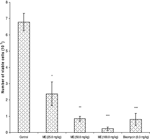
Effects of ME and bleomycin on EAC cell growth on day 6, after tumor inoculation are shown in . Treatment with ME resulted in maximum cell growth inhibition at the doses of 100 and 50 mg/kg (i.p.), as they showed 96% and 87% inhibition respectively, whereas bleomycin showed cell growth inhibition by 88% (0.3 mg/kg/day).
Figure 1. Effects of ME on EAC cell growth inhibition. Results are shown as mean ± SEM (standard error of mean), where significant values are *p < 0.05, **p < 0.01 and ***p < 0.001 when (EAC + ME) treated mice compared with EAC bearing control mice (EAC bearing only).
Transplantability of EAC cells receiving ME decreased remarkably as 72.4% reduction in EAC cell growth was observed when EAC cells from mice treated for five days (at the dose 100 mg/kg; i.p.) were re-inoculated into fresh mice and sacrificed on day 5.
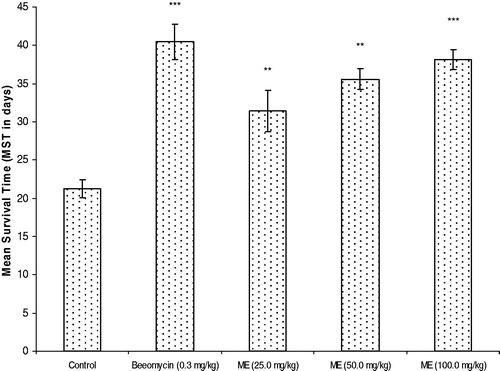
All effective anticancer drugs show a significant effect on survival time of tumor bearing mice. The effect of ME on survival time at different doses is shown in . It has been observed that tumor-induced mice treated with ME at doses 25, 50 and 100 mg/kg resulted in a significant increase of lifespan, which were 47.41, 67.13 and 79.34%, respectively, compared to that of control mice. On the other hand, bleomycin increased lifespan by 90.14% when compared to control.
Figure 2. Effects of ME on survival time of tumor bearing mice. Results are shown as mean ± SEM (standard error of mean), where significant values are *p < 0.05, **p < 0.01 and ***p < 0.001 when (EAC + ME) treated mice compared with EAC bearing control mice (EAC bearing only).
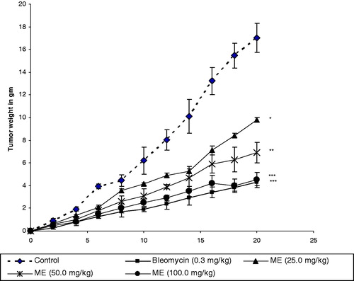
The effect of ME at doses 25, 50, 100 mg/kg (i.p.) and the antitumor drug bleomycin (0.3 mg/kg) on the average tumor weight due to tumorgenesis is shown in . Supplementation of the extract given to the mice, which were previously inoculated with EAC cells, resulted in the inhibition of tumor growth. In the case of the control (EAC-bearing) group, the body weight increased by 75.34% on 20th day when compared to the normal. The body weight of mice treated with ME at doses 25, 50 and 100 mg/kg (i.p.) increased by 55.23, 40.54 and 26.2%, respectively, on the 20th day. In contrast, with the use of bleomycin as standard at a dose of 0.3 mg/kg (i. p.), body weight increased by 25% on the 20th day.
Figure 3. Tumor weight of EAC bearing mice treated with ME and bleomycin. Results are shown as mean ± SEM (standard error of mean), where significant values are *p < 0.05, **p < 0.01 and ***p < 0.001 when (EAC + ME) treated mice compared with EAC bearing control mice (EAC bearing only).
The hematological parameters like RBC, WBC, Hb content, differential counts, etc. of both treated and non-treated mice were studied. For normal mice receiving ME at 100 mg/kg/day, all the examined parameters were found to be slightly changed during the treatment period from normal level. After 25 days of the initial treatment, they were found to have almost normal values. In the case of parallel treatment of EAC-bearing mice, these parameters were found to be significantly deteriorated as compared to those of the normal mice. It is hypothesized that this was due to toxic effects of tumorogenesis. However, these deteriorated parameters reversed themselves when ME supplementation was given at a dose of 100 mg/kg (i.p.). All the experimental data are presented in .
Table 2. Effects of ME on hematological parameters in experimental mice.
Effects of ME on the enzymes such as GPT, GOT and ALP are presented in . With normal mice, activities of these enzymes were found to be moderately increased during the treatment period (14 consecutive days at a dose of 100 mg/kg/day i.p.) and then these values gradually returned to almost normal levels. For EAC-bearing untreated mice, all such values increased almost linearly with time. In EAC-bearing mice treated with ME, however, the increment of GPT, GOT and ALP values was significantly less. After treatment, the GPT values returned to normal levels with time () while the ALP and GOT values remained reduced.
Table 3. Effect of ME on biochemical parameters in experimental mice.
also shows the effects of ME on serum biomolecules such as glucose, cholesterol, urea, triglyceride and creatinine content of both normal and EAC-bearing mice. All these parameters, except glucose, increased in both normal and EAC-bearing mice. However, they returned toward the normal level in EAC-bearing mice treated with the extract. The glucose content increased a bit during the treatment period in normal mice. However, after the treatment period, it slowly returned to normal level. For EAC-bearing mice, the glucose content was found to have been reduced abruptly from normal level due to the hypoglycemic effects of EAC cells. The supplementation of ME increased the value close to the normal level.
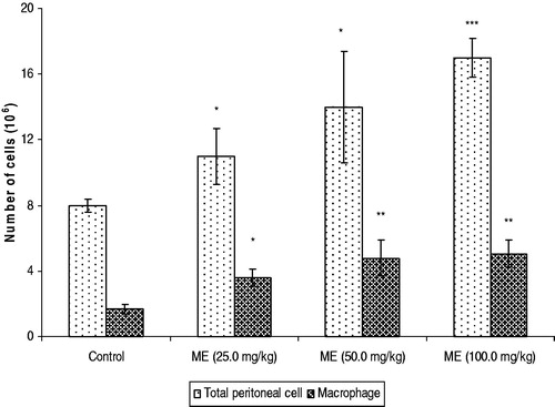
The average number of peritoneal exudate cells of normal mice treated with ME was found to be (17 ± 3.4) × 106 and the macrophage count was (4.8 ± 1.1) × 106 at 100 mg/kg/day, respectively. Treatment with the extract at a dose of 100 mg/kg/day for three consecutive days significantly enhanced the number of macrophages. Results are shown in .
Figure 4. Effects of ME on the enhancement of macrophages and peritoneal cells. Results are shown as mean ± SEM (standard error of mean), where significant values are *p < 0.05, **p < 0.01 and ***p < 0.001 when (EAC + ME) treated mice compared with EAC bearing control mice (EAC bearing only).
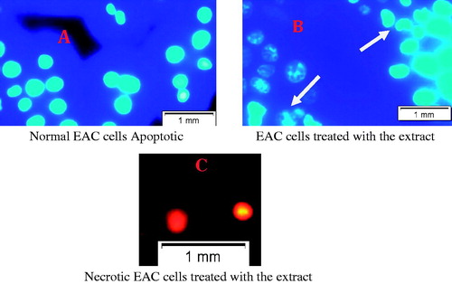
Morphological changes in EAC cells were examined by Hoechst 33342 staining, after treating the cells with and without extract (100 µg/ml) for 24 h. EAC nuclei were round, regular and homogeneously stained with Hoechst 33342 in the control group, as shown in . Extract-treated EAC cells showed manifest fragmented DNA in nuclei as shown in . Apoptotic morphologic alterations such as membrane blebbing and nuclear condensation were also observed clearly by fluorescence microscopy. These results indicate that the extract could induce apoptosis of EAC cells. Necrotic or late apoptotic cell death caused by extract was also observed by staining with PI as shown in . Here, the number of necrotic cells was found to be much lower than the number of apoptotic cells.
Figure 5. Fluorescence microscopic view of control and treated EAC cells. (A) EAC of normal mice shown no apoptotic features. (B) EAC cells treated with extract shown nuclear condensation, fragmentation, cell membrane blebbing, apoptotic bodies, etc, (indicated by arrows). (C) Cells undergone late apoptosis shown in PI staining. EAC cells were collected from control and treated mice on day 6. After washing with PBS all cells were stained with 0.1 µg/ml of Hoechst 33342 at 37 °C for 20 min. Then the cells were washed with phosphate buffer saline (PBS) and re-suspended in PBS for observation of morphological changes under fluorescence microscopy (Olympus IX71, Korea). Cells also stained with propidium iodide (PI) to observe necrotic or late apoptotic death.
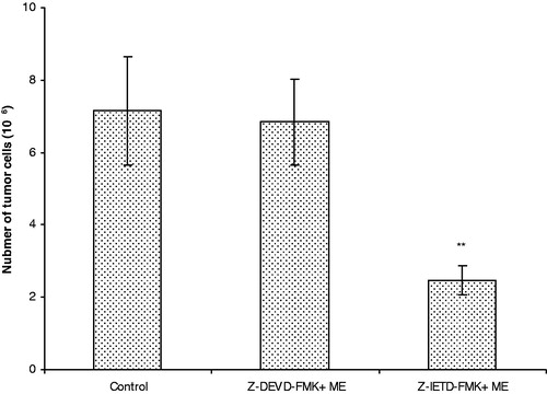
To examine the involvement of specific caspase in the apoptosis of EAC, the treatment of extract was investigated using caspase inhibitors Z-DEVD-FMK (caspase-3 inhibitor) and Z-IETD-FMK (caspase-8 inhibitor). The cytotoxicity of extract towards Z-IETD-FMK-pretreated EAC cells was significantly reduced to 65.3%, but Z-DEVD-FMK-pretreated cells did not show any reduction of cytotoxicity in comparison to the control group ().
Figure 6. Effect of caspase inhibitors on EAC cell. Results are shown as mean ± SEM (standard error of mean), where significant value **p < 0.01 when (EAC + ME + Z-IETD-FMK) treated mice compared with EAC bearing control mice (EAC bearing only).
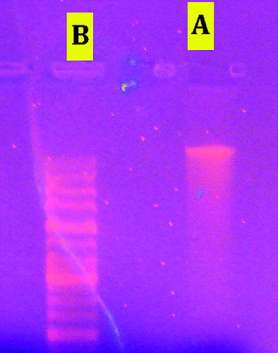
The activation of the endogenous Ca2+/Mg2+-dependent endonuclease is the most distinctive biochemical hallmark of apoptosis. This activated endonuclease-mediated cleavage of internucleosomes and generated oligonucleotide fragments of about 180–200 base-pair length or their polymers. DNA ladder bands were obtained in agarose gel electrophoresis of DNA preparation extracted from extract-treated EAC cells which, is a characteristic feature of apoptosis induction. In the control group, smear-like DNA degradation was obtained, as shown in .
Figure 7. In vivo effects of extract on DNA fragmentation of EAC cells. DNA run and detected on 1.5% agarose gel electrophoresis. (A) DNA from control EAC cells, (B) DNA from Eucalyptus treated EAC cells (DNA fragmentation detected from treated EAC cells).
Discussion
Reduction of tumor weight, enhancement of lifespan of tumor-bearing mice, tumor cell growth inhibition, as well as hematological profile, determined the potency of an anticancer agent. The efficiency of the extract was compared with data obtained by running a parallel experiment with a known clinically-used anticancer drug, bleomycin, at the dose of 0.3 mg/kg (i.p.). Results were also compared with those obtained from similar types of extracts reported in the literature (Muhammad et al., Citation2011; Perveen et al., Citation2012). The average tumor weight reducing ability of the extract (ME) has been examined. For tumor-bearing mice, body weight was found to be increased gradually. The treatment of such mice with this extract reduced the tumor burden remarkably. The extract also inhibited the cell growth rate effectively; more than 96% inhibition was achieved at a dose of 100 mg/kg (i.p.) which is quite comparable to that of bleomycin (0.3 mg/kg). So, a methanol extract used in a much higher dose can be compared to a commercial drug is a little precipitate.
The extract supplementation increased the lifespan of tumor-bearing mice very effectively. The potency was found to increase with the enhancement of dose. In the present study, a dose up to 100 mg/kg (i.p.) was used and further enhancement of the lifespan will therefore be expected using higher doses. It is noted that the enhancement of lifespan has been assigned as a very important parameter for judging the suitability of a compound as anticancer agent (Price & Greenfield, Citation1958). Numerous animal studies have been published demonstrating decreased tumor size and/or increased longevity with the combination of chemotherapy and antioxidants (Chinery et al., Citation1997). Resistance to chemotherapy and chemotherapeutic agents is thought to be due to reduced accumulation in tumor cell (Robert, Citation1999). Recent research has focused on the ability of flavonoids-type antioxidant compound to increase the concentration of chemotherapeutics in tumor cells. Thus, the presence of flavonoids and the cytotoxic nature of the extract could produce the anticancer effect of E. camaldulensis against EAC cells.
One of the major problems in cancer chemotherapy is myelosuppression, followed by anemia (Maseki et al., Citation1981), due to the reduction of RBC and hemoglobin content. This is probably owing to the deficiency of iron in a hemolytic or myelopathic condition (Hogland, Citation1982). Recovery of hemoglobin content, and the RBC/WBC cells count in the experimental mice, indicates the protective action of ME on the hemopoetic system.
The development of hypoglycaemia and hyperlipidaemia in experimental animals with carcinoma has been previously reported (Silverstein et al., Citation1988). In this experiment, the reduced glucose level and elevated cholesterol, triglycerides, creatinine, urea, etc., were returned to more-or-less normal levels in ME-treated mice, thereby indicating a potent antitumour efficacy of ME (). It is well-known (Muhammad et al., Citation2011) that there are significant elevations in the levels of GPT, GOT and ALP in liver diseases and disorders and in hepatocellular damage caused by a number of agents. An increase in these enzymes levels is observed in patients with cardiac damage due to myocardial infarction and liver disorders (Muhammad et al., Citation2011). Biochemical measurements of these parameters showed that strong hepatotoxicity was associated with the inoculation of EAC in mice. After the treatment with ME, these values remain near the normal range in the treated groups (). From this it follows that the tumor cells induce hepatotoxicity and the damage was partly prevented by ME supplementation.
It has been found that the extract of E. camaldulensis has a significant effect on the enhancement of normal peritoneal cells in normal mice. Treatment with the plant extract at a dose of 100 mg/kg (i.p.) increased the number of macrophages to some extent. This is considered to be a very important parameter for acquiring self destroying ability of the animals or living beings towards cancer cells (Fernandes & Klubes, Citation1979). Enhancement of macrophages might produce some cytokines such as tumor necrosis factors (TNF), interleukins, etc., inside the peritoneal cavity, which in turn may be responsible for killing of tumor cells (Lee et al., Citation1982).
Conventional screening models for anticancer agents are geared toward the selection of cytotoxic drugs. It is highly desirable to have compounds that can cause cancer cell death via apoptosis. Apoptosis eliminates malignant or cancer cells without damaging normal cells and surrounding tissues (Ashkenazi, Citation2008). Apoptosis is characterized by cell morphological changes, chromatin condensation, DNA cleavage, and nuclear fragmentation. Typical morphological features of apoptotic cells can be observed through microscopic studies such as those using inverted phase contrast and fluorescence microscopes. Other features, such as chromatin condensation and nuclear fragmentation, can be better observed through double staining with Hoechst 33342 and propidium iodide (PI) using fluorescence microscopic analysis. This is a convenient and rapid assay, widely used to identify living and dead cells. Hoechst 33342 is a blue fluorescing dye that stains chromatin DNA. The red fluorescing dye, PI, is only permeable to dead cells and cannot enter the intact plasma membrane of living cells. Thus, the staining pattern which resulted from the simultaneous use of these dyes makes it possible to distinguish normal, apoptotic, and dead cell population by fluorescence microscopy (Brown & Attardi, Citation2005; Moongkarndi et al., Citation2004; Thuret et al., Citation2003; Wahab et al., Citation2009). EAC cells treated with extract (ME) showed nuclear condensation, fragmentation, cell membrane blebbing, and apoptotic bodies, etc., under the fluorescence microscope, which implies that the extract induced EAC cell apoptosis. PI staining of EAC cells also indicates late phase apoptosis induction after treatment with the extract (). The integrity of the DNA was also assessed by agarose gel electrophoresis. DNA isolated from cells showed a ‘‘ladder’’ pattern in apoptosis (Chun-Ping et al., Citation2012). Genomic DNA isolated from treated and untreated cells showed an apoptotic pattern in agarose (1.5%) electrophoresis (). These characteristics, such ladder-like DNA band in the gel, further confirm the induction of apoptosis in EAC cells to the treatment of ME.
Cytotoxic compounds trigger apoptosis through two signaling mechanisms: the extrinsic (death receptor) pathway is activated by binding of tumor necrosis factor (TNF) ligands such as TRAIL to corresponding cell surface receptors, for example, TRAIL receptors, triggering the assembly of the death-inducing signaling complex (DISC) that drives caspase-8 activation (Ashkenazi, Citation2008). The intrinsic (mitochondrial) pathway involves mitochondrial outer membrane permeabilization, accompanied by the release of cytochrome C and second mitochondria-derived activator of caspases (Smac) into the cytosol. Cytochrome C initiates the formation of a caspase-9, caspase-3 activation platform, that is, the apoptosome, while Smac relieves the inhibition of caspases by neutralizing inhibitor of apoptosis (IAP) proteins (Fulda et al., Citation2010).
To understand, how ME triggers the apoptotic signaling pathway in EAC cells, specific inhibitors of candidate molecules such as caspase-8 and caspase-3 were used. Data obtained from our experiment shows that EAC cells pretreated with caspase-8 inhibitor (caspase-8 blocked/inactivated and caspase-3 remain active) showed 65.3% growth inhibition in comparison to control whereas, cells pretreated with caspase-3 (caspase-3 inactive and caspase-8 active) shown very little or no cell growth inhibition (). This result leads one to conclude that the caspase-3 mediated signaling pathway is involved in EAC cell apoptosis by the treatment of E. camaldulensis.
Conclusions
In light of the above observations, it can be concluded that the methanol extract of E. camaldulensis shows potential anticancer activity, through apoptosis and stimulation of host antitumor immunity. Therefore, it might be considered as a promising resource in cancer chemotherapy with a better host safety profile. However, before assuming so, it is necessary to carry out extensive research on this plant at a more advanced level, using other cell lines and higher animal models.
Declaration of interest
The authors declare no conflicts of interest. The authors alone are responsible for the content and writing of this article.
Acknowledgements
The authors are grateful to IICB, Kolkata, India, for providing the EAC cells and ICDDR’B, for supplying the experimental mice and standard mouse pellets. The authors are also thankful to the Ministry of Education, the People’s Republic of Bangladesh for providing financial support of this research work.
References
- Abbott BJ. (1976). Bioassay of plant extracts for anticancer activity. Cancer Treat Rep 60:1007–10
- Abdel-Nasser S, Nahla A, Eman AS, et al. (2011). Phenolic constituents of Eucalyptus camaldulensis Dehnh, with potential antioxidant and cytotoxic activities. Rec Nat Prod 5:271–80
- Abdullaev FI, Luna RR, Roitenburd BV, Espinosa AJ. (2000). Pattern of childhood cancer mortility in Mexico. Arch Med Res 31:526–31
- Al-Fatimi M, Friedrich U, Jenett-Siems K. (2005). Cytotoxicity of plants used in traditional medicine in Yemen. Fitoterapia 76:355–8
- Ali MM, Jesmin M. (2010). Hepatotoxicity of Schiff bases derived from benzoin salicylaldehyde, aminophenol and 2,4-dinitrophenyl hydrazine. J Natn Sci Foundation Sri Lanka 38:145–9
- Ashkenazi A. (2008). Directing cancer cells to self-destruct with proapoptotic receptor agonists. Nat Rev Drug Discov 7:1001–12
- Atta AH, Alkofahi A. (1998). Anti-nociceptive and anti-inflammatory effects of some Jordanian medicinal plant extracts. J Ethnopharmacol 60:117–24
- Brown MM, Attardi LD. (2005). The role of apoptosis in cancer development and treatment response. Nat Rev Cancer 5:231–7
- Cassady JM, Douros JD. (1980). Anticancer Agents Based on Natural Product Models. New York, NY: Academic Press
- Chinery R, Brockman JA, Peeler MO, et al. (1997). Antioxidant enhances the cytotoxicity of chemotherapeutic agents in colorectal cancer: A p53-independent induction p21 via C/EBP-β. Nat Med 3:1233–41
- Chun-Ping J, Hui D, Da-Hua S, et al. (2012). Pro-apoptotic effects of tectorigenin on human hepatocellular carcinoma HepG2 cells. World J Gastroenterol 18:1753–64
- Coelho-de-Souza LN, Leal-Cardoso JH, de Abreu Matos FJ, et al. (2005). Relaxant effects of the essential oil of Eucalyptus tereticornis and its main constituent 1,8-cineole on guinea-pig tracheal smooth muscle. Planta Med 71:1173–5
- El-Ghorab AH, El-Massry KF, Marx F, Fadel HM. (2003). Antioxidant activity of Egyptian Eucalyptus camaldulensis var. brevirostris leaf extracts. Nahrung 47:41–5
- Fernandes DJ, Klubes P. (1979). A biochemical and pharmacological study of therapeutic system with 5-fluorouracil plus cyclophosphamide in murine L1210 leukemia. Can Res 39:1396–404
- Fulda S, Galluzzi L, Kroemer G. (2010). Targeting mitochondria for cancer therapy. Nat Rev Drug Discov 9:447–64
- Ghani A. (2003). Medicinal Plants of Bangladesh. Dhaka, Bangladesh: Asiatic Society of Bangladesh
- Harborne JB. (1976). Phytochemical Methods: A Guide to Modern Technique of Plant Analysis. London, UK: Chapman and Hall Ltd
- Hogland HC. (1982). Heamatological complications of cancer chemotherapy. Sem Oncol 9:95–102
- Islam F, Khatun H, Soby G, et al. (2012). Bioassay of Eucalyptus extracts for anticancer activity against Ehrlich ascites carcinoma (EAC) cells in Swiss albino mice. Asian Pac J Trop Biomed 5:394–8
- Ito H, Koreishi M, Tokuda H, Nishino H, Yoshida T. (2000). Cypellocarpins A-C, phenol glycosides esterified with oleuropeic acid, from Eucalyptus cypellocarpa. J Nat Prod 63:1253–7
- Khan MN, Ngassapa O, Matee MIN. (2000). Antimicrobial activity of Tanzanian chewing sticks against oral pthogenic microbes. Pharm Biol 38:235–40
- Kinghorn AD, Balandrin MF. (1993). Human Medical Agents from Plants. Am Chem Soc Symp Series 534. Washington, DC: American Chemical Society
- Lee NN, Cadman EC, Michael IN, et al. (1982). Randomized study comparing doxorubicin, cyclophosphamide, vincristine, methotrexate with leucovorin rescue, and cytarabine (ACOMLA) with cyclophosphamide, doxorubicin, vincristine, prednisone, and bleomycin (CHOP-B) in the treatment of diffuse histiocytic lymphoma. Cancer Treat Rep 66:1279–84
- Litchfield JR, Wilcoxon F. (1949). A simplified method of evaluating dose-effect experiments. J Pharmacol Exp Ther 96:99–113
- Lorke D. (1983). A new approach to practical acute toxicity testing. Arch Toxicol 54:275–87
- Maseki M, Nishiagaki I, Hagishara M, et al. (1981). Lipid peroxidation levels and lipid content of serum lipoprotein fractions of pregnant subjects with or without preeclamsia. Clin Chim Acta 41:424–6
- Mehmet O, Isik DK, Ibrahim HK, Muhammed EG. (2011). Ehrlich ascites carcinoma. Afr J Biotechnol 10:2375–8
- Meyer BN, Ferringni NR, Putnam JE, et al. (1982). Convenient general bioassay for active plant constituents. Planta Med 45:34–9
- Moongkarndi P, Kosem N, Kaslungka S, et al. (2004). Antiproliferation, antioxidation and induction of apoptosis by Garcinia mangostana (mangosteen) on SKBR3 human breast cancer cell line. J Ethnopharmacol 90:161–6
- Mukherjee KL. (1988). Medical Laboratory Technology. New Delhi, India: Tata Mc Graw Hill Pub. Com. Ltd
- Muhammad RH, Muhammad AA, Muhammad RK. (2011). Inhibition of Ehrlich’s ascites carcinoma by ethyl acetate extract from the flower of Calotropis gigantia L. in mice. J Appl Biomed 8:47–54
- Perveen R, Islam F, Khanum JA, Yeasmin T. (2012). Preventive effect of ethanol extract of Alpinia calcarata Rosc. on Ehrlich’s ascitic carcinoma cell induced malignant ascites in mice. Asian Pac J Trop Med 5:121–5
- Price VE, Greenfield RE. (1958). Anemia in cancer. Adv Cancer Res 5:199–200
- Rahman SNSA, Norhanom AW, Nurestri AMS. (2013). In vitro morphological assessment of apoptosis induced by antiproliferative constituents from the rhizomes of Curcuma zedoaria. Evid Based Complement Alternat Med 2013:1–14
- Robert J. (1999). Multi drug resistance in oncology: Diagnostic and therapeutics approaches. Eur J Clin Invest 29:536–45
- Shariff ZU. (2001). Modern Herbal Therapy for Common Ailments. Ibadan, Nigeria: Spectrum Books Limited in Association with Safari Books (Export) Limited, UK
- Silverstein H, Dervot K, Oscar D. (1988). Studies on carbohydrate metabolism and different types of tumors bearing animals. Lancet 22:40–5
- Sur P, Ganguli DK. (1994). Tea plant extract (TRE) as an antineoplastic agent. Planta Med 60:106–9
- Tayeb K, William TB. (1999). Merbarone, a catalytic inhibitor of DNA topoisomerase II, induces apoptosis in CEM cells through activation of ICE/CED-3-like protease. Mol Pharmacol 55:548–56
- Thuret G, Chiquet C, Herrag S, et al. (2003). Mechanisms of staurosporine induced apoptosis in a human corneal endothelial cell line. Br J Ophthalmol 87:346–52
- Wahab SIA, Abdul AB, Alzubairi AS, et al. (2009). In vitro ultramorphological assessment of apoptosis induced by Zerumbone on (HeLa). J Biomed Biotechnol 2009:1–10
- Yinyuan W, Dianjun W, Xiaodong W, et al. (2011). Caspase-3 is activated through caspase-8 instead of caspase-9 during H2O2-induced apoptosis in HeLa cells. Cell Physiol Biochem 27:539–46