
Abstract
Context: Polygonum cuspidatum Sieb et Zucc. (Polygonaceae) possesses various pharmacological activities and has been widely using as one of the most popular and valuable Chinese herbal medicines in clinics. Its usage has increasingly attracted much of our attention and urges investigation on its bioactive components.
Objective: To establish a rapid and valid approach for screening potential neuroprotective components from P. cuspidatum.
Materials and methods: Potential neuroprotective components from P. cuspidatum were screened utilizing liposome equilibrium dialysis followed by high-performance liquid chromatography (HPLC) analysis. Their neuroprotective effects on modulation of protein expression of α7 nAChR, α3 nAChR and synaptophysin (SPY) on SH-SY5Y human neuroblastoma cell line (SH-SY5Y) were evaluated by means of Western blotting.
Results: Two potential compounds, polydatin (C1) and emodin-8-O-β-d-glucoside (C2), were detected and identified in our study. The biological tests showed that both compounds C1 and C2, respectively, at concentrations of 0.1 and 0.25 mg/mL significantly increased protein expression of α7 and α3 nicotinic acetylcholine receptors (nAChRs) in SH-SY5Y cells. Moreover, C1 and C2 at 0.1 mg/mL significantly reversed the Aβ1-42-induced decrease of α7 and α3 nAChRs protein expression in SH-SY5Y cells. In addition, C2 at 0.1 mg/mL significantly increased protein expression of SPY in SH-SY5Y cells and Aβ1-42-induced SH-SY5Y cells whereas C1 did not provide any positive effects.
Discussion and conclusion: In conclusion, our approach utilizing liposome equilibrium dialysis combined with HPLC analysis and cell-based assays is a prompt and useful method for screening neuroprotective agents.
Introduction
Alzheimer’s disease (AD) is the most common subtype of dementia. It was predicted to affect 36 million people worldwide in 2000 and is expected to increase to 115 million by 2050, which has resulted in a significant quality of life burden on patients and cost burden on the healthcare system (Prince et al., Citation2011). Due to the specific molecular initiators of AD remain unknown in most patients, current medical therapies are limited to provide modest symptomatic relief. Therefore, the research and development of effective drugs for the prevention and treatment of AD is of significant importance.
Extensive research suggests that aberrant amyloid-β (Aβ) peptide accumulation along with altered expression and function of nicotinic acetylcholine receptors (nAChRs) and synaptic damage stand prominently in the etiology of AD (Parri et al., Citation2011). nAChRs are members of the super-family of ligand-gated ion channels which respond to acetylcholine to participate in a wide range of brain activities and functions (Tang et al., Citation2008). nAChRs are enriched in the cholinergic target areas; however, the cholinergic deficit in AD is due in part to a depletion of these receptors on both projection and target neurons (Dineley, Citation2007). In AD, the cholinergic system is affected, and a reduction in the number of nAChRs has been reported, and administration of nAChR agonists such as nicotine and 3-(2, 4)-dimethoxybenzylidene anabaseine could significantly enhance their cognitive ability (Shimohama & Kihara, Citation2001). In addition, significantly reduced synaptic connectivity was also detected in the neocortex and hippocampus of AD patients, and reductions in synaptic densities had showed a strong correlation with cognitive decline in AD (Davies et al., Citation1987; Scheff & Price, Citation2003). This evidence suggested that down-regulation of nAChRs and loss of synapses are involved in the intellectual dysfunction in AD and thus nAChRs and synaptic density may be important therapeutic targets for the treatment of AD.
Polygonum cuspidatum Sieb et Zucc. (Polygonaceae) is widely distributed in Asia and North America. In Traditional Chinese Medicine (TCM), P. cuspidatum, also known as Hu-Zhang, is used as an analgesic, antipyretic, diuretic and expectorant agent in treatments of diseases such as arthralgia, chronic bronchitis, jaundice, amenorrhea and high blood pressure (Bralley et al., Citation2008). In our study, we screened neuroprotective effects of alcohol extract of P. cuspidatum and its different polarity fractions (petroleum ether, ethyl acetate, n-butanol and remaining H2O fractions), and found that n-butanol extract provides neuroprotective effect as alcohol extract of P. cuspidatum to significantly reduce Aβ-induced neurotoxicity and up-regulate protein expression of α7 nAChR and α3 nAChR in SH-SY5Y cells (data not shown). However, the bioactive components in this active fraction responsible for its therapeutic effects are unknown. Unfortunately, this active fraction comprises up to hundreds of different components, which makes the purification and screening of bioactive components processes extremely difficult.
It is well known that the majority of TCMs are orally delivered drugs, and their active components should be firstly absorbed by the gut before getting into the bloodstream. In other words, active components must be able to pass through the lipid bilayer or epithelial cell linings of the intestinal wall prior to their circulation in the bloodstream. Therefore, intestinal absorption is prerequisite for active components of Chinese herbal medicines. Liposomes are structurally similar to biological membranes because of their lipid bilayer structure. This similarity allows liposomes to mimic the biological membrane; therefore, the penetrability of the compounds into cells can be predicted. More recently, a new approach has been proposed for screening and analyzing bioactive components in TCM using liposome equilibrium dialysis and high-performance liquid chromatography-mass spectrometry (HPLC-MS) analysis, which was successfully applied to simultaneously predict the potential bioactivities of multiple compounds in Dang-gui-bu-xue decoction (Qi et al., Citation2006); nevertheless, their bioactivities were not further confirmed and the feasibility of this approach needs further evaluation.
In this study, liposome equilibrium dialysis combined with HPLC method was established to rapidly identify potential active components in the n-butanol extract of P. cuspidatum, and their neuroprotective effects were evaluated in vitro to confirm the feasibility of this approach.
Materials and methods
Polygonum cuspidatum was obtained from Zunyi city of Guizhou province, China, in July 2010. The identity was confirmed by Professor Qing-de Long, and the voucher specimen was deposited in Laboratory of Medicinal Plant and Pharmacognosy, School of Pharmacy, Guiyang Medical University, Guiyang, China. Dulbecco’s Modified Eagle Medium, trypsin-ethylenediaminetetraacetic acid and fetal bovine serum were purchased from Gibco/BRL Life Technologies (Paisley, UK). 3-(4, 5-Dimethyl-thiazol-2-yl)-2, 5-diphenyl tetrazolium bromide (MTT), streptomycin, penicillin, dimethylsulphoxide and Aβ1–42 were obtained from Sigma (St. Louis, MO). Goat polyclonal anti-α3 nAChR antibody, goat polyclonal anti-α7 nAChR antibody, mouse monoclonal anti-β-actin, donkey anti-goat IgG-horseradish peroxidase (HRP), goat anti-rabbit IgG-HRP and goat anti-mouse IgG-HRP were supplied by Santa Cruz Biotechnology (Santa Cruz, CA), and rabbit monoclonal anti-synaptophysin (SPY) antibody was supplied by Epitomics (Burlingame, CA).
Preparation of n-butanol extract of P. cuspidatum (PCB)
The P. cuspidatum sample was powdered to homogeneous size in a mill and sieved through a 60-mesh filter. The collected powder (100 g) was extracted twice by 10-fold 75% ethanol (w/v) for 60 min under reflux. The ethanol extract was obtained by combining the filtrates dried in a vacuum at 45 °C. Subsequently, the ethanol extract was dissolved in water and then extracted by ethyl acetate in order to remove some non-polar compounds. At a consecutive stage, n-butanol extract (PCB) was obtained after the further extraction by n-butanol and vacuuming at 45 °C. Eventually, PCB (0.5 g) was dissolved in 50 mL of 5 mM phosphate buffer saline (PBS, pH 7.4) and its fingerprint was determined using HPLC.
Preparation of liposome membranes
Liposomes were prepared by solvent injection method as described previously by Qi et al. (Citation2006). Briefly, 0.3 g of phosphatidylcholine was dissolved in 3 mL of ethanol and poured into an injector with 5 mL capacity. Then, the solution of phosphatidylcholine was drop-wisely added to a beaker containing 10 mL of warmed PBS (60 ± 1 °C) with continuous stirring. After removal of the remaining ethanol, a liposomal dispersion was obtained. These lipid vesicles were pressed through 0.22 μm pore size polycarbonate membrane, and liposomes of desired size were generated with the help of a hand extrusion device (Millipore Corp., Billerica, MA). The mean particle size of the resulting liposomes was found to be between 90 and 150 nm.
Permeation experiment
One milliliter of liposome solution and 1 mL of PCB extract were transferred to a dialysis tubing of 7 cm length. The ends of the dialysis tubing were tied tightly with thin cotton thread and the tubing was placed in a glass tube containing 8 mL of buffer solution for dialysis at 4 °C. After equilibration for 24 h, the “interaction dialysate” of the solution outside the tubing was obtained for HPLC analysis. A “blank dialysate” was also obtained by exactly the same procedure as the “interaction dialysate” was substituted with 1 mL of buffer solution instead of the liposome solution. The “interaction dialysate” and “blank dialysate” represent the extract of PCB after and before interaction with liposome membranes, respectively.
HPLC analysis and identification of permeable components
Multiple permeable components were simultaneously assessed by comparison of chromatograms of PCB extract before and after interaction with liposome membranes. Their chromatographic analyses were performed on an Agilent Series 1100 liquid chromatography (Agilent Technologies, Palo Alto, CA), equipped with a vacuum degasser, a quaternary pump, an autosampler and a DAD detector, connected to an Agilent ChemStation software (Agilent Technologies, Palo Alto, CA). The system was operated at 30 °C and a Hyperisil octadecylsilyl C18 column (4.6 × 250 mm i.d., 5 μm) was used. The injection volume was 10 μL and the mobile phase flow rate was 1.0 mL/min. Solvents that constituted the mobile phase were (A) 1% aqueous phosphoric acid and (B) acetonitrile. The elution conditions were as follows: 0–30 min, linear gradient 20–38% B; 30–70 min, linear gradient 38–100% B. Peaks were detected at 284 nm. As dialysis liposome membranes have certain cut-off molecular weights, the components of PCB cannot pass through the dialysis tubing when combined with liposome; therefore, a decrease in peak areas was observed and which were considered as permeable components. The strength of interaction between permeable components and liposome could be evaluated by binding degree and calculated as follows:
where Aa and Ab are the peak areas in chromatograms of a compound before and after interaction with liposome membranes, respectively. The stronger the strength of interaction between compound and liposome, the greater the binding degree.
Cell culture and treatments
The SH-SY5Y cells (German Collection of Microorganisms and Cell Cultures, Braunschweig, Germany) were cultured as described previously by Guan et al. (Citation2003) and Tang et al. (Citation2008). In order to examine the protective effect of permeable components against Aβ-induced down-regulation of nAChRs and SPY expression, the cells were treated with permeable components overnight, prior to the exposure to Aβ1–42 for 24 or 48 h.
Assay of MTT reduction
To determine the appropriate physiological concentrations of permeable components, the SH-SY5Y cells were exposed to permeable components at concentrations ranging from 0 to 10 mg/mL for 48 h. Cytotoxicity of the permeable components was evaluated by means of MTT assay as described previously by Guan et al. (Citation2003) and Tang et al. (Citation2008).
Western blot analysis
The levels of the α3 and α7 nAChR subunits, SPY and internal control β-actin were semi-quantified by Western blotting as described previously by Goto and Hirano (Citation1990), Guan et al. (Citation2003) and Tang et al. (Citation2008). The intensity of each band on the film was quantitated by employing a computer-assisted program and expressed as a ratio with respect to the corresponding value of β-actin. Thereafter the experimental values were expressed as percentage of the corresponding control values.
Statistical analysis
The data were expressed as means ± SD, and Student's t-test was performed using SPSS software (version 11.5 from Statistical Product and Service Solutions, Chicago, IL), and a p value (two-tailed) less than 0.05 was regarded as significant.
Results and discussion
Liposome membrane-permeable components in PCB
In this study, we used liposome equilibrium dialysis combined with HPLC analysis for screening and analyzing bioactive components in TCM. According to the procedure described in the “Materials and methods” section, the “interaction dialysate” and “blank dialysate” of PCB extract were collected and analyzed by HPLC under the same conditions. The permeable property of compounds in PCB extract could be deduced from comparison of the two obtained chromatograms. Primary test showed that the fingerprints of PCB detected at 220, 254 and 284 nm were similar (data not shown). However, the signal was more sensitive at 284 nm. Therefore, the components were set monitored at 284 nm for HPLC analysis. HPLC chromatograms of PCB and eluate of “interaction dialysate” and “blank dialysate” under the experimental conditions described above were shown in . There were 11 principal peaks detected at 284 nm, among which two peaks, designated peak P2 and P5, obviously decreased in peak area when liposome solution was added. The binding degrees for these two peaks are shown in . Obviously, both permeable components have strong interaction with liposome membranes, and the strengths were 43.78 and 67.23%, respectively.
Figure 1. HPLC chromatograms of dialysate of n-butanol extract of P. cuspidatum (PCB): (1) “blank dialysate” of PCB; (2) “interaction dialysate” of PCB and (3) blank control.
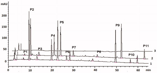
Table 1. Binding degrees of two permeable components in PCB.
Identification of permeable components
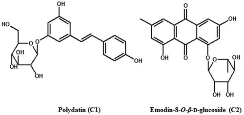
The liposome equilibrium dialysis study identified peaks P2 and P5 as potential active candidates. In order to identify their structures and test their bioactivities, compounds of peaks P2 and P5 were rapidly isolated and purified using semi-preparative HPLC, and their structures were resolved by nuclear magnetic resonance (NMR) spectroscopic techniques. Briefly, P. cuspidatum sample (1 kg) was extracted to prepare PCB as described in the “Materials and methods” section. Subsequently, the concentrated PCB extract was chromatographed on D-101 macroporous adsorption resin (Shanghai Hualing Resin Co., Ltd, Shanghai, China) and eluted with distilled water and 20% ethanol, respectively. Afterwards, the 20% ethanol elute was subjected semi-preparative HPLC for isolation and purification of permeable compounds. Semi-preparative HPLC was conducted on the Agilent 1100 liquid chromatography system (Semi-preparative system) which is similar to analytical HPLC, except fitted with a Zorbax SB C18 column (21.2 × 150 mm i.d., 5 µm). The mobile phase was the same as analytical HPLC, but the elution condition was adjusted to 0–40 min, with a linear gradient 20–38% B at a flow rate of 3 mL/min. Peaks eluting from the column were collected and further analyzed by analytical HPLC for locating the target compounds. Finally, both compounds of P2 and P5 were successfully purified, and their chemical structures were identified as polydatin (C1) and emodin-8-O-β-d-glucoside (C2) (as shown in ) based on their NMR results (1H NMR and 13C NMR) comparing to the references in the literature (Chu et al., Citation2005; Manila et al., Citation1993; Yang et al., Citation2001).
Figure 2. Chemical structures of polydatin (C1) and emodin-8-O-β-d-glucoside (C2).
Effects of compounds C1 and C2 on the expression of α7 nAChR, α3 nAChR and SPY in SH-SY5Y cells and Aβ1–42-induced SH-SY5Y cells
The direct cytotoxicity of compounds C1 and C2 was measured by MTT assays (). The results showed that the concentrations of C1 and C2 below 0.5 mg/mL did not induce any changes in cell viability. Therefore, 0.1 and 0.25 mg/mL of compounds C1 and C2 were used in experiments. In our previous study, n-butanol fraction from P. cuspidatum was found to significantly up-regulate the protein expression of α7 and α3 nAChR in SH-SY5Y cells (data not shown). To confirm whether compounds C1 and C2 can up-regulate the protein expression of α7 and α3 nAChRs, SH-SY5Y cells were treated with compound C1 or C2 at 0.1 or 0.25 mg/mL. The Western blotting results showed that both compounds C1 and C2 significantly increased protein expression of α7 and α3 nAChRs in a dose-dependent manner (as shown in ). Previous investigations have revealed that exposure of neuronal cells in vitro to various concentrations of Aβ results in decreased levels of the nAChR protein subunits (Guan et al., Citation2001, Citation2003; Liu et al., Citation2001). To test whether compounds C1 and C2 can reverse the decrease in protein expression of α7 and α3 nAChRs induced by Aβ, SH-SY5Y cells were treated with 5 μM Aβ1–42 and with or without 0.1 mg/mL of compounds C1 and C2, respectively. As shown in and , Aβ1–42 significantly reduced the protein expression of α7 and α3 nAChRs, but this decrease was reversed by both compounds C1 and C2. Accordingly, the present study suggests that neuroprotective effects of compounds C1 and C2 may be associated with up-regulation of α7 and α3 nAChRs expression.
Figure 3. Effects of polydatin (C1) and emodin-8-O-β-d-glucoside (C2) on cell viability of SH-SY5Y cells. Compared with the control group, *p < 0.05 and **p < 0.01.
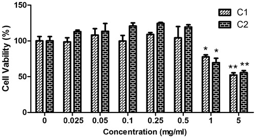
Figure 4. Effects of polydatin (C1) and emodin-8-O-β-d-glucoside (C2) on protein expression of α7 nAChR, α3 nAChR and SPY in SH-SY5Y cells. Protein expression of α3 nAChR in SH-SY5Y cells treated with 0.1 and 0.25 mg/mL C1 (A) or C2 (B); protein expression of α7 nAChR in SH-SY5Y cells treated with 0.1 and 0.25 mg/mL C1 (C) or C2 (D); protein expression of SPY in SH-SY5Y cells treated with 0.1 mg/mL C2 (E); compared with the control group, *p < 0.05 and **p < 0.01. The image shown is representative of three independent experiments.
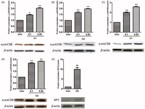
Figure 5. Effects of polydatin (C1) and emodin-8-O-β-d-glucoside (C2) on protein expression of α7 nAChR, α3 nAChR and SPY in Aβ1-42-induced SH-SY5Y cells. Protein expression of α3 nAChR in Aβ1–42-induced SH-SY5Y cells treated with 0.1 mg/mL C1 or C2 (A); protein expression of α7 nAChR in Aβ1-42-induced SH-SY5Y cells treated with 0.1 mg/mL C1 or C2 (B); protein expression of SPY in Aβ1–42-induced SH-SY5Y cells treated with 0.1 mg/mL C2 (C); compared with the control group, ###p < 0.001; compared with Aβ1–42-treated model group, *p < 0.05, **p < 0.01 and ***p < 0.001. The image shown is representative of three independent experiments.
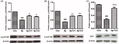
SPY is a synaptic vesicle glycoprotein, and is a marker for quantification of synaptic density (Calhoun et al., Citation1996). To investigate the synaptic protective effects of compounds C1 and C2, SH-SY5Y cells were treated with 0.1 mg/mL of compounds C1 and C2 and with or without 5 μM Aβ1–42. Proteins were harvested 24 h after treatment. The results showed that compound C2 significantly increased protein expression of SPY in SH-SY5Y cells without Aβ treatment (). When SH-SY5Y cells were treated with Aβ, the protein level of SPY was significantly suppressed, and compound C2, but not compound C1 (data not shown), significantly reversed this inhibitory effect of Aβ1–42 (). These evidences indicated that the neuroprotective effect of compound C2 involves in the regulation of synaptic density.
It has been demonstrated that both compounds C1 and C2 have the ability to penetrate the blood–brain barrier and achieve wide distribution in the brain, and exert multiple central nervous system effects, such as cerebrovascular effects, neuroprotection, anti-inflammation and anti-oxidation, etc. (Du et al., Citation2013; Kim et al., Citation2009; Wang et al., Citation2007; Xiang et al., Citation2005). In the present study, it was found that compounds C1 and C2 could significantly reverse decreased expression of α7 and α3 nAChRs and C2 also could significantly increase expression of SPY in Aβ1–42-induced SH-SY5Y cells, which indicating that compounds C1 and C2 effectively have protective roles against Aβ-induced neurotoxicity. These findings are compatible with previous studies that compound C1 could significantly and dose-dependently inhibit Aβ polymerization, and compound C2 exerts a comparable effect with huperzine A to ameliorate Aβ-induced never cell damage and scopolamine-induced memory function disturbance in mice (Cheng et al., Citation2001; Du et al., Citation2013). Therefore, it can be speculated that compounds C1 and C2 are at least partially responsible for neuroprotective effects of P. cuspidatum, and suggest that these two compounds could be good candidates to further develop neuroprotective agents for prevention and treatment of AD.
Conclusions
In this study, we used liposome equilibrium dialysis combined with the HPLC analysis method to rapidly identify two neuroprotective components, polydatin and emodin-8-O-β-d-glucoside, from the n-butanol extract of P. cuspidatum, which suggests that this method is prompt and useful for screening neuroprotective agents from TCM.
Declaration of interest
This study was supported by grant J20092143 from Science and Technique Foundation of Guizhou Province, China. The authors report no conflicts of interest.
References
- Bralley EE, Greenspan P, Hargrove JL, et al. (2008). Topical anti-inflammatory activity of Polygonum cuspidatum extract in the TPA model of mouse ear inflammation. J Inflamm (Lond) 5:1--7
- Calhoun ME, Jucker M, Martin LJ, et al. (1996). Comparative evaluation of synaptophysin-based methods for quantification of synapses. J Neurocytol 25:821–8
- Cheng WS, Xu JP, Yin XP, et al. (2001). Application of emodin-8-O-beta-d-glucoside. Chinese Patent, publication number: CN1114413C
- Chu X, Sun A, Liu R. (2005). Preparative isolation and purification of five compounds from the Chinese medicinal herb Polygonum cuspidatum Sieb. et Zucc by high-speed counter-current chromatography. J Chromatogr A 1097:33–9
- Davies CA, Mann DM, Sumpter PQ, et al. (1987). A quantitative morphometric analysis of the neuronal and synaptic content of the frontal and temporal cortex in patients with Alzheimer’s disease. J Neurol Sci 78:151–64
- Dineley KT. (2007). beta-Amyloid peptide – nicotinic acetylcholine receptor interaction: The two faces of health and disease. Front Biosci 12:5030–8
- Du QH, Peng C, Zhang H. (2013). Polydatin: A review of pharmacology and pharmacokinetics. Pharm Biol. [Epub ahead of print]. doi:10.3109/13880209.2013.792849
- Goto S, Hirano A. (1990). Neuronal inputs to hippocampal formation in Alzheimer’s disease and in parkinsonism-dementia complex on Guam. Acta Neuropathol 79:545–50
- Guan ZZ, Miao H, Tian JY, et al. (2001). Suppressed expression of nicotinic acetylcholine receptors by nanomolar beta-amyloid peptides in PC12 cells. J Neural Transm 108:1417–33
- Guan ZZ, Yu WF, Shan KR, et al. (2003). Loss of nicotinic receptors induced by beta-amyloid peptides in PC12 cells: Possible mechanism involving lipid peroxidation. J Neurosci Res 71:397–406
- Kim SH, Jang SD, Lee KY, et al. (2009). Chemical constituents isolated from Polygala japonica leaves and their inhibitory effect on nitric oxide production in vitro. J Enzyme Inhib Med Chem 24:230–3
- Liu Q, Kawai H, Berg DK. (2001). Beta-amyloid peptide blocks the response of alpha 7-containing nicotinic receptors on hippocampal neurons. Proc Natl Acad Sci USA 98:4734–9
- Manila E, Talvitie A, Kolehmainen E. (1993). Anti-leukaemic compounds derived from stilbenes in Picea abies bark. Phytochemistry 33:813–16
- Parri HR, Hernandez CM, Dineley KT. (2011). Research update: Alpha7 nicotinic acetylcholine receptor mechanisms in Alzheimer’s disease. Biochem Pharmacol 82:931–42
- Prince M, Bryce R, Ferri C. (2011). World Alzheimer Report 2011: The Benefits of Early Diagnosis and Intervention. London: Alzheimer’s Disease International
- Qi LW, Li P, Li SL, et al. (2006). Screening and identification of permeable components in a combined prescription of Danggui Buxue decoction using a liposome equilibrium dialysis system followed by HPLC and LC-MS. J Sep Sci 29:2211–20
- Scheff SW, Price DA. (2003). Synaptic pathology in Alzheimer’s disease: A review of ultrastructural studies. Neurobiol Aging 24:1029–46
- Shimohama S, Kihara T. (2001). Nicotinic receptor-mediated protection against beta-amyloid neurotoxicity. Biol Psychiatry 49:233–9
- Tang Z, An Y, Qi XL, et al. (2008). Inhibiting gene expression of alpha3 nicotinic receptor in SH-SY5Y cells with the effects on APP metabolism and antioxidation in Alzheimer’s disease. Neurochem Int 53:112–17
- Wang C, Zhang D, Ma H, et al. (2007). Neuroprotective effects of emodin-8-O-beta-d-glucoside in vivo and in vitro. Eur J Pharmacol 577:58–63
- Xiang L, Lei F, Xing DM, et al. (2005). Neuron protective constituents from rheum nanum and rheum sublanceolatum. Tsinghua Sci Technol 10:426–9
- Yang F, Zhang T, Ito Y. (2001). Large-scale separation of resveratrol, anthraglycoside A and anthraglycoside B from Polygonum cuspidatum Sieb. et Zucc by high-speed counter-current chromatography. J Chromatogr A 919:443–8