
Abstract
Context: Cancer prevention remains a high priority for the scientific world. Magnolia dealbata Zucc (Magnoliaceae), a Mexican endemic species, is used for the empirical treatment of cancer.
Objective: To evaluate the cytotoxic and cancer chemopreventive effects of an ethanol extract of Magnolia dealbata seeds (MDE).
Materials and methods: The cytotoxic effect of MDE, at concentrations ranging from 1 to 200 µg/ml, on human cancer cells and human nontumorigenic cells was evaluated using the MTT assay for 48 h. The apoptotic activities of MDE 25 μg/ml on MDA-MB231 breast cancer cells were evaluated using the TUNEL assay and the detection of caspase 3 using immunofluorescence analysis for 48 h, each. The chemopreventive effect was evaluated by administrating different doses of MDE, between 1 and 50 mg/kg, injected intraperitoneally daily into athymic mice which were implanted with MDA-MB231 cells during 28 days. The growth and weight of tumors were measured.
Results: MDE showed cytotoxic effects on MDA-MB231 cells (IC50 = 25 µg/ml) and exerted pro-apoptotic activities as determined by DNA fragmentation in MDA-MB231 cells. MDE 25 µg/ml also induces the activation of caspase 3 in MDA-MB231 cells. These results suggest that Magnolia dealbata may be an optimal source of the bioactive compounds: honokiol (HK) and magnolol (MG). MDE 50 mg/kg i.p. exerted chemopreventive effects by inhibiting the growth of MDA-MB231 tumor by 75% in athymic mice, compared to the control group.
Conclusions: MDE exerts cytotoxic, apoptotic and chemopreventive activities on MDA-MB231 human cancer cells.
Introduction
Cancer is a global health problem with high morbidity and mortality and poses severe economic challenges. Cancer treatment and prevention remain a high priority for the scientific community as well as health systems across the world.
México has great knowledge in the use of plant extracts for the empirical treatment of cancer (Alonso-Castro et al., Citation2011a). Up to now, 300 plant species belonging to 90 botanical families used for cancer treatment have been recorded in México, but only 181 of them have been experimentally analyzed (Alonso-Castro et al., Citation2011a). The mechanisms by which many plant extracts and active compounds exert their cytotoxic activities remain to be studied (Alonso-Castro et al., Citation2011a). In addition, it is necessary to evaluate the in vivo cancer chemopreventive and antitumor effects of many plant extracts and their active compounds.
Magnoliaceae family is composed of 220 species and approximately 20% of its members are endemic of America. In Mexico, there are 12 species and 3 subspecies of Magnolia (Sánchez-Velásquez et al., Citation2010). Magnolia dealbata Zucc (Magnoliaceae), a Mexican endemic species, which grows in cloud forests, is distributed in the states of Oaxaca, Queretaro, Veracruz, Hidalgo, San Luis Potosi, and Nuevo Leon (Sánchez-Velásquez et al., Citation2010). Magnolia dealbata is used in Centre Mexico as a diuretic, as well as for the empirical treatment of anxiety, hypertension, dysentery, stomachache, flu, asthma and cancer (De la Cruz, Citation1964; Domínguez-Yescas, Citation2012). Scientific reports have indicated that Magnolia dealbata exerts anxiolytic, anticonvulsant, antimicrobial, antidiabetic and insecticidal effects (Alonso-Castro et al., Citation2011b; Flores-Estévez et al., Citation2013; Jacobo-Salcedo et al., Citation2011; Martinez et al., Citation2006). Magnolia dealbata produces honokiol (HK) and magnolol (MG), two antitumor compounds (Lee et al., Citation2011). In addition, an efficient protocol for the production of HK and MG for in vitro tissue cultures of Magnolia dealbata has been performed (Dominguez et al., Citation2009). However, the cytotoxic effects of Magnolia dealbata remain to be studied. The aim of this investigation was to evaluate the cytotoxic and cancer chemopreventive effects of Magnolia dealbata
This study shows for the first time that the ethanol extract of Magnolia dealbata seeds exerts cytotoxic and apoptotic effects against MDA-MB231 cells as well as chemopreventive activities.
Materials and methods
Materials
Dulbecco's modified Eagle's medium (DMEM), Roswell Park Memorial Institute (RPMI) medium and fetal bovine serum (FBS) were from GIBCO BRL (Grand Island, NY). Cisplatin (CDDP) was from Accord Farma (Distrito Federal, México). MTT (3-(4,5-dimethylthiazol-2-yl)-2,5-diphenyl tetrazolium bromide, MG (5,5′-diallyl-2,2′-biphenyldiol) and HK (5,3′-diallyl-2,4′-biphenyldiol), 95% purity according to the manufacturer, were from Sigma Chemical Co. (St. Louis, MO). The anti-cleaved caspase 3 (sc-22171) and its secondary antibody for immunofluorescence assays donkey anti-goat IgG FITC (sc-2024) were purchased from Santa Cruz Biotechnology Inc. (Santa Cruz, CA).
Cell lines and culture conditions
Cell lines of immortal human keratinocytes (HaCaT) and cell lines of human cervical carcinoma (HeLa), hepatocarcinoma (HepG2) colorectal adenocarcinoma (SW-480), breast carcinoma (MDA-MB231) and erythromyeloblastoid leukemia (K562) were maintained in DMEM supplemented by 7% FBS and antibiotics (100 units/ml penicillin and 100 pg/ml streptomycin). Cell lines of ovarian carcinoma (SKOV-3) and prostate carcinoma (DU-145) were maintained in RPMI medium supplemented by 7% FBS and antibiotics. All cell lines were obtained from ATCC (Manassas, VA). All cell cultures were grown at 37 °C, in a humidified atmosphere of 5% CO2.
Animals
Six-week-old nu/nu mice, weighing 18–23 g, from the Instituto Nacional de Ciencias Médicas y Nutrición Salvador Zubirán, were used. The experiments were performed following the NIH Guide for Treatment and Care for Laboratory Animals and the Mexican Official Norm for Animal Care and Handling (NOM-062-ZOO-1999). All the procedures carried out in this study were approved by the Research Ethic Committee from Instituto Nacional de Cancerología (Distrito Federal, México). The mice, having free access to food and water, were housed in cages with filtered air in a climate and light controlled room with a 12 h light/dark cycle.
Preparation of Magnolia dealbata extract and quantitation of HK and MG
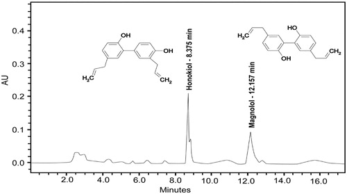
Samples of Magnolia dealbata, collected in Veracruz, México in August 2008, were identified by specialist (Dr. Martin Mata) and preserved at the herbarium of Instituto de Ecología (XAL, Xalapa México) for future reference (voucher number 12520). Magnolia dealbata dried seeds and leaves (3 g) were extracted, each with ethanol as described previously (Martinez et al., Citation2006). Detection and quantification of HK and MG were performed by reverse-phase HPLC (Tsai & Chen, Citation1992) in a Waters 2795 (Waters Corp., Milford, MA) instrument equipped with autosampling and a 996 photodiode array detector. Briefly, Magnolia dealbata seeds (MDE), HK and MG were separated on Kromasil C-18 (150 mm × 4.6 mm) (Metachem Technologies Inc, Torrance, CA) column. The mobile phase was acetonitrile–water–phosphoric acid (65:35:0.1, v/v/v) of pH 2.4–2.7 at a flow rate of 1.0 ml/min. Maximum detection wavelengths were 254 nm for HK and 290 nm for MG with retention times of 8.8 min and 12.2 min, respectively. Retention times and UV spectra of HK and MG from MDE were compared with those of high-purity commercial standards. HK and MG concentrations in the extract were calculated from calibration plots obtained from dilutions of the commercial standards with known concentrations.
MTT assay
Human cancer cell lines were seeded in 96-well microplates at a density of 5000 cells/well. After 24 h of incubation, MDE at concentrations ranging from 1 to 200 µg/ml were added to the cells. Then, the assay was carried out as described by Jacobo-Salcedo et al. (Citation2011) and the optical density (OD) was measured at 590 nm using an ELISA reader (Biorad Laboratories, Hercules, CA). The wells without cells were considered as blank. The viability of treated cells was estimated from the relative growth as follows:
The concentration leading to 50% inhibition of viability (IC50) was also calculated by regression analysis (percent survival versus log concentration).
Proliferation assay
MDA-MB231 and HaCaT cells were plated (1 × 10⁴ cells/well) in 24-well plates (Corning Glass Works, Corning, NY) with DMEM supplemented with 7% FBS. Two days later, cultures were fed with MDE concentrations ranging 1–200 µg/ml. Cell viability was monitored for several days of culture by direct cell counting in a hemacytometer. Cell cultures were maintained at 37 °C in a humidified 5% CO2 atmosphere with medium changes every other day.
TUNEL assay
The in situ DNA fragmentation was detected by Dead End Flurometric TUNEL system (Promega Corporation, Madison, WI) according to the manufacturer’s instructions. MDA-MB231 cells, seeded at a density of 5 × 10⁵ on 60 mm Petri dishes, were allowed to adhere and grow on glass coverslips for 24 h. Cells were treated with IC50 of MDE or CDDP, or the vehicle (DMSO 0.01%) for 24 or 48 h. After incubation, cells were treated as described previously (González-Sánchez et al., Citation2011). The samples were three-times rinsed with PBS and analyzed by fluorescence microscopy.
Immunofluorescence assay
MDA-MB231 cells grown on coverslips were treated with IC50 of MDE. After 24 or 48 h of treatment, cells were washed with PBS and fixed with paraformaldehyde (4%) in PBS for 20 min. The cells were permeabilized with methanol 100% and washed with PBS. Cells were incubated with caspase 3 antibody for 1 h at 37 °C, washed and incubated with a secondary FITC-conjugated anti-goat antibody for 45 min at room temperature. After washing, cells were mounted on glass slides using Vectashield (Vector Laboratories, Burlingame, CA). Images were acquired with an Axiovert 40 CFL epifluorescence microscope (Carl Zeiss, Thornwood, NY) by the use of a 100 × objective.
Chemopreventive effect
The nu/nu mice were injected subcutaneously in their backs with MDA-MB231 cells (5 × 10⁶). Four hours after tumor implantation, groups of five mice received doses of MDE between 1 and 50 mg/kg, dissolved in 0.1 ml of 0.9% saline solution, CDDP 1 mg/kg or PCX 1 mg/kg injected intraperitoneally daily over a period of 28 days. The animal control group received 0.1 ml of vehicle solution. Tumors were measured using a Vernier caliper, and their size in millimeter cube was calculated as follows (Looney et al., Citation1971):
At the end of the experiments, animals were sacrificed and their tumors were excised and weighed.
Statistical analysis
Experimental values are expressed as mean ± standard deviation of at least two experiments in triplicate. Data were analyzed by using Student’s t-test or a one-way ANOVA when indicated. The level of p ≤ 0.05 was used as criterion of statistical significance. All calculations were done using the JMP 5.1 program (SAS Institute Inc., Cary, NC).
Results
HK and MG quantitation in MDE
The HPLC analysis showed that HK and MG were the two major components in MDE (). The content of these compounds in MDE was 23 mg/g for HK and 37 mg/g for MG. Therefore, MDE concentrations assayed contained 3.5 μM HK and 3.9 μM MG (MDE 25 μg/ml). In the ethanol extract of Magnolia dealbata leaves, it was not possible to detect HK or MG (results not shown). Therefore, further studies were performed with the ethanol extract of Magnolia dealbata seeds.
Figure 1. HPLC elution pattern of honokiol and magnolol from an ethanol extract of Magnolia dealbata seeds (MDE).
MDE exerts cytotoxic and antiproliferative effects against MDA-MB231 breast cancer cells
Cisplatin [cis-diamminedichloroplatinum(II) (CDDP)], the positive cytotoxicity control, exerted strong toxic effects on all human cancer cell lines, whereas MDE showed the highest cytotoxic effects against MDA-MB231 cells (). In nontumorigenic cells (HaCaT), CDDP exerted high cytotoxic effects, whereas MDE showed low toxic effects ().
Table 1. Cytotoxic activity of MDE on human cancer and non-tumorigenic cells.
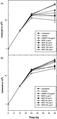
The vehicle (DMSO 0.01%) and MDE 5 µg/ml lacked antiproliferative effects on MDA-MB231 and HaCaT cells (). In contrast, CDDP tested at 2.9 µg/ml decreased cell growth by 61% in MDA-MB231 cells () and by 47% in HaCaT cells (), compared to untreated cells. In MDA-MB231 cells, MDE tested at 25 and 50 µg/ml decreased cell growth by 40 and 56%, respectively, whereas in HaCaT cells, the same concentrations decreased cell growth by 13 and 20%, respectively (). In MDA-MB231 cells, MDE tested at 100 and 200 µg/ml decreased cell growth higher than 70% (). In HaCaT cells, MDE tested at 100 and 200 µg/ml decreased cell growth by 40 and 55%, respectively ().
Figure 2. Effect of MDE on cell growth. MDA-MB231 (A) and HaCaT (B). After 2 days, cultures were fed with DMEM supplemented with 7% FBS added with different MDE concentrations. At the indicated days, the cell number was determined by direct cell counting in a hemacytometer. Figure represents the mean value of three independent experiments in triplicate ± standard deviation (SD).
MDE exerts apoptotic effects in MDA-MB231 cells
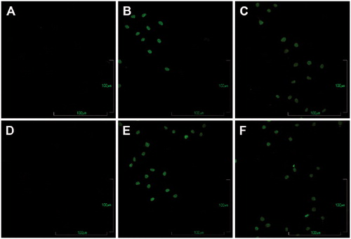
MDA-MB231 cells incubated with the vehicle showed no DNA damage (, panels A and D). In contrast, cells treated with CDDP 2.9 μg/ml were positive to TUNEL reaction at 24 h and 48 h (, panels B and E), showing typical signs of apoptosis such as an increase in DNA fragmentation and appearance of apoptotic bodies. MDA-MB231 cells treated with MDE 25 μg/ml showed TUNEL positive cells in a time-dependent manner with a similar pattern than those treated with CDDP (, panels C and F).
Figure 3. Determination of DNA fragmentation in MDA-MB231 cells treated with MDE by the TUNEL assay. Cells incubated with DMSO 0.01% for 24 h (A) and 48 h (D), cisplatin (CDDP) 2.9 μg/ml, as the positive control of apoptosis, for 24 h (B) and 48 h (E) or MDE 25 μg/ml for 24 h (C) and 48 h (F). Cells were subjected to the TUNEL assay. All photographs are shown at 20 × magnification. Results represent three independent experiments in duplicate.
MDE induces the activation of caspase 3 in MDA-MB231 cells
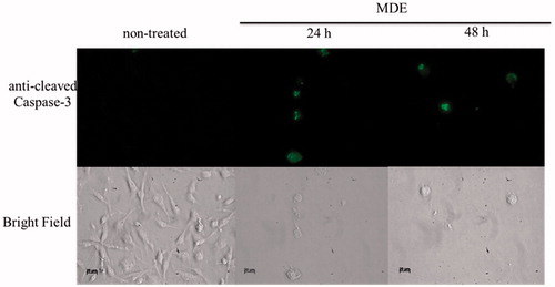
Immunofluorescent analysis showed that MDE at 25 mcg/ml induced the proteolytic cleavage of caspase 3 at 24 and 48 h in MDA-MB231 cells compared to untreated cells (). This indicates that MDE exerts its apoptotic effects on breast cancer cells via a caspase-dependent manner.
Figure 4. MDE induces the activation of caspase 3 in MDA-MB231 cells. Detection of caspase 3 using immunofluorescence analysis. MDA-MB231 cells grown on coverslips were treated with MDE 25 μg/ml. After 24 h or 48 h of treatment, cells were treated as described in Materials and methods and incubated with caspase 3 antibody. All photographs are shown at 40 × magnification. Data are representative of three independent experiments in duplicate.
MDE induces chemopreventive effects
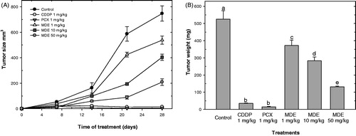
MDE administrated at 1, 10 and 50 mg/kg inhibited significantly (p ≤ 0.05) the MDA-MB231 tumor growth by 28, 46 and 72%, respectively, compared to untreated mice, whereas PCX and CDDP 1 mg/kg, each, inhibited the tumor growth by 98 and 99%, respectively (). MDE decreased significantly (p ≤ 0.05) tumor weight by 29% (1 mg/kg), 48% (10 mg/kg) and 75% (25 mg/kg), whereas PCX and CDDP, 1 mg/kg each, decreased the tumor weight by 99 and 97% ().
Figure 5. MDE induces chemopreventive effects in nu/nu mice bearing MDA-MB231 tumor. Mice were injected with MDA-MB231 cells (5 × 10⁶) and 4 h after tumor implantation, mice were treated with doses of MDE between 1 and 50 mg/kg, CDDP 1 mg/kg or PCX 1 mg/kg daily over a period of 28 days. At the end of the experiment, tumors were measured (A) and weighed (B) as described in the section “Materials and methods”. Data are representative of three independent experiments in quintuplicate. Results represent the mean ± standard deviation (SD). Lower case letters indicate significant differences according to ANOVA test (p ≤ 0.05), followed by the post hoc Tukey test.
Discussion
Magnolia dealbata is used in Centre Mexico for the empirical treatment of cancer (De la Cruz, Citation1964; Domínguez-Yescas, Citation2012). However, the cytotoxic effects of Magnolia dealbata are yet to be studied.
Phytochemical analysis showed that the contents of HK and MG in MDE were 11.6-fold higher and 3.5-fold higher, respectively, than those reported for Magnolia obovata (Ikarashi et al., Citation2001), and 2.9-fold higher (HK) and 0.2-fold lower (MG) than those reported for Magnolia officinalis (Karki et al., Citation2012). These results suggest that Magnolia dealbata may be an optimal source of the bioactive compounds HK and MG.
In previous work (Martinez et al., Citation2006), we evaluated the neuropharmacological effects of an ethanol extract from Magnolia dealbata leaves. In the present study, we found good quantities of HK and MG in an ethanol extract of Magnolia dealbata seeds (see above). In this study, we also quantified the levels of HK and MG in an ethanol extract of Magnolia dealbata leaves. However, we could not detect, by the HPLC analysis, HK or MG in Magnolia dealbata leaves (results not shown). Therefore, in this study, the cytotoxic and chemopreventive effects of Magnolia dealbata seeds were assayed. Plants synthesize and store secondary metabolites, usually in reproduction sites such as seeds, as a mechanism of protection against pathogen attack (Wink, Citation1988). This could explain, in part, why HK or MG was not detected in the ethanol extract of Magnolia dealbata leaves. In addition, other scientific groups have also used Magnolia dealbata seeds to analyze its pharmacological effects. Flores-Estévez et al. (Citation2013) showed that ethanol extract of Magnolia dealbata seed coat showed insecticidal activity against the Mexican fruit fly Anastrepha ludens.
The results indicated that MDE exerted high toxic effects on the viability and growth of human breast cancer cells. Other members of the Magnoliaceae family such as Magnolia denudate Desr, Magnolia officinalis Rehder &Wils, Magnolia grandiflora L and Magnolia obovata Thunb (IC50 = 84 µg/ml, HepG2) have shown cytotoxic and apoptotic effects against human cancer cells (Karki et al., Citation2012; Marin et al., Citation2010; Park et al., Citation2002). The results indicate that MDE (IC50 = 25 µg/ml, MDA-MB231 cells) showed similar or higher cytotoxic effects against human cancer cells, compared to other Magnolia species. The results also suggest that Magnolia species are an important source of cytotoxic compounds. In addition, here we showed that Magnolia dealbata exerts low toxic effects on non-tumorigenic cells. This is in accordance with our previous studies, which indicated that MDE showed low cytotoxic effects on human peripheral blood mononuclear cells, primary cultures of human fibroblasts and murine fibroblasts (Alonso-Castro et al., Citation2011b; Jacobo-Salcedo et al., Citation2011).
Induction of apoptosis is considered as one of the key mechanisms for the targeted therapy of various cancers (Constantini et al., Citation2000). To analyze whether MDE has the potential effect to induce apoptosis in cancer cells, MDA-MB231 cells were treated with MDE and analyzed by the TUNEL assay. MDE showed similar pro-apoptotic potency on MDA-MB231 cells compared to CDDP. The results show that Magnolia dealbata exerts cytotoxic and apoptotic effects against cancer cells.
Activation of caspases is a crucial point when the cells become committed to apoptosis (Elmore, Citation2007). The cleavage of caspase 3 induces DNA fragmentation, degradation of cytoskeletal and nuclear proteins, cross-linking of proteins, formation of apoptotic bodies, expression of ligands for phagocytic cell receptors and finally uptake by phagocytic cells (Elmore, Citation2007). In this study, we observed an activation of caspase 3 induced by MDE, by immunofluorescence assay, which was cleaved at 24 h and 48 h of treatment. This indicates that the apoptotic effects of MDE are caspase dependent.
Cancer chemoprevention is defined as an intervention in the carcinogenic process by a chemical that either blocks neoplastic induction or prevents transformed cells from progressing to a malignant phenotype (Amin et al., Citation2005). Cancer chemoprevention is regarded as one of the most efficient strategies for cancer control. In the last three decades, phytochemicals have been proposed as an option for reducing the risk of human population for developing cancer (Amin et al., Citation2005). The MDE doses used to evaluate chemopreventive effects were selected based on their lack of toxic effects in rodents, as evaluated in acute toxicity test (Martinez et al., Citation2006) and on preliminary studies carried out in our laboratory. In this study, we showed that MDE decreased the growth of MDA-MB231 tumor xenograft in nude mice. This effect suggested that MDE exerts chemopreventive effects on the in vivo growth of MDA-MB231 breast cancer cells. Although the capability of MDE 50 mg/kg to decrease tumor growth and weight was lower compared to CDDP 1 mg/kg, this plant extract did not have any effect on body weight in mice (results not shown). In contrast, CDDP reduced body weight by 11% (results not shown).
Conclusions
The results indicate for the first time that the ethanol extract of Magnolia dealbata seeds exerts cytotoxic and apoptotic effects, in a caspase-dependent manner, on MDA-MB231 cells. MDE also exerted chemopreventive effects on the growth of MDA-MB231 tumor xenograft in nude mice. Further experiments are necessary to be carried out in order to evaluate the mechanisms by which MDE exerts its chemopreventive effects.
Declaration of interest
The authors declare that there are no conflicts of interest.
Acknowledgements
A.J.A.C. (174493) and I.G.S. (226748) were provided with graduate fellowships from CONACYT. We acknowledge generous grant support from Instituto de Ciencia y Tecnología del Gobierno del Distrito Federal (ICyT-GDF; GI/PIFUTP08-142 to A.G.C.), and Consejo Nacional de Ciencia y Tecnología- México (CONACYT-México Grant 127822 to A.G.C.).
References
- Alonso-Castro AJ, Villarreal ML, Salazar-Olivo LA, et al. (2011a). Mexican medicinal plants used for cancer treatment: Pharmacological, phytochemical and ethnobotanical studies. J Ethnopharmacol 133:945–72
- Alonso-Castro AJ, Zapata-Bustos R, Domínguez F, et al. (2011b). Magnolia dealbata Zucc and its active principles honokiol and magnolol stimulate glucose uptake in murine and human adipocytes using the insulin-signaling pathway. Phytomedicine 18:926--33
- Amin A, Alkaabi A, Al-Falasi S, Daoud SA. (2005). Chemopreventive activities of Trigonella foenum graecum (Fenugreek) against breast cancer. Cell Biol Int 29:687–94
- Constantini P, Jocotot E, Decaudin D, Kroemer G. (2000). Mitochondrion as a novel target of anticancer chemotherapy. J Natl Cancer Inst 92:1042–53
- De la Cruz M. (1964). Libellus de medicinalibus indorum herís. México: IMSS
- Dominguez F, Chavez M, Garduño-Ramirez ML, et al. (2009). Production of honokiol and magnolol in suspension cultures of Magnolia dealbata Zucc. Nat Prod Commun 4:939–43
- Domínguez-Yescas R. (2012). Estudio etnobiológico de Magnolia dealbata Zucc en San Juan Juquila Vijanos, Oaxaca [Ethnobiological study of Magnolia dealbata Zucc in San Juan Juquila Vijanos, Oaxaca] [bachelor thesis]. Universidad de la Sierra Juarez
- Elmore S. (2007). Apoptosis: A review of programmed cell death. Toxicol Pathol 35:495–516
- Flores-Estévez N, Vasquez-Morales SG, Cano-Medina T, et al. (2013). Insecticidal activity of raw ethanolic extracts from Magnolia dealbata Zucc on a tephritid pest. J Environ Sci Health B 48:582–6
- González-Sánchez I, Solano JD, Loza-Mejia MA, et al. (2011). Antineoplastic activity of the thiazolo [5,4-b] quinoline derivative D3CLP in K562 cells is mediated through effector caspase activation. Eur J Med Chem 46:2102–8
- Ikarashi Y, Yuzurihara M, Sakakibara I, et al. (2001). Effects of the extract of the bark of Magnolia obovata and its biphenolic constituents magnolol and honokiol on histamine release from peritoneal mast cells in rats. Planta Med 67:709–13
- Jacobo-Salcedo MdR, Gonzalez-Espindola LA, Alonso-Castro AJ, et al. (2011). Antimicrobial activity and cytotoxic effects of Magnolia dealbata and its active compounds. Nat Prod Commun 6:1121–4
- Karki R, Jeon ER, Kim DW. (2012). Magnoliae Cortex inhibits intimal thickening of carotid artery through modulation of proliferation and migration of vascular smooth muscle cells. Food Chem Toxicol 50:634–40
- Lee YJ, Lee YM, Lee CK, et al. (2011). Therapeutic applications of compounds in the Magnolia family. Pharmacol Ther 130:157–76
- Looney WB, Mayo AA, Janners MY, et al. (1971). Cell proliferation and tumor growth in hepatomas 3924A. Cancer Res 31:821–5
- Marin GH, Mansilla E. (2010). Apoptosis induced by Magnolia grandiflora extract in chlorambucil-resistant B-chronic lymphocytic leukemia cells. J Cancer Res Ther 6:463–5
- Martinez AL, Dominguez F, Orozco S, et al. (2006). Neuropharmacological effects of an ethanol extract of the Magnolia dealbata Zucc leaves in mice. J Ethnopharmacol 106:250–5
- Park KJ, Yang S, Eun YA, et al. (2002). Cytotoxic effects of Korean medicinal herbs determined with hepatocellular carcinoma cell lines. Pharm Biol 40:189–95
- Sánchez-Velásquez LR, PinedaLópez MdR. (2010). Comparative demographic analysis in contrasting environments of Magnolia dealbata: An endangered species from Mexico. Pop Ecol 52:203–10
- Tsai TH, Chen CF. (1992). Identification and determination of honokiol and magnolol from Magnolia officinalis by high-performance liquid chromatography with photodiode-array UV detection. J Chromatogr 598:143–6
- Wink M. (1988). Plant breeding: Importance of plant secondary metabolites for protection against pathogens and herbivores. Theor Appl Genet 75:225–33