
Abstract
Context: Atorvastatin is a member of the drug class known as statins, which is used for lowering blood cholesterol.
Objective: The present study investigates the effect and mechanism of atorvastatin on neointimal hyperplasia after carotid artery injury (CAI) of rat.
Materials and methods: Fifty male rats were randomly divided into four groups: control group, sham-operated group, model group, and atorvastatin treatment group. The treatment group was fed with atorvastatin (10 mg/kg) with gastro-gavage at 5 p.m. every day for 28 d after surgery. The control group, model group, and sham-operated group were fed with the same volume of distilled water instead. The proliferations of intimal and medial layers were evaluated by hematoxylin & eosin (H&E) staining. The apoptosis of vascular smooth muscle cells (VSMCs) was determined by terminal deoxynucleotidyl transferased UTP nick end labeling (TUNEL) staining. Plasma concentrations of survivin and sFas were detected by enzyme-linked immunosorbent assay (ELISA).
Results: Atorvastatin reduced neointimal formation and increased apoptosis of VSMCs in neointima. VSMCs apoptosis emerged at 3 d (8.42 ± 0.449 μm) and the intimal proliferation peaked by the end of 14 d (41.58 ± 1.64 μm). The plasma levels of survivin and sFas were gradually increased with the neointimal hyperplasia and increasingly decreased after atorvastatin treatment. The plasma levels of survivin and sFas in rats were elevated at 3 d (464.80 ± 105.27 pg/ml and 3256.00 ± 478.20 pg/ml, respectively), reached the peak of survivin at 14 d (1089.20 ± 232.32 pg/ml) and sFas at 7 d (4362.00 ± 639.92 pg/ml) and decreased at 28 d (562.00 ± 90.11 pg/ml and 2148.00 ± 257.14 pg/ml, respectively) in the model group. Compared with the model group, the atorvastatin treatment group has significantly less neointimal hyperplasia and more apoptosis of VSMCs.
Conclusions: Atorvastatin can inhibit neointimal hyperplasia and promote SMCs apoptosis in neointimal layers, which may be mainly associated with down-regulation of survivin and Fas expression after CAI of rat.
Introduction
The use of drug-eluting stent for preventing restenosis has created a stir in interventional cardiology. Although preclinical studies showed modest improvements in restenosis, the first human trials of drug-eluting stents promised its complete demise. The first experience in humans from the RAVEL trial (Morice et al., Citation2002) showed no restenosis and pointed mechanistically to almost complete inhibition of neointimal formation. However, a large number of recent studies showed that restenosis was markedly reduced rather than eliminated. Previous studies demonstrated that the rate of restenosis is 7.6–18.9% in patients implanted with paclitaxel–eluting stents (Dawkins et al., Citation2005; Lansky et al., Citation2005; Mehilli et al., Citation2006) and 6.9–14.2% in patients treated with sirolimus-eluting stents (Dibra et al., Citation2005; Kastrati et al., Citation2005; Mehilli et al., Citation2006). These results indicated that we need to study more the effects of drug eluting, and it is necessary to seek a new method to prevent restenosis.
Recently, numerous studies suggested that statins have a variety of beneficial effects on cardiovascular disease. The study by Preusch et al. (Citation2010) showed that rosuvastatin attenuates neointima formation without affecting early MMP-9 activity in a rat model of vascular injury (Preusch et al., Citation2010). Another investigation by Makuuchi et al. (2005) found that pravastatin can potentially reduce atherosclerotic progression in both the bypass graft and native coronary arteries of patients after coronary artery bypass surgery. The atheroma study indicates that pravastatin 10–20 mg/d for 3 years improves hyperlipidemia, then suppresses progression, and induces regression of focal coronary atherosclerosis in Japanese patients diagnosed as coronary artery disease with elevated serum cholesterol (Yokoi et al., Citation2005). Although this beneficial effect of statins has been suggested to be independent of lipid-lowering properties, the possible mechanisms remain largely unknown.
The present study investigates the effects and mechanisms of atorvastatin on neointimal hyperplasia after carotid artery injury (CAI) in a rat model by detecting the neointimal formation and the apoptosis of vascular smooth muscle cells (VSMCs), the plasma levels of survivin and sFas, as well as the protein and mRNA levels of survivin and Fas in injury site. We found that atorvastatin may inhibit neointimal hyperplasia after vascular injury via down-regulation of survivin and Fas expression, and provides a new explanation for beneficial effects of statins on cardiovascular diseases.
Materials and methods
Animals
The experiment was performed in the National Key Laboratory for Medical Genetics, Central Laboratory of Xiangya Second Hospital of Central South University from April 2004 to March 2005. A total of 50 male rats aged 4–6 weeks, with a body weight of 250–300 g, were purchased from the Central Laboratory of Xiangya Second Hospital of Central South University (Changsha, China). Animals were randomly divided into four groups: control group (n = 5), sham-operated group (n = 5), model group (n = 20), and atorvastatin treatment group (n = 20). Rats were housed in a humidified room with a constant temperature (25 °C), free access to water, and food for at least 1 week before the experiment. All procedures were approved by the Central South University Institutional Animal Care and Use Committee and in accordance with the Institutional Guidelines for Care and Use of Laboratory Animals of Central South University of China.
Rat carotid injury model establishment, administration, and specimen collection
The rats, apart from the control group, were intraperitoneally anesthetized with 1% pentobarbital sodium (30 mg/kg). Surgical procedures were performed by the sterile surgical technique. The right carotid artery was surgically exposed, bluntly dissected at approximately 1 cm, clipped in both the ends, and injected the air three times with a 20-ml syringe. Then the needle was pulled out, blood flow was restorated by declamping the carotid artery, and the wound was sutured layer by layer. The sham-operated group was performed the same surgery except injecting the air. All rats were intramuscularly injected penicillin sodium (120 mg/kg) for 3 d after surgery to prevent infection. The treatment group was fed with atorvastatin (10 mg/kg, manufacturer Pfizer, New York, NY) with gastro-gavage at 5 p.m. every day for 28 d after surgery. The control group, model group, and sham-operated group were fed with the same volume of distilled water instead.
The rats underwent CAI (n = 5 in each group) were sacrificed by intraperitoneal injection with over-dose pentobarbital sodium (60 mg/kg) at 3, 7, 14, and 28 d after surgery, whereas the rats in the control group and the sham-operated group were euthanized at 14 d after surgery. Both the injured and the contralateral uninjured carotid arteries were harvested. Half of them were designated for the isolation of total RNA. Another half was used for paraffin embedding, cut into 4 µm thick slices, and performed hematoxylin & eosin (H&E) as well as terminal deoxynucleotidyltransferase (TdT)-mediated dUTP nick-end labeling (TUNEL) staining.
Morphological analysis
Four-micrometer thick sections were deparaffinized, rehydrated, stained with H&E, and then measured using SPOT advanced software (v. 3.2.5, Diagnostic Instruments, Sterling Heights, MI). For each measurement, three separate sections from different parts of the vessel were analyzed, and the thickness of the neointima, the media, as well as intima/media ratio was quantified. In order to determine the cell type in neointima, we performed α-actin immunohistochemitry and found that 99% cells in neointima are positive for α-actin, indicating that these cells are VSMCs.
Analysis of apoptosis and proliferation
Apoptosis was analyzed by using a TUNEL kit (Jingmei Company, Zhejiang, China) according to the manufacturer’s protocol. The 4% paraformaldehyde fixed and paraffin-embedded vessel sections were deparaffinized, rehydrated, and then treated with 20 µg/ml proteinase K for 30 min. Both terminal deoxynucleotidyltransferase (TdT) enzyme and fluorescein-dUTP were added to the tissue sections in accordance with the manufacturer’s instructions. Nuclei were counterstained with hematoxylin. The total nuclei for TUNEL-positive were counted in three separate sections from different regions of each vessel. The rate of apoptosis was expressed as the percentage of TUNEL-positive nuclei.
Measurement of survivin and sFas by enzyme-linked-immunosorbent assay (ELISA)
At the time of sacrifice, blood samples (4 ml for each rat) were collected by cardiac puncture and centrifuged for 5 min at 3000g/min, and then the plasma supernatant was removed and stored frozen at −70 °C until assayed. The plasma levels of survivin and sFas in rat were detected by ELISA with survivin ELISA kit (R&D systems, Minneapolis, MN) and sFas ELISA kit (GeneMay Company, San Diego, CA) according to the manufacturer’s instructions, respectively.
Detection of survivin and Fas mRNA expression by RT-PCR
The mRNA expression of survivin and Fas were detected by RT-PCR. The following primers were used: survivin forward 5′-TAAGCCACTTGTCCCAGCTT-3′ and reverse 5′-AGGATGGTACCCCATTACCT-3′ (product 391 bp); Fas forward 5′-CCTCCTGTTACAGACCTC-3′ and reverse 5′-CGCCTATGGTTGTTGACC-3′ (product 477 bp); β-actin forward 5′-CGCTGCGCTGGTCGTCGACA-3′ and reverse 5′-GTCACGCACGATTTCCCGCT-3′ (product 612 bp). Total RNA was isolated by the TRI reagent (MBI) method with RT-PCR kit (GeneMay Company, San Diego, CA). The RT reaction system involved 1 µg of total RNA, 4 µl of 5× RT buffer, 1 µl of oligo (dt) primer (0.5 µg/µl), 1 µl of RibolockTMRNAase inhibitor (20 µg/µl), 2 µl of 2.5 µM of each deoxynucleotidetriphosphate, and 1 µl of RevertaidTM M-MULV reverse transcriptase (200 µg/µl), reaching a final volume of 20 µl by adding ddH2O. The RT reactions were incubated at 42 °C for 1 h. PCR amplifications were performed on a PTC-100 Programmable Thermal Controller (Biozym Diagnostic, Hess, Oldendorf, Germany). The 3 µl of cDNA mixture was amplificated in a total of 25 µl mixture containing 2.5 units of Taq polymerase, 2.5 µl of 10× PCR buffer, 2.5 mM of each of dATP, dCTP, dGTP, and dTTP, and 25 pmol of each of 5′ and 3′ primers. PCR conditions were as follows: initial denaturation at 95 °C for 2 min, followed by 35 cycles of denaturation at 94 °C for 30 s, annealing at 62 °C (for survivin) or 58 °C (for Fas) or 60 °C (for β-actin) for 1 min, extension at 72 °C for 1 min, and a final extension at 72 °C for 10 min.
Western blotting analysis of protein expression of survivin and Fas
The total protein was isolated using TRI reagent (MBI, Sigma-Aldrich Chemie Gmbh Munich, Germany). The concentration of protein was determined using Bio-Rad protein assay solution. Up to 50 µg of total protein was denatured in 2× SDS sample loading buffer at 100 °C for 5 min, separated on 12% SDS-PAGE gels, and electrotransferred to AC membranes (Millipore, Bedford, MA) using semidry electrophoretic transfer. After then specific binding sites on the membranes were blocked with 5% skimmed milk in TBS-T (20 mM Tris–HCl, pH 7.5, 0.137 M NaCl, and 0.01% Tween 20) at room temperature for 1 h, the membranes were incubated in TBS-T containing survivin (1:1000, Novus Biologicals, Inc., Littleton, CO) and Fas (1:200, Santa Cruz Biotechnology, Santa Cruz, CA) antibody at room temperature for 2 h. After washed with TBS-T three-times, the membrane was incubated in 5% skimmed milk in TBS-T buffer containing the appropriate second anti-IgG antibody (1:5000, NeoMarkers, Fremont, CA) at room temperature for 1 h. The target protein was detected by the ECL protein detection kit (Amersham Biosciences) following the manufacturer’s instructions and visualized by autoradiography with the Kodak X-Omat AR film (Roche Applied Science, Branford, CT). For normalization of protein loading, the same procedure with a monoclonal antibody against tubulin (1:1000, NeoMarker) was performed as an internal standard.
Statistical analysis
All numeric data were expressed as the mean ± SD. The means were statistically compared by ANOVA, followed by the Student t-test. All data analyses were performed with SPSS v 20.0 (SPSS, Chicago, IL). A value of p < 0.05 was considered statistically significant.
Results
Reduced neointimal formation after treatment with atorvastatin
In the model group, the neointima formed at 3 d (8.42 ±0.449 μm), significantly proliferated at 7 d (31.87 ± 3.10 μm), and peaked at 14 d (41.58 ± 1.64 μm) after injury. The neointimal hyperplasia in the atorvastatin treatment group was significantly lower at 7, 14, and 28 d (13.72 ± 1.25 μm, 12.30 ± 0.99 μm, and 13.62 ± 1.56 μm, respectively) than that in the model group (all p < 0.01; and ). Therefore, compared with the model group, the atorvastatin treatment group has a markedly lower intima/media (I/M) ratio at 7, 14, and 28 d (all p < 0.01; and ).
Figure 1. The intimal hyperplasia after vascular injury. A1, 7 d after vascular injury in group without treatment with significant intimal hyperplasia, right carotid artery. B1, 7 d after vascular injury and with 10 mg/kg atorvastatin treatment group with less intimal hyperplasia, right carotid artery. A2, 14 d after vascular injury in group without atorvastatin treatment with significant intimal hyperplasia, right carotid artery. B2, 14 d after vascular injury and with 10 mg/kg atorvastatin treatment group, right carotid artery, less intimal hyperplasia than without treatment group. A3, 28 d after vascular injury in group without atorvastatin treatment with significant intimal hyperplasia, right carotid artery. B3, 28 d after vascular injury in group with 10 mg/kg atorvastatin treatment, right carotid artery, less intimal hyperplasia than group without treatment. The directions of arrow are neointima.
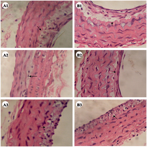
Table 1. The change of intimal media and intimal/media I/M ratio after vascular injury with or without treatment with atorvastatin.
Increased apoptosis of VSMCs in neointima treated with atorvastatin
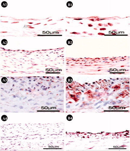
There were different degrees of VSMCs apoptosis determined by TUNEL staining in neointima from the atorvastatin treatment group. Interestingly, accompanied by the reduction of neointimal formation, the TUNEL-positive cells were notably increased in neointima treated with atorvastatin, indicating that atorvastatin can induce VSMCs apoptosis and may contribute to inhibit neointimal formation ().
Figure 2. The results of TUNEL staining. A1, 2, 3, and 4 are 3, 7, 14, and 28 d after vascular injury in groups without treatment, respectively. We can see significant intimal hyperplasia, and less TUNEL-positive cells. B1, 2, 3, and 4 are 3, 7, 14, and 28 d after vascular injury in groups with atorvastatin treatment, respectively. We can see less intimal hyperplasia and more TUNEL-positive cells, indicated that atorvastatin can inhibit neointimal formation and induce SMCs apoptosis.
Atorvastatin can drastically reduce the plasma levels of survivin and sFas
The plasma levels of survivin and sFas in rats were elevated at 3 d (464.80 ± 105.27 pg/ml and 3256.00 ± 478.20 pg/ml, respectively), reached the peak of survivin at 14 d (1089.20 ± 232.32 pg/ml) and sFas at 7 d (4362.00 ±639.92 pg/ml), and decreased at 28 d (562.00 ± 90.11 pg/ml and 2148.00 ± 257.14 pg/ml, respectively) in the model group (). Compared with the rats in the model group, those in the atorvastatin treatment group have significantly lower plasma concentrations of survivin and sFas at 7, 14, and 28 d (all p < 0.01; ), respectively.
Table 2. The changes of levels of survivin and sFas in the plasma of rats after treated with atorvastatin.
Fas protein and mRNA expression persisted while survivin expression is down-regulated after treatment with atorvastatin
Persisted Fas protein and mRNA expression can be detected in both the model and atorvastatin treatment groups but not in the control and sham-operated groups. However, the protein and mRNA expression of survivin at 7 d or earlier after vascular injury were not detected in the model group. Notably, the protein and mRNA levels of survivin can be measured in the model group but still not in the atorvastatin treatment group at 14 and 28 d. This indicated that atorvastatin can down-regulate survivin mRNA level when it suppressed the neointimal proliferation ( and ).
Figure 3. The expression of Fas in local vascular injury sites. (A) Relative levels of mRNAs encoding Fas in local vascular injury sites. Lane 1, β-actin. Lane 2, normal control artery with no Fas mRNA expression. Lanes 3, 4, and 5 are 28, 14, and 7 d after vascular injury and with atorvastatin treatment groups, respectively, there are Fas mRNA expressions. Lanes 6, 7, 8, and 9 is 28, 14, 7, and 3 d after vascular injury but without atorvastatin treatment, respectively, we can also see various types of Fas mRNA expression. Lane 10, marker. (B) Relative level of Fas expression was analyzed by Western Blotting. (B1) Tubulin, as an internal standard; (B2) Fas western blotting analysis. Lanes 1, 2, and 3 are 28, 14, and 7 d after vascular injury and with atorvastatin treatment groups, respectively. Lanes 4, 5, 6, and 7 are 28, 14, 7, and 3 d after vascular injury but without atorvastatin treatment, respectively. Fas protein expression is similar to the Fas mRNA expression.
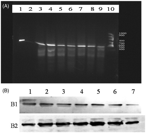
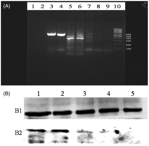
Figure 4. The expression of survivin in local vascular injury site. (A) Relative levels of mRNAs encoding survivin expression. Lane 1, blank control. Lane 2, the normal control group, no survivin mRNA expression. Lanes 3 and 4 were β-actin. Lanes 5 and 6 is 28 and 14 d after vascular injury in group without treatment, respectively, these are survivin mRNA expression. Lane 7 is 7 d after vascular injury in group without treatment, no survivin mRNA expression. Lanes 8 and 9 is 28 and 14 d after vascular injury in groups with atorvastatin treatment, respectively, no survivin mRNA expression. Lane 10, marker. (B) Relative level of survivin expression was analyzed by Western blotting. (B1) Tubulin, as an internal standard; (B2) survivin protein. Lanes 1, 2, and 3 are 28, 14, and 7 d after vascular injury in group without treatment, respectively. Lanes 4 and 5 are 28 and 14 d after vascular injury in group with atorvastatin treatment. Survivin expression is similar to the survivin mRNA.
Discussion
The current study demonstrated that atorvastatin can significantly inhibit the neointimal hyperplasia and promote VSMCs apoptosis in neointimal layers at 7, 14, and 28 d after vascular injury. This result is consistent with the conclusions from previous studies. Indolfi et al. (Citation2000) reported that simvastatin potently affected in vitro VSMC proliferation and in vivo prevented neointimal formation after vascular injury. This beneficial effect was abolished using local administration of mevalonate. Schafer et al. (Citation2005) found that the neointimal area and the severity of luminal stenosis were significantly reduced in rosuvastatin-treated mice after endothelial injury; these effects are independent of systemic lipid lowering. Komukai et al. (Citation1999) revealed that carvastatin inhibited dose-dependently VSMC migration and proliferation with IC50 values of 0.5 μM and 1 μM, respectively, suggesting that carvastatin may be useful in rabbits as an anti-atherogenic drug by inhibiting VSMC migration or proliferation. Therefore, these findings indicated that statins have a significant effect on the inhibition of neointimal formation after vascular injury.
However, the mechanism of statins in the reduction of neointimal formation after vascular injury remains incompletely elucidated. A study in a rabbit model demonstrated that balloon injury caused upregulation of C×40 and C×43 inneointimal VSMC. Lovastatin and fluvastatin suppressed upregulated C×40 and C×43 expression and reduced neointimal proliferation, suggesting that C×40 and C×43 may play a role in statin-induced antiproliferative effect (Wang et al., Citation2005b). In addition, an in vitro experiment showed that pitavastatin induced NO production by vascular endothelial cells and protected vascular endothelial cells from injury (Tokoro et al., Citation2004). Similarly, another in vitro study revealed that a new statins drug R780 can protect the vascular endothelium from oxidant stress and inhibit the migration and proliferation of VSMC (Wajima et al., Citation2003). In this study, we observed that atorvastatin can simultaneously decrease survivin levels and neointimal formation, especially in the earlier stage. The effects of atorvastatin on reduction of neointimal formation may contribute to the increase of VSMCs apoptosis in neointimal layers and the down-regulation of survivin expression, which can be further confirmed by a significant positive correlation between plasma survivin level and neointimal thickness. To our knowledge, this is the first study to elucidate the mechanism of atorvastatin in the inhibition of neointimal formation after CAI.
Survivin, a unique member of the inhibitor of apoptosis proteins family, is a bifunctional protein that regulates cell division and suppresses apoptosis (Altieri & Marchisio, Citation1999). Although highly expressed in fetal tissues (Adida et al., Citation1998; Altieri & Marchisio, Citation1999; Li et al., Citation1998) and a variety of human cancers (Xia et al., Citation2006; Yonesaka et al., Citation2006), survivin is undetectable in most normal and terminally differentiated adult tissues (Adida et al., Citation1998). It has been demonstrated that inhibition of survivin expression or interference of surivin function induces cancer cell death (Takashima et al., Citation2005; Yonesaka et al., Citation2006). Thus, survivin appears to be an important cancer therapeutic target, and modulation of survivin expression and/or function may provide effective strategies for cancer therapeutics (Takashima et al., Citation2005; Yonesaka et al., Citation2006). However, its potential role is poorly understood in cardiovascular proliferation diseases such as restenosis after PCI.
Blanc-Brude et al. reported that balloon-mediated arterial injury in rabbits resulted in the expression of survivin in vascular cells. Serum or PDGF-AB can stimulate survivin expression, which prevented caspase-3,7 activation and suppressed the apoptosis of cultured VSMCs. Adenoviral delivery of a phosphorylation-defective survivin mutant reversed the cytoprotective effect of PDGF in VSMCs without affecting mitotic progression, suppressed neointimal formation in wire-injured mouse femoral arteries, and induced VSMCs apoptosis in vivo. These data identify survivin as a critical regulator of VSMC apoptosis after acute vascular injury. Disrupting the survivin pathway may provide a novel therapy to limit pathological vessel-wall remodeling (Blanc-Brude et al., Citation2002). Wang et al. (Citation2005a) observed that survivin is an important regulator of multiple processes, including proliferation, apoptosis, and angiogenesis, that determines the remodeling response of vein grafts following arterializations. Similar to the result by Simosa et al. (Citation2005) that delivery of survivin mutants (AdT34A) after balloon injury attenuated neointimal hyperplasia, Daniel et al. (Citation2012) revealed that survivin played a crucial role in inhibiting the proliferative response of VSMCs and neointima formation mediated by a highly potent inhibitor of transcription-3. In addition, our previous in vitro experiment found that VSMCs apoptosis induced by atorvastatin may be mainly associated with down-regulation of survivin expression (Xu et al., Citation2007). In line with the above investigations, the present in vivo study identified that the inhibition of neointimal proliferation by atorvastatin may closely be related to VSMCs apoptosis and down-regulation of survivin levels after vascular injury.
Likewise, our knowledge about the effects of Fas on neointimal formation after vascular injury is very limited. Studies have showed that Fas can induce cell apoptosis by binding Fas ligand (FasL). Soluble Fas (sFas) is an anti-apoptotic factor because it can combine with FasL and prohibited the combination of Fas and FasL and its effects. A clinical study revealed that lower serum levels of sFas seemed to be associated with the development of instent restenosis (ISR) and determination of serum serum levels before and after percutaneous coronary intervention (PCI) might help in identifying patients at higher risk of ISR (Katsaros et al., Citation2011). Matter et al. (Citation2006) reported that proapoptotic and anti-inflammatory effects of endogenous FasL are of much importance in the process of neointimal lesion formation after balloon injury, indicating that the activation of FasL may decrease neointimal thickening after PCI. Luo et al. (Citation2001) found that coexpression of p35 in FasL-transduced VSMCs is more potent at inhibiting neointimal formation and as such represents an improved gene therapy approach for restenosis. Wang et al. (Citation2002) identified that blocking Fas/Fas-L interaction by overexpression of sFas in graft endothelium inhibited vascular cell apoptosis as well as CD45+ mononuclear cell infiltration, and significantly attenuated the disruption of the arterial wall and transplant arteriosclerosis.
Our data showed that protein and mRNA expression of Fas were persistent after vascular injury in the model and the atorvastatin treatment groups. Therefore, the effect of atorvastatin on Fas protein and mRNA expression needs further study. However, sFas plasma levels were significantly decreased after atorvastatin treatment, suggesting that sFas expression was inhibited after treatment with atorvastatin. So, inhibition of neointimal formation by atorvastatin is partially associated with down-regulation of sFas expression because sFas is an anti-apoptosis factor (Kamihira & Yamada, Citation2001).
Our study may have two limitations. One is that the effect of different doses of atorvastatin on the neointimal hyperplasia was not observed. Another is that the detailed mechanism on the inhibition of neointima proliferation such as the signal pathway involved still needs further investigation.
Conclusions
Our data showed that Fas expression was persistent in vascular injury sites while survivin expression emerged only in a later stage after vascular injury. Atorvastatin treatment can down-regulate survivin expression in vascular injury sites and induce VSMCs apoptosis in neointimal layers, and suppress the neointimal formation after vascular injury. The mechanism of atorvastatinon inhibition of neointimal formation after vascular injury may be mainly associated with down-regulation survivin and Fas expression in vascular injury sites. Inhibiting the survivin and Fas expression with atorvastatin may provide a novel therapy to prevent neointimal hyperplasia and restenosis.
Declaration of interest
The authors report no declarations of interest.
Acknowledgements
We wish to express our warm thanks to all the authors who contributed to the study.
References
- Adida C, Crotty PL, McGrath J, et al. (1998). Developmentally regulated expression of the novel cancer anti-apoptosis gene survivin in human and mouse differentiation. Am J Pathol 152:43–9
- Altieri DC, Marchisio PC. (1999). Survivin apoptosis: An interloper between cell death and cell proliferation in cancer. Lab Invest 79:1327–33
- Blanc-Brude OP, Yu J, Simosa H, et al. (2002). Inhibitor of apoptosis protein survivin regulates vascular injury. Nat Med 8:987–94
- Daniel JM, Dutzmann J, Bielenberg W, et al. (2012). Inhibition of STAT3 signaling prevents vascular smooth muscle cell proliferation and neointima formation. Basic Res Cardiol 107:261
- Dawkins KD, Grube E, Guagliumi G, et al. (2005). Clinical efficacy of polymer-based paclitaxel-eluting stents in the treatment of complex, long coronary artery lesions from a multicenter, randomized trial: Support for the use of drug-eluting stents in contemporary clinical practice. Circulation 112:3306–13
- Dibra A, Kastrati A, Mehilli J, et al. (2005). Paclitaxel-eluting or sirolimus-eluting stents to prevent restenosis in diabetic patients. N Engl J Med 353:663–70
- Indolfi C, Cioppa A, Stabile E, et al. (2000). Effects of hydroxymethylglutaryl coenzyme A reductase inhibitor simvastatin on smooth muscle cell proliferation in vitro and neointimal formation in vivo after vascular injury. J Am Coll Cardiol 35:214–21
- Kamihira S, Yamada Y. (2001). Soluble Fas (APO-1/CD95) isoform in adult T-cell leukemia. Leuk Lymphoma 41:169–76
- Kastrati A, Dibra A, Eberle S, et al. (2005). Sirolimus-eluting stents vs paclitaxel-eluting stents in patients with coronary artery disease: Meta-analysis of randomized trials. JAMA 294:819–25
- Katsaros KM, Wiesbauer F, Speidl WS, et al. (2011). High soluble Fas and soluble Fas ligand serum levels before stent implantation are protective against restenosis. Thromb Haemost 105:883–91
- Komukai M, Wajima YS, Tashiro J, et al. (1999). Carvastatin suppresses intimal thickening of rabbit carotid artery after balloon catheter injury probably through the inhibition of vascular smooth muscle cell proliferation and migration. Scand J Clin Lab Invest 59:159–66
- Lansky AJ, Costa RA, Mooney M, et al. (2005). Gender-based outcomes after paclitaxel-eluting stent implantation in patients with coronary artery disease. J Am Coll Cardiol 45:1180–5
- Li F, Ambrosini G, Chu EY, et al. (1998). Control of apoptosis and mitotic spindle checkpoint by survivin. Nature 396:580–4
- Luo Z, Garron T, Palasis M, et al. (2001). Enhancement of Fas ligand-induced inhibition of neointimal formation in rabbit femoral and iliac arteries by coexpression of p35. Hum Gene Ther 12:2191–202
- Makuuchi H, Furuse A, Endo M, et al. (2005). Effect of pravastatin on progression of coronary atherosclerosis in patients after coronary artery bypass surgery. Circ J 69:636–43
- Matter CM, Chadjichristos CE, Meier P, et al. (2006). Role of endogenous Fas (CD95/Apo-1) ligand in balloon-induced apoptosis, inflammation, and neointima formation. Circulation 113:1879–87
- Mehilli J, Kastrati A, Wessely R, et al. (2006). Randomized trial of a nonpolymer-based rapamycin-eluting stent versus a polymer-based paclitaxel-eluting stent for the reduction of late lumen loss. Circulation 113:273–9
- Morice MC, Serruys PW, Sousa JE, et al. (2002). A randomized comparison of a sirolimus-eluting stent with a standard stent for coronary revascularization. N Engl J Med 346:1773–80
- Preusch MR, Vanakaris A, Bea F, et al. (2010). Rosuvastatin reduces neointima formation in a rat model of balloon injury. Eur J Med Res 15:461–7
- Schafer K, Kaiser K, Konstantinides S. (2005). Rosuvastatin exerts favourable effects on thrombosis and neointimal growth in a mouse model of endothelial injury. Thromb Haemost 93:145–52
- Simosa HF, Wang G, Sui X, et al. (2005). Survivin expression is up-regulated in vascular injury and identifies a distinct cellular phenotype. J Vasc Surg 41:682–90
- Takashima H, Nakajima T, Moriguchi M, et al. (2005). In vivo expression patterns of survivin and its splicing variants in chronic liver disease and hepatocellular carcinoma. Liver Int 25:77–84
- Tokoro T, Wang J, Kitajima I. (2004). The novel HMG-CoA reductase inhibitor, pitavastatin, induces a protective action in vascular endothelial cells through the production of nitric oxide (NO). Yakugaku Zasshi 124:121–6
- Wajima T, Makita S, Oshima K. (2003). Direct vascular effects of HR780, a novel 3-hydroxy-3-methylglutaryl coenzyme A reductase inhibitor. Clin Exp Pharmacol Physiol 30:958–62
- Wang GJ, Sui XX, Simosa HF, et al. (2005a). Regulation of vein graft hyperplasia by survivin, an inhibitor of apoptosis protein. Arterioscler Thromb Vasc Biol 25:2081–7
- Wang L, Chen J, Sun Y, et al. (2005b). Regulation of connexin expression after balloon injury: Possible mechanisms for antiproliferative effect of statins. Am J Hypertens 18:1146–53
- Wang T, Dong C, Stevenson SC, et al. (2002). Overexpression of soluble Fas attenuates transplant arteriosclerosis in rat aortic allografts. Circulation 106:1536–42
- Xia W, Bisi J, Strum J, et al. (2006). Regulation of survivin by ErbB2 signaling: Therapeutic implications for ErbB2-overexpressing breast cancers. Cancer Res 66:1640–7
- Xu YG, Zhou SH, Li YG, et al. (2007). The mechanism underlying vascular smooth muscle cell apoptosis induced by atorvastatin may be mainly associated with down-regulation of survivin expression. Cardiovasc Drugs Ther 21:145–53
- Yokoi H, Nobuyoshi M, Mitsudo K, et al. (2005). Three-year follow-up results of angiographic intervention trial using an HMG-CoA reductase inhibitor to evaluate retardation of obstructive multiple atheroma (ATHEROMA) study. Circ J 69:875–83
- Yonesaka K, Tamura K, Kurata T, et al. (2006). Small interfering RNA targeting survivin sensitizes lung cancer cell with mutant p53 to adriamycin. Int J Cancer 118:812–20