
Abstract
Context In clinical practice, the promotion of neuron survival is necessary to recover neurological functions after the onset of stroke.
Objective This study aimed to investigate the post-ischaemic neuroprotective effect of SMND-309, a novel metabolite of salvianolic acid, on differentiated SH-SY5Y cells.
Materials and methods SH-SY5Y cells were differentiated by pre-treating with 5 μM all-trans-retinoic acid for 6 d. The differentiated SH-SY5Y cells were exposed to oxygen–glucose deprivation (OGD) for 2 h and reperfusion (R) for 24 h to induce OGD/R injury. After OGD injury, differentiated SH-SY5Y cells were treated with or without SMND-309 (5, 10, 20 μM) for another 24 h. Cell viability was detected through Cell counting kit-8 assay and lactate dehydrogenase leakage assay. Apoptosis was evaluated through flow cytometry, caspase-3 activity assay. Changes in protein levels were assessed through Western blot.
Results SMND-309 ameliorated the degree of injury in the differentiated SH-SY5Y cells by increasing cell viabilities (5 μM, 65.4% ± 4.1%; 10 μM, 69.8% ± 3.7%; 20 μM, 75.3% ± 5.1%) and by reducing LDH activity (20 μM, 2.5 fold) upon OGD/R stimulation. Annexin V-fluorescein isothiocyanate/propidium iodide staining results suggested that apoptotic rate of differentiated SH-SY5Y cells decreased from 43.8% induced by OGD/R injury to 19.2% when the cells were treated with 20 μM SMND-309. SMND-309 significantly increased the Bcl-2 level of the injured differentiated SH-SY5Y cells but decreased the caspase-3 activity of these cells by 1.6-fold. In contrast, SMND-309 did not affect the Bax level of these cells. SMND-309 evidently increased the protein expression of BDNF when Akt and CREB were activated. This function was antagonized by the addition of LY294002.
Conclusion SMND-309 can prevent neuronal cell death in vitro. This process may be related to the activation of the PI3K/Akt/CREB-signalling pathway.
Introduction
Stroke is the primary cause of long-term disability and death worldwide (Iadecola & Anrather Citation2011; Lee et al. Citation2014). Ischemic stroke caused by arterial occlusions constitutes approximately 80% of all stroke cases (Wang et al. Citation2010a). Moreover, ischaemia and reperfusion induce serious brain injury as a result of reactive oxygen species (ROS) overproduction, calcium overload and blood–brain barrier injury (Lee et al. Citation2000; Wilson et al. Citation2003). Repair and functional recovery after brain damage are major challenges for current clinical and basic research. The inhibition of cell death or the activation of cell survival can protect cells after ischaemia/reperfusion injury occurs (Song et al. Citation2014; Wang et al. Citation2014b); thus, agents that can prevent neuronal apoptosis likely exhibit therapeutic potential against brain ischaemia/reperfusion.
Medicinal plant-derived natural compounds, such as ginsenosides (Rb1 and Rg1) and curcumin, are beneficial for the treatment of ischaemic cerebrovascular disorders (Mook-Jung et al. Citation2001; Kim et al. Citation2008; Shang et al. Citation2013). Salvianolic acid (Sal B) is a major aqueous compound found in Salvia miltiorrhiza Bunge(Lamiaceae), which is a commonly used traditional Chinese herb. Sal B is also widely applied in experimental and clinical research to limit ischaemic cerebrovascular disease (Jiang et al. Citation2015a; Tang et al. Citation2006). SMND-309, with the chemical name of (2E)-2-{6-[(E)-2-carboxyvinyl]-2,3-dihydroxyphenyl}-3-(3,4-dihydroxyphenyl) propenoic acid, is a novel metabolite produced in the brains and hearts of rats after Sal B is orally administered. This metabolite has also been obtained in vitro by scientists from the Department of Chemistry, Shandong Engineering Research Center for Natural Drugs (Shandong, China) (Chinese patent: CN 200710015108.8). This metabolite elicits strong neuroprotective effects by promoting angiogenesis and by inhibiting cell death and cerebral oedema (Tian et al. Citation2009; Zhu et al. Citation2013). However, the mechanism of SMND-309 post-conditioning against cerebral ischaemia–reperfusion injury remains unclear. In this study, a model of ischaemia/reperfusion-like damage was used to elucidate the potential molecular mechanism of the neuroprotective effect of SMND-309.
Materials and methods
Cell culture and treatment
Human neuroblastoma cell line SH-SY5Y was purchased from the Cell Bank of the Chinese Academy of Sciences (Shanghai, China). The cells were maintained at 37 °C in Dulbecco’s modified Eagle’s Medium (DMEM) supplemented with 10% newborn calf serum under a humidified atmosphere of 5% CO2. The SH-SY5Y cells were treated with 5 μM all-trans-retinoic acid (ATRA) (Sigma, St. Louis, MO) in DMEM in the dark for 6 d to induce neuronal differentiation. The cells with axon lengths that were twice those of their neuronal cell bodies were regarded as positive cells. Six visual fields were randomly selected from each slide by two blinded observers. At least 200 cells were counted. The differentiated cells were divided into four groups: control group, model group, SH-SY5Y group, and SH-SY5Y + LY294002 (10 μM) group (n = 6 per group).
Oxygen–glucose deprivation/reperfusion (OGD/R) model
The OGD/R model was constructed in accordance with a previously described method (Liu et al. Citation2011) with slight modifications. In brief, the neurons were washed thrice with Earle’s balanced salt solution without glucose. The neurons were then cultured with 0.5 mmol/L sodium dithionite in glucose-free Earle’s balanced salt solution for 2 h to induce deoxygenated injury. The medium was subsequently replaced with basal medium without or with different SMND-309 concentrations in 5% CO2 and 95% air. This condition was maintained for another 24 h. The control cells were treated with Earle’s balanced salt solution containing glucose for 2 h and incubated in basal medium under treatments similar to those of the OGD/R cells.
Cell counting kit-8 (CCK-8) assay
Cell viability was detected through CCK-8 assay in accordance with the manufacturer’s instructions. CCK-8 solution (10 μL) was added to each well of a 96-well plate and then incubated for 4 h at 37°C. The absorbance was determined using a microplate reader (Molecular Devices, Silicon Valley, CA) at a reference wavelength of 540 nm.
Lactate dehydrogenase (LDH) leakage assay
The amount of LDH that leaked into the culture supernatants was detected using an LDH diagnostic kit (Jiancheng Bioengineering Institute, Nanjing, China) in accordance with the manufacturer’s instructions to detect neuron injury. The absorbance was determined using a microplate reader (Molecular Devices, Silicon Valley, CA) at 490 nm. LDH was calculated using the following equation:
Caspase-3 activity assay
Caspase-3 activity was determined using commercially available caspase-3 colorimetric assay kits (Sigma, St. Louis, MO). In brief, total protein was extracted from the cells, and protein concentration was quantified using a bicinchoninic acid protein assay kit (Beyotime Institute of Biotechnology, Haimen, Jiangsu, China). Protein samples were incubated with the caspase-3 substrate N-acetyl-Asp-Glu-Val-Asp p-nitroanilide for 1.5 h at 37 °C. p-Nitroanilide was detected at 405 nm using a microplate reader (Molecular Devices, Silicon Valley, CA).
Measurement of apoptosis through flow cytometry
After the treatment was administered, the suspended and adherent cells were collected and washed with cold phosphate-buffered saline. Binding buffer (500 μL) was added to re-suspend the cells. Annexin V-fluorescein isothiocyanate (FITC; 5 μL) and propidium iodide (PI) staining solution (5 μL) were also added; the cells were then incubated for 20 min in the dark. The apoptosis rate was measured through flow cytometry (Beckman, Brea, CA).
Western blot analysis
The proteins in the treated cells were extracted, and the protein concentration was quantified using a bicinchoninic acid protein assay kit. Later on, the protein extracts were boiled in the sample buffer in a water bath for 5 min. The protein samples were separated through 15% sodium dodecyl sulphate-polyacrylamide gel electrophoresis for 2 h. The samples were subsequently transferred onto a polyvinylidene difluoride membrane and blocked with 5% non-fat milk for 1.5 h. The blots were incubated overnight at 4 °C with the following specific primary antibodies: BDNF, Akt, Bax, Bcl2 and CREB. Species-specific horseradish peroxidase-conjugated secondary antibodies containing IgG were then added to the membrane. The membrane was further incubated for 1 h at room temperature. Immunoreactive bands were visualized using an ECL-assay kit.
Statistical analysis
Data were presented as mean ± SD. Unpaired Student’s t-test was applied in the experiments that compared two groups; one-way ANOVA was performed with the Student–Newman–Keuls post hoc test for the experiments that compared three or more groups. Analyses were performed using SPSS version 11.0 for Windows (SPSS Inc., Chicago, IL). p < 0.05 was considered statistically significant.
Results
Attenuation of OGD/R-induced cytotoxicity in the differentiated SH-SY5Y cells by SMND-309
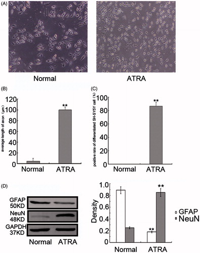
The SH-SY5Y cells were differentiated by pre-treating with 5 μM ATRA for 6 d to determine the neuroprotective effect of SMND-309. The morphological characteristics of the ATRA-treated cells were significantly modified by the appearance of axon-like processes with long axons, which are a typical neuronal phenotype (). Furthermore, almost 90% of the SH-SY5Y cells were successfully induced by ATRA, and the neurites of the treated SH-SY5Y cells were 22.9-fold longer than those of the control cells (, p < 0.01). Furthermore, the differentiated cells also exhibited a neuronal characteristic with the increased expression of the neuronal marker NeuN; in contrast, the astrocytic marker GFAP was almost not expressed in the differentiated cells (, p < 0.01).
Figure 1. ATRA-induced differentiation of SH-SY5Y cells. (A) Representative morphological images of undifferentiated and differentiated SH-SY5Y cells. (B) Change in the axonal length of the cells. (C) Bar graph representing the percentage of differentiated cells relative to the total number of cells counted and summarized. At least 500 cells were counted per parallel hole. (D) Protein levels of NeuN and GFAP in undifferentiated and differentiated SH-SY5Y cells were detected by Western blot analysis. Data are shown as mean ± SD, n = 6, **p < 0.01 versus the normal group as measured by unpaired Student’s t-tests.
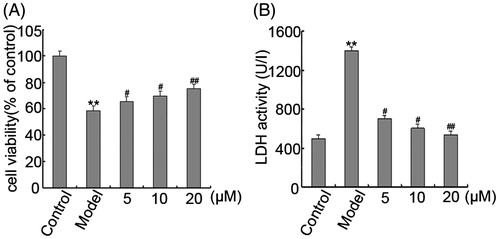
The cytotoxicity of SMND-309 on the differentiated SY5Y cells was investigated through the CCK-8 assay. The resultant growth curve indicated that the viability of the differentiated SY5Y cells was not affected by SMND-309 from 0 mM to 0.2 mM (, p < 0.05). The OGD/R model was applied to the differentiated SH-SY5Y cells to simulate ischaemic injury. The viability of the differentiated SH-SY5Y cells exposed to 2 h of OGD and 24 h of reperfusion was lower than that of the non-stimulated control cells. In contrast, SMND-309-pretreated cells exhibited increased viabilities of 65.4 ± 4.1%, 69.8 ± 3.7% and 75.3 ± 5.1% at 5, 10 and 20 μM, respectively (, p < 0.05 and p < 0.01).
The protective effect of SMND-309 against OGD/R-induced cytotoxicity was further confirmed through the LDH leakage assay. The results showed that OGD/R induced a 2.8-fold increase in the LDH activities of the media; conversely, 20 μM SMND-309 significantly reduced the OGD/R-induced LDH leakage by 2.5-fold. Thus, 20 μM was the most appropriate SMND-309 concentration for the subsequent experiments (, p < 0.05 and p < 0.01).
Figure 2. Growth curve of the differentiated SH-SY5Y cells treated with different SMND-309 concentrations. Data are shown as mean ± SD, n = 6, *p < 0.05 versus the control group as measured by unpaired Student’s t-tests.
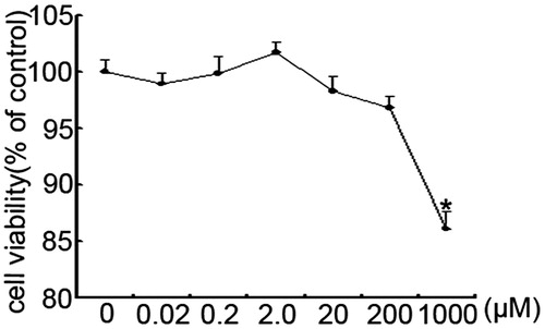
Figure 3. Attenuation of OGD/R-induced cytotoxicity in differentiated SH-SY5Y cells by SMND-309. (A) SMND-309 increased the viability of the OGD/R-injured differentiated SH-SY5Y cells. (B) LDH release by differentiated SH-SY5Y cells was detected by LDH leakage assay. Data are shown as mean ± SD, n = 6, **p < 0.01 versus the control group; #p < 0.05 and ##p < 0.01 versus the model group as measured by the Student–Newman–Keuls test.
Inhibition of the OGD/R-induced-apoptosis of differentiated SH-SY5Y cells by SMND-309
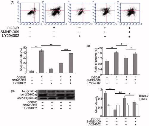
We determined whether or not the long-term survival of the SMND-309-treated SH-SY5Y cells was associated with the inhibition of apoptosis. Annexin V-FITC/PI staining was performed to quantify cell apoptosis. Further, the involvement of PI3K/Akt pathway in the anti-apoptotic mechanism of SMND-309 was checked using LY294002 (10 μM), a PI3K-specific inhibitor. Flow cytometry analysis showed that the apoptotic rate of cells increased from 4.6% representing the basal levels of apoptosis to 43.8% in differentiated SH-SY5Y cells injured by OGD/R; in contrast, the apoptosis was strongly suppressed by treating with 20 μM of SMND-309. Moreover, treatment with LY294002 evoked apoptosis in differentiated SH-SY5Y cells (, p < 0.01). Western blot analysis results indicated that SMND-309 upregulated the expression of the antiapoptotic protein Bcl-2 but failed to downregulate the Bax level (, p < 0.05 and p < 0.01). The activity and the level of caspase-3 were also ameliorated by SMND-309 treatment, these results confirmed the Annexin-V-FITC/PI staining assay findings (, p < 0.05 and p < 0.01). However, this anti-apoptotic effect was reversed when the cells were incubated with LY294002 (p < 0.05 and p < 0.01).
Figure 4. Inhibition of OGD/R-mediated apoptosis in differentiated SH-SY5Y cells by SMND-309. (A) SMND-309 reduced the apoptosis rate of differentiated SH-SY5Y cells as determined by Annexin V-FITC/PI assay. (B) SMND-309 inhibited the activity of caspase-3. (C) Immunoblot analysis highlighted the changes in the expression of apoptotic proteins Bcl2 and Bax. Data are shown as mean ± SD, n = 6, **p < 0.01 versus the control group; #p < 0.05 and ##p < 0.01 versus the OGD/R group; △p < 0.05, △△p < 0.01 versus the OGD/R + SMND-309 group as measured by the Student–Newman–Keuls test.
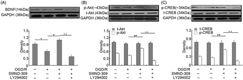
Activation of the neuroprotective pathways of PI3K/Akt/CREB/BDNF by SMND-309
Brain-derived neurotrophic factor (BDNF) is implicated in neuronal survival, synaptic plasticity and cognitive processes (Linares et al. Citation2013). Thus, we investigated the effect of SMND-309 on the BDNF expression. Western blot analysis demonstrated that the BDNF levels of the OGD/R-injured differentiated SH-SY5Y cells were lower than those of the control cells. Conversely, SMND-309 significantly restored the BDNF expression. This positive effect was partly abrogated when the cells were incubated with LY294002 (, p < 0.05 and p < 0.01). Afterwards, we detected the effect of SMND-309 on PI3K-related proteins, namely Akt and CREB. Under OGD/R conditions, the expression of intracellular t-Akt and t-CREB did not change significantly. In contrast, p-Akt and p-CREB were considerably downregulated in the treated cells compared with those in the control cells. In the PI3K-blocked cells, SMND-309 did not show any significant increase in the p-Akt and p-CREB expression levels under OGD/R conditions; in contrast, the upregulated effect was not influenced in the non-blocked cells (, p < 0.01).
Figure 5. Modulation of SMND-309 on PI3K pathway-related proteins with or without LY294002. (A) Representative immunoblot of the protein levels of BDNF in OGD/R-injured differentiated SH-SY5Y cells treated with SMND-309 with or without LY294002. (B) Immunoblot result of the protein levels of Akt and p-Akt. (C) Protein levels of CREB and p-CREB. Quantification analysis results are also presented. Data are shown as mean ± SD, n = 6, *p < 0.05 versus the control group; #p < 0.05 and ##p < 0.01 versus the OGD/R group; △△p < 0.01 versus the OGD/R + SMND-309 group as measured by the Student–Newman–Keuls test.
Discussion
Ischaemia/reperfusion injury is a serious pathological condition that induces various cellular and molecular responses in the brain. The system of cortical neuron cultures subjected to OGD/R has been widely used as an in vitro model to mimic the effects of ischaemia/reperfusion injury in ischemic cerebrovascular disease (Zhang et al. Citation2007). ATRA, a vitamin A derivative, plays an essential role in the development of the nervous system (Wenker et al. Citation2010). ATRA is considered as an efficient inducer of neuronal differentiation in neuroblastoma cells; ATRA also induces these cells to obtain phenotypes, such as neurite outgrowth and morphological changes functionally and electrically similar to neurons (Sakane & Shidoji Citation2011; Wang et al. Citation2014c). Among several human neutoblastoma cell lines, SH-SY5Y is the most widely used in vitro neuronal differentiation (Sallmon et al. Citation2010). Hence, the in vitro model of OGD/R injury in ATRA-treated SH-SY5Y cells is suitable to investigate the neurophysiological changes associated with ischemic stroke; this model can also facilitate the development of therapeutic drugs or potential targets. Consistent with previous research, our study revealed the marked changes in the morphological characteristics of the cells and the increased number of the cells exhibiting a neuron-like phenotype after ATRA was administered. The highly expressed NeuN and non-detectable GFAP, which are specific differentiation markers, further confirmed the course of neuronal differentiation indicating ATRA-induced neuronal differentiation in SH-SY5Y cells.
Neuronal degeneration and death are two major pathophysiological changes that occur after the onset of cerebral ischaemia. Apoptosis is the main form of neurocyte death; this process plays a major role in the ischaemic penumbra during the progression of ischaemic stroke (Zhang et al. Citation2013a; Li et al. Citation2014a,Citationb). This damage is aggravated by reperfusion. The concept of post-conditioning through the modulation of reperfusion rather than ischaemia has been proposed in the treatment of ischemic diseases (Cheng et al. Citation2012; Sun et al. Citation2013; Li et al. Citation2014a,Citationb). Sal B can cross the blood–brain barrier and act directly on neuronal cells. Furthermore, Sal B reduces OGD/R-induced injury in primary rat cortical neurons by reducing the amount of ROS and by increasing the activities of Mn-superoxide dismutase, catalase and glutathione peroxidase (Wang et al. Citation2010b). In our research, SMND-309 can protect the differentiated SH-SY5Y cells against OGD/R-induced injury by activating the PI3K/Akt/CREB/BDNF pathway.
BDNF is a member of the neurotrophin superfamily. This factor is implicated in synaptic plasticity, neuronal survival and development, and normal neuronal function in the nervous system (Croce et al. Citation2014; Xia et al. Citation2014). BDNF can protect different types of neurons from ischemic, traumatic and toxic brain injury (Weishaupt et al. Citation2012). BDNF can also inhibit caspase-3 activation and cell apoptosis caused by hypoxia–ischemic injury in vivo (Wang et al. Citation2013b). Moreover, BDNF affects the apoptotic processes of Raji cells, especially Bcl-2 and caspase-signalling pathways.
Sal B promotes the survival and differentiation of bone marrow-derived neural stem cells in unfavourable environments by inducing the BDNF production (Zhang et al. Citation2012). Consistent with previous findings, our results revealed that the protective effect of SMND-309 on OGD/R-induced neuron injury is correlated with the BDNF upregulation.
The PI3K/Akt/CREB pathway plays a critical role in neuronal cell survival and cell death; thus, this pathway has been investigated in the stroke research (Sun et al. Citation2008). Yu et al. (Citation2014) reported that the proliferation of folic acid-stimulated neural stem cell is dependent on the PI3K/Akt/CREB pathway. Activated PI3K recruits Akt (protein kinase B), as well as the kinases that act on Akt, to the cell membrane and is responsible for the activation of Akt, which directly or indirectly affects CREB (Choi Citation2011a,Citationb).
As the downstream target of PI3K, Akt is a good candidate that mediates cell survival responses via the phosphorylation of multiple downstream signalling molecules, including the inactivation of the Bcl-2 family member Bad and the inhibition of the apoptotic enzyme caspase-9 (Seo & Choi Citation2012). Huang et al. (Citation2015a) noted that 4-(2-(1H-imidazole-1-yl)ethoxy)-3-methoxybenzoic acid strongly prevents apoptotic cell death by regulating Bax and Bcl-2 expression in the PI3K/Akt pathway. PI3K/Akt contains numerous downstream effectors, including mTOR, CREB and CDK. CREB is a ubiquitous nuclear transcription factor in higher eukaryotes and is abundant in the brain, particularly in neurons. The number of surviving neurons is closely correlated with the upregulation of the p-CREB expression in the penumbra regions (Nakajima et al. Citation2002). Moreover, the activation of CREB triggers the transcription of many genes associated with memory, learning, neuroprotection, synaptic transmission, neuronal survival, cell differentiation and axonal growth (Freitas et al. Citation2013). Huang et al. (Citation2015b) reported that mice die immediately after birth as a result of CREB knockout. CREB is positively regulated by the PI3K/Akt pathway in PC12 cells, neonatal cardiomyocytes, striatal neurons and osteoblastic MC3T3-E1 cells (Brami-Cherrier et al. Citation2002; Choi & Lee Citation2011). The Akt/CREB-signalling pathway contributes to the protective effect of nobiletin on neurological impairment and tissue injury following cerebral ischaemia (Zhang et al. Citation2013b). Sal B can protect H9c2 cells against arsenic trioxide-induced myocardial toxicity; however, this protection is dependent on the PI3K/Akt-signalling pathway (Wang et al. Citation2013a). Zhuang et al. (Citation2012) revealed that Sal B can maintain self-renewal and promote the proliferation of neural stem/precursor cells by activating the PI3K/Akt signal pathway. In our study, LY294002 antagonized the SMND-309-induced phosphorylation of Akt and CREB.
The protective effect of BDNF on hippocampal neurons against apoptosis is mediated by PI3K and Ras/MAPK-signalling pathways; this mediation involves a long-term change in protein synthesis (Novkovic et al. Citation2015). BDNF is a CREB-responsive gene controlled by the cAMP response element and regulated by CREB in neurons (Jin et al. Citation2009; Wang et al. Citation2014a). Melemedjian et al. (Citation2014) reported that CREB acts upstream of BDNF in DRG neurons at 3 h after IL-6 treatment. Consistent with previous findings, our results demonstrated that p-CREB levels were strongly correlated with and could be responsible for the observed changes in the BDNF expression upon SMND-309 treatment possibly through the activation of the PI3K/Akt pathway. PI3K/Akt activity can be affected by multiple regulators, such as phosphatase and tensin homolog, PH domain leucine-rich repeat protein phosphatases and the transcriptional factor FOXO3 (Jiang et al. Citation2015b). Nonetheless, further studies must be conducted to explore the underlying molecular mechanisms of SMND-309 in the regulation of the PI3K/Akt-signalling pathway.
In conclusion, the pharmacological protective effect of SMND-309 on OGD/R-induced neuron damage was elucidated in this study. This protection required an intracellular increase in BDNF partly via the PI3K/Akt/CREB-dependent signalling pathways. This finding provides scientific evidence for the pharmacological mechanism of SMND-309.
Funding information
This study was supported by Taishan Scholar Project to Qingyin Zheng and National Natural Science Foundation of China (Nos. 81271085 and 31300288) and Natural Science Foundation of Shandong Province (ZR2011CM021, ZR2012HQ042, ZR2013HM051 and 2014GSF119014).
Disclosure statement
The authors report that they have no conflicts of interest. The authors alone are responsible for the content and writing of this article.
References
- Brami-Cherrier K, Valjent E, Garcia M, Hipskind RA, Caboche J. 2002. Dopamine induces a PI3-kinase-independent activation of Akt in striatal neurons: a new route to cAMP response element-binding protein phosphorylation. J Neurosci. 22:8911–8921.
- Cheng F, Zhong X, Lu Y, Wang X, Song W, Guo S, Wang X, Liu D, Wang Q. 2012. Refined Qingkailing protects MCAO mice from endoplasmic reticulum stress-induced apoptosis with a broad time window. Evid Based Complement Alternat Med. 2012:567872.
- Choi EM. 2011a. Luteolin protects osteoblastic MC3T3-E1 cells from antimycin A-induced cytotoxicity through the improved mitochondrial function and activation of PI3K/Akt/CREB. Toxicol In Vitro. 25:1671–1679.
- Choi EM. 2011b. Kaempferol protects MC3T3-E1 cells through antioxidant effect and regulation of mitochondrial function. Food Chem Toxicol. 49:1800–1805.
- Choi EM, Lee YS. 2011. Involvement of PI3K/Akt/CREB and redox changes in mitochondrial defect of osteoblastic MC3T3-E1 cells. Toxicol In Vitro. 25:1085–1088.
- Croce N, Bernardini S, Caltagirone C, Angelucci F. 2014. Lithium/valproic acid combination and l-glutamate induce similar pattern of changes in the expression of miR-30a-5p in SH-SY5Y neuroblastoma cells. Neuromol Med. 16:872–877.
- Freitas AE, Machado DG, Budni J, Neis VB, Balen GO, Lopes MW, de Souza LF, Dafre AL, Leal RB, Rodrigues AL. 2013. Fluoxetine modulates hippocampal cell signaling pathways implicated in neuroplasticity in olfactory bulbectomized mice. Behav Brain Res. 237:176–184.
- Huang J, Kodithuwakku ND, He W, Zhou Y, Fan W, Fang W, He G, Wu Q, Chu S, Li Y. 2015a. The neuroprotective effect of a novel agent N2 on rat cerebral ischemia associated with the activation of PI3K/Akt signaling pathway. Neuropharmacology. 95:12–21.
- Huang W, Cao J, Liu X, Meng F, Li M, Chen B, Zhang J. 2015b. AMPK plays a dual role in regulation of CREB/BDNF pathway in mouse primary hippocampal cells. J Mol Neurosci. 56:782–788.
- Iadecola C, Anrather J. 2011. The immunology of stroke: from mechanisms to translation. Nat Med. 17:796–808.
- Jiang YF, Liu ZQ, Cui W, Zhang WT, Gong JP, Wang XM, Zhang Y, Yang MJ. 2015b. Antioxidant effect of salvianolic acid B on hippocampal CA1 neurons in mice with cerebral ischemia and reperfusion injury. Chin J Integr Med. 21:516–522.
- Jiang L, Wang C, Lei F, Zhang L, Zhang X, Liu A, Wu G, Zhu J, Song L. 2015a. miR-93 promotes cell proliferation in gliomas through activation of PI3K/Akt signaling pathway. Oncotarget. 6:8286–8299.
- Jin CH, Shin EJ, Park JB, Jang CG, Li Z, Kim MS, Koo KH, Yoon HJ, Park SJ, Choi WC, et al. 2009. Fustin flavonoid attenuates beta-amyloid (1-42)-induced learning impairment. J Neurosci Res. 87:3658–3670.
- Kim SJ, Son TG, Park HR, Park M, Kim MS, Kim HS, Chung HY, Mattson MP, Lee J. 2008. Curcumin stimulates proliferation of embryonic neural progenitor cells and neurogenesis in the adult hippocampus. J Biol Chem. 283:14497–14505.
- Lee JM, Grabb MC, Zipfel GJ, Choi DW. 2000. Brain tissue responses to ischemia. J Clin Invest. 106:723–731.
- Lee KF, Chen JH, Teng CC, Shen CH, Hsieh MC, Lu CC, Lee KC, Lee LY, Chen WP, Chen CC, et al. 2014. Protective effects of Hericium erinaceus mycelium and its isolated erinacine A against ischemia-injury-induced neuronal cell death via the inhibition of iNOS/p38 MAPK and nitrotyrosine. Int J Mol Sci. 15:15073–15089.
- Linares M, Marín-García P, Pérez-Benavente S, Sánchez-Nogueiro J, Puyet A, Bautista JM, Diez A. 2013. Brain-derived neurotrophic factor and the course of experimental cerebral malaria. Brain Res. 1490:210–224.
- Liu R, Zhang L, Lan X, Li L, Zhang TT, Sun JH, Du GH. 2011. Protection by borneol on cortical neurons against oxygen-glucose deprivation/reperfusion: involvement of anti-oxidation and anti-inflammation through nuclear transcription factor κappaB signaling pathway. Neuroscience. 176:408–419.
- Li B, Sun J, Lv G, Yu Y, Wang G, Xie K, Jiao Y, Yu Y. 2014a. Sevoflurane postconditioning attenuates cerebral ischemia-reperfusion injury via protein kinase B/nuclear factor-erythroid 2-related factor 2 pathway activation. Int J Dev Neurosci. 38:79–86.
- Li Z, Hu H, Lin R, Mao J, Zhu X, Hong Z, Tao J, Zhang Y, Chen L. 2014b. Neuroprotective effects of Gua Lou Gui Zhi decoction against glutamate-induced apoptosis in BV-2 cells. Int J Mol Med. 33:597–604.
- Melemedjian OK, Tillu DV, Moy JK, Asiedu MN, Mandell EK, Ghosh S, Dussor G, Price TJ. 2014. Local translation and retrograde axonal transport of CREB regulates IL-6-induced nociceptive plasticity. Mol Pain. 10:45.
- Mook-Jung I, Hong HS, Boo JH, Lee KH, Yun SH, Cheong MY, Joo I, Huh K, Jung MW. 2001. Ginsenoside Rb1 and Rg1 improve spatial learning and increase hippocampal synaptophysin level in mice. J Neurosci Res. 63:509–515.
- Nakajima T, Iwabuchi S, Miyazaki H, Okuma Y, Inanami O, Kuwabara M, Nomura Y, Kawahara K. 2002. Relationship between the activation of cyclic AMP responsive element binding protein and ischemic tolerance in the penumbra region of rat cerebral cortex. Neurosci Lett. 331:13–16.
- Novkovic T, Mittmann T, Manahan-Vaughan D. 2015. BDNF contributes to the facilitation of hippocampal synaptic plasticity and learning enabled by environmental enrichment. Hippocampus. 25:1–15.
- Sakane C, Shidoji Y. 2011. Reversible upregulation of tropomyosin-related kinase receptor B by geranylgeranoic acid in human neuroblastoma SH-SY5Y cells. J Neurooncol. 104:705–713.
- Sallmon H, Hoene V, Weber SC, Dame C. 2010. Differentiation of human SH-SY5Y neuroblastoma cells by all-trans retinoic acid activates the interleukin-18 system. J Interferon Cytokine Res. 30:55–58.
- Seo MS, Choi EM. 2012. The effects of dehydrocostus lactone on osteoblastic MC3T3-E1 cells in redox changes and PI3K/Akt/CREB. Immunopharmacol Immunotoxicol. 34:810–814.
- Shang YH, Tian JF, Hou M, Xu XY. 2013. Progress on the protective effect of compounds from natural medicines on cerebral ischemia. Chin J Nat Med. 11:588–595.
- Song J, Kang SM, Lee WT, Park KA, Lee KM, Lee JE. 2014. The beneficial effect of melatonin in brain endothelial cells against oxygen-glucose deprivation followed by reperfusion-induced injury. Oxid Med Cell Longev. 2014:639531.
- Sun JJ, Liu Y, Ye ZR. 2008. Effects of P2Y1 receptor on glial fibrillary acidic protein and glial cell line-derived neurotrophic factor production of astrocytes under ischemic condition and the related signaling pathways. Neurosci Bull. 24:231–243.
- Sun L, Zhuang W, Xu X, Yang J, Teng J, Zhang F. 2013. The effect of injection of EGb 761 into the lateral ventricle on hippocampal cell apoptosis and stem cell stimulation in situ of the ischemic/reperfusion rat model. Neurosci Lett. 555:123–128.
- Tang M, Feng W, Zhang Y, Zhong J, Zhang J. 2006. Salvianolic acid B improves motor function after cerebral ischemia in rats. Behav Pharmacol. 17:493–498.
- Tian J, Fu F, Li G, Wang Y, Gao Y, Liu Z, Zhang S. 2009. SMND-309, a novel derivate of salvianolic acid B, ameliorates cerebral infarction in rats: characterization and role. Brain Res. 1263:114–121.
- Wang BY, Zhong Y, Zhao Z, Miao Y. 2014a. Epigenetic suppression of hippocampal BDNF mediates the memory deficiency induced by amyloid fibrils. Pharmacol Biochem Behav. 126:83–89.
- Wang CP, Li GC, Shi YW, Zhang XC, Li JL, Wang ZW, Ding F, Liang XM. 2014b. Neuroprotective effect of schizandrin A on oxygen and glucose deprivation/reperfusion-induced cell injury in primary culture of rat cortical neurons. J Physiol Biochem. 70:735–747.
- Wang K, Zhou F, Zhu X, Zhang K, Huang B, Zhu L, Zhu L. 2014c. Neuroprotective properties of ciliary neurotrophic factor on retinoic acid (RA)-predifferentiated SH-SY5Y neuroblastoma cells. Folia Neuropathol. 52:121–127.
- Wang M, Sun GB, Sun X, Wang HW, Meng XB, Qin M, Sun J, Luo Y, Sun XB. 2013a. Cardioprotective effect of salvianolic acid B against arsenic trioxide-induced injury in cardiac H9c2 cells via the PI3K/Akt signal pathway. Toxicol Lett. 216:100–107.
- Wang N, Wu L, Cao Y, Wang Y, Zhang Y. 2013b. The protective activity of imperatorin in cultured neural cells exposed to hypoxia re-oxygenation injury via anti-apoptosis. Fitoterapia. 90:38–43.
- Wang Q, Gong Q, Wu Q, Shi J. 2010a. Neuroprotective effects of Dendrobium alkaloids on rat cortical neurons injured by oxygen-glucose deprivation and reperfusion. Phytomedicine. 17:108–115.
- Wang Y, Jiang YF, Huang QF, Ge GL, Cui W. 2010b. Neuroprotective effects of salvianolic acid B against oxygen-glucose deprivation/reperfusion damage in primary rat cortical neurons. Chin Med J (Engl). 123:3612–3619.
- Weishaupt N, Blesch A, Fouad K. 2012. BDNF: the career of a multifaceted neurotrophin in spinal cord injury. Exp Neurol. 238:254–264.
- Wenker SD, Chamorro ME, Vota DM, Callero MA, Vittori DC, Nesse AB. 2010. Differential antiapoptotic effect of erythropoietin on undifferentiated and retinoic acid-differentiated SH-SY5Y cells. J Cell Biochem. 110:151–161.
- Wilson FC, Harpur J, Watson T, Morrow JI. 2003. Adult survivors of severe cerebral hypoxia-case series survey and comparative analysis. NeuroRehabilitation. 18:291–298.
- Xia D, Li W, Zhang L, Qian H, Yao S, Qi X. 2014. RNA interference-mediated knockdown of brain-derived neurotrophic factor (BDNF) promotes cell cycle arrest and apoptosis in B-cell lymphoma cells. Neoplasma. 61:523–532.
- Yu M, Li W, Luo S, Zhang Y, Liu H, Gao Y, Wang X, Wilson JX, Huang G. 2014. Folic acid stimulation of neural stem cell proliferation is associated with altered methylation profile of PI3K/Akt/CREB. J Nutr Biochem. 25:496–502.
- Zhang J, Lu W, Lei Q, Tao X, You H, Xie P. 2013a. Salvianolate increases heat shock protein expression in a cerebral ischemia-reperfusion injury model. Neural Regen Res. 8:2327–2335.
- Zhang L, Zhao H, Zhang X, Chen L, Zhao X, Bai X, Zhang J. 2013b. Nobiletin protects against cerebral ischemia via activating the p-Akt, p-CREB, BDNF and Bcl-2 pathway and ameliorating BBB permeability in rat. Brain Res Bull. 96:45–53.
- Zhang N, Kang T, Xia Y, Wen Q, Zhang X, Li H, Hu Y, Hao H, Zhao D, Sun D, et al. 2012. Effects of salvianolic acid B on survival, self-renewal and neuronal differentiation of bone marrow derived neural stem cells. Eur J Pharmacol. 697:32–39.
- Zhang ZG, Zhang QZ, Cheng YN, Ji SL, Du GH. 2007. Antagonistic effects of ultra-low-molecular-weight heparin against cerebral ischemia/reperfusion injury in rats. Pharmacol Res. 56:350–355.
- Zhu H, Zou L, Tian J, Tian J, Du G, Gao Y. 2013. SMND-309, a novel derivative of salvianolic acid B, protects rat brains ischemia and reperfusion injury by targeting the JAK2/STAT3 pathway. Eur J Pharmacol. 714:23–31.
- Zhuang P, Zhang Y, Cui G, Bian Y, Zhang M, Zhang J, Liu Y, Yang X, Isaiah AO, Lin Y, et al. 2012. Direct stimulation of adult neural stem/progenitor cells in vitro and neurogenesis in vivo by salvianolic acid B. PLoS One. 7:e35636.