Abstract
Objectives. Minimal access Aortic Valve Replacement (AVR) has been demonstrated to have beneficial effects over median sternotomy. Minimal extracorporeal circulation (MECC) has been shown to have less deleterious effects than conventional cardiopulmonary bypass. We describe for the first time AVR via upper J-shaped partial sternotomy compared to median sternotomy using MECC. Methods. Prospectively collected pre-operative, intra-operative, post-operative and follow-up data from 104 consecutive patients who underwent minimal access AVR were compared to 72 consecutive patients undergoing median sternotomy using MECC during the same period (January 2007 to December 2009). Results. No significant differences were found in patient's characteristics or intra-operative data with the exception of pre-existing pulmonary disease. The mean cardiopulmonary bypass (86 ± 18 min vs. 78 ± 15 min, p = 0.0079) and cross-clamp times (65 ± 13 min vs. 59 ± 12 min, p = 0.0013) were significantly shorter in the median sternotomy group. Mediastinal blood loss (397 ± 257 ml vs. 614 ± 339 ml, p < 0.0001) and ventilation time (8 ± 6.9 h vs. 11 ± 16.5 h, p = 0.0054) were significantly less in the minimal access group. No differences were seen in transfusion requirements, inotropic support, intensive care unit (ICU) stay, total hospital stay, post-operative haemoglobin drop, major events or mortality. Quality of life scores after discharge demonstrated less pain with a quicker recovery and return to daily activities in patients receiving J-shaped sternotomy. Conclusions. Minimal access AVR using MECC is feasible and provides excellent clinical results. Less pain and quicker recovery was experienced among patients in this group.
Median sternotomy has been the standard surgical approach for replacement of diseased aortic valves since the late 1950s (Citation1). Minimal access techniques for aortic valve replacement (AVR) are gaining popularity over the past 10 years. Better clinical outcomes by reducing pain and surgical trauma, improving cosmetics, decreasing blood loss, earlier functional recovery and shorter hospital stay reducing total costs have been demonstrated (Citation2–5). The majority of these operations have been performed with standard cardiopulmonary bypass (CPB). Many studies have demonstrated the deleterious effects of standard CPB like haemodilution, cytokine response and initiation of coagulation cascade (Citation6,Citation7). To overcome these drawbacks of CPB Wiesenack et al. in 2004, presented a retrospective series of patients undergoing coronary artery bypass surgery with minimal extracorporeal circulation (MECC) demonstrating reduction in post-operative complications and blood loss (Citation8). Other studies also have demonstrated the beneficial effects of MECC in lowering the post-operative inflammatory responses (Citation9,Citation10). In addition, AVR via standard median sternotomy utilising MECC has been shown to have better outcomes than AVR with conventional perfusion techniques (Citation11). Recently we have described our initial experience in minimal access AVR with the use of the MECC (Citation12). We now present a comparison of minimal access AVR to median sternotomy using MECC.
Material and methods
Data collection
One hundred and four patients were operated on using minimal access AVR using MECC from January 2007 to December 2009. These patients were compared to patients (n = 72) undergoing AVR using median sternotomy and MECC during the same period. The choice of technique was based on surgeon's preference. The local Ethical Committee approved this study and waived the need for individual consent. Data were collected prospectively. Patient characteristics for both groups are summarised in . Follow-up assessing patient satisfaction and post-operative quality of life (QOL) was performed by SF-12® health questionnaires at one, four, eight and 12 months (Citation12).
Table I. Baseline patient characteristics.
Operative technique
Anaesthetic management. Patients in both groups received standard premedication (diazepam 10 mg) one hour before arrival to the operating room. Induction of anaesthesia was performed with intravenous sufentanyl and propofol. Muscle relaxation was achieved with pancuronium (0.1 mg/kg). Anaesthesia was maintained with a combination of propofol (2–3 mg/kg/h) and isoflurane. A full dose of heparin (300 IU/kg intravenously) was given and activated clotting time was maintained above 400 seconds. On completion of the procedure heparin was reversed with protamine at 1:1 equivalent dosage.
All patients were transferred to the intensive care unit (ICU) remaining sedated and mechanically ventilated based on our local protocol. Criteria for the post-operative use of blood products did not differ between both groups and were: administration of packed red blood cells when haemoglobin less than 8.0 g/dl, fresh frozen plasma when International Normalised Ratio more than 2.0, thrombocytes when thrombocytes count was less than 100 × 109 per litre.
Cardiopulmonary bypass. Maquet HL30 heart lung machines (Maquet Cardiopulmonary, Hirrlingen, Germany) were used in both groups. MECC consisted of a totally closed Bioline heparin coated system circuit with rotaflow centrifugal pump, Quadrox-i microporeus membrane oxygenator and venous bubble trap (VBT) (Maquet Cardiopulmonary, Hirrlingen, Germany). A blood collection reservoir connected to the VBT was integrated in the circuit. No open venous reservoir was present. Autologus retrograde priming of the MECC was performed, reducing priming volume to 250 cm3. Cell saver drainage was used for intrapericardially blood shed. A pulmonary artery vent (Medtronic Inc, DLP catheter 13 Fr, Minneapolis, USA) was inserted via the main pulmonary trunk distal to the pulmonary valve. Optional sump suction directly through the aortotomy was used when necessary. Pulmonary artery vent was directly connected to the venous bubble trap maintaining the same level of vacuum suction. Aortic root vent ran via a drip chamber and was also connected to the venous bubble trap. Continuous carbon dioxide (CO2) field flooding (6 l/min) was maintained during the entire procedure. Antegrade warm blood cardioplegia (Calafiore, 1.7 mmol/ml potassium) was administered via the aortic root and repeated every 15–20 minutes thereafter selectively via the coronary ostia. Nasopharyngeal temperature was kept at 34°C.
Surgical procedure. Minimal access AVR using MECC was performed as previously described by Yilmaz et al. (Citation13). In short; the patient was in supine position with access to the groin for arterial and venous femoral canulation. A 4–5cm median, subjugular skin incision was performed in the upper sternal region. This was followed by a J-shaped partial sternotomy into the right third intercostal space, performed with an oscillating saw. First insertion of an appropriate sized femoral artery cannula (Medtronic Inc.) using Seldinger technique was performed. This was followed by insertion of a dual stage venous 21 to 25 Fr cannula (Medtronic Inc.) under transeosophageal echocardiography (TEE) guidance in a bicaval position till the tip of the cannula was visible in the superior vena cava using Seldinger technique via the femoral vein. The cross clamp was applied through the sternal incision. Patients receiving a full sternotomy were cannulated using venous access through the right atrial appendage and arterial access in the ascending aorta. A hockey stick aortotomy was performed to access the aortic valve in both groups. Excision of the diseased aortic valve was followed by placement of interrupted sutures (Ticron 2-0, Ethicon) with the pledgets subannular (see for operative data). The aortic incision was closed using a double layer technique (Blalock) and a ventricular pacemaker was placed. De-airing was performed by cessation of the pulmonary artery venting, filling of the heart, ventilation of the lungs in trendelenburg position and suction on the aortic root vent before release of the aortic clamp. The patients were weaned from CPB, when they were haemodynamically stable and no significant air was seen on TEE in the left-sided chambers of the heart. After achieving adequate haemostasis, the sternotomy was closed using sternal wires and the incision was closed in layers.
Table II. Intraoperative data.
Statistical analysis. Data were collected prospectively. Standard descriptive statistics were computed. For the comparison between MECC and the standard CPB group we used χ2 tests or Fisher's exact test for categorical data when appropriate, unpaired t-tests or Mann-Whitney tests for continuous data. All statistical analyses were performed using R (www.R-project.org). Continuous variables are expressed as mean ± standard deviation (SD).
Results
Patient demographics displayed a wide array of co-morbidities. Significantly higher prevalence of chronic obstructive pulmonary disease in the minimal invasive group was seen (). Aortic valve stenosis was present in 87% of the patients in the minimal invasive group (mean valve area of 0.74 ± 0.17 cm2, peak gradient of 84.5 ± 24.4 mmHg) compared to 86% in the standard median sternotomy group (mean valve area of 0.73 ± 0.16 cm2 and peak gradient of 88.8 ± 28.2 mmHg) (p = 0.3269 resp. 0.7149). Associated aortic valve incompetence, mostly Grade I to II was present in 38% of the patients in both groups. Intra-operative problems encountered in the minimal invasive group consisted of two conversions to a full sternotomy. In one patient femoral canulation was impossible due to calcifications and a second patient had a bleeding from the right atrium during canulation. In the full sternotomy group procedure was complicated due to air lock through leakage of the venous cannula. This occurred during removal of the aortic cross clamp with ventricular rhythm and adequate cardiac output. There were no consequences for the patient.
The mean cardiopulmonary bypass and cross-clamp times were significantly shorter in the standard median sternotomy group compared to the minimal invasive group. No other differences between the two groups regarding intra-operative data were seen (). The post-operative mediastinal blood loss (397 ml ± 257 ml vs. 614 ml ± 339 ml) and the post-operative ventilation time (8 h ± 6.9 h vs. 11 h ± 16.5 h) were significantly less in the minimal access group compared to the full sternotomy. Post-operative pain (as measured by visual analogue scale at two days post-operative) was significantly less in the partial sternotomy group. No differences were seen in post-operative haemoglobin drop, transfusion requirements, inotropic needs, ICU stay, total hospital stay, major adverse cardiac events or mortality ().
Table III. Postoperative complications and transfusion requirements.
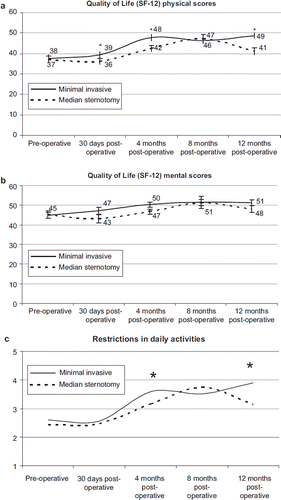
Average follow-up period was 25 ± 9 months in the minimal invasive group compared to 25 ± 12 months in the median sternotomy group with an average overall response rate of 50%. In the standard median sternotomy group no correlation between responders and age or gender were found. In the minimal invasive group responders were younger and predominantly male compared to the non- responders. During follow-up at one, four, eight and 12 months post-operative no deaths occurred. Pre-operative SF-12 questionnaires revealed no clinically relevant differences between groups. In both groups psychological and functional QOL improved significantly after surgery compared to pre-operative values. Post-operative functional QOL scores in the minimal invasive group were significantly higher than in the median sternotomy group at 30 days and four months. This difference is not present at eight months but seen again after 12 months. Post-operative psychological QOL demonstrated the same trend as functional testing however significance was not reached. No differences were noted in pain experience after discharge. Patients in the minimal invasive group were more contend with their wound (healing) at eight months post-operative as compared to the median sternotomy group (p = 0.015). Patients in the minimal invasive group experienced significantly less restrictions in their daily activities at four and 12 months follow-up compared to the median sternotomy group ().
Figure 1. Figure 1. a, b. Quality of life (SF-12) scores. c. Restrictions in daily activities, 1 = always, 2 = usually, 3 = sometimes, 4 = seldom, 5 = never. *significant difference between scores.
Discussion
In the last decade minimal invasive cardiac surgery is emerging and challenging traditional operative access. Recent developments in percutaneous AVR have spurred interest in other less-invasive approaches. Minimal invasive AVR by partial J-shaped, V-shaped or transverse upper sternotomy as well as parasternal right mini-thoracotomy approaches have been described by several investigators with some discrepancies in benefits over the years. To our knowledge minimal access AVR has never been compared with AVR via median sternotomy with the use of MECC. Our series of minimal access AVR using MECC shows that post-operative mediastinal blood loss, post-operative ventilation time and post-operative pain is significantly less when compared to the median sternotomy group. No differences were seen in transfusion, inotropic requirements, ICU stay, length of hospital stay, post-operative haemoglobin drop, major events or mortality. Patients in the minimal invasive group have a quicker recovery and return to daily activities as measured by quality of life scores.
The baseline characteristics in minimal access and median sternotomy groups were similar with the exception of presence of chronic obstructive pulmonary disease which was more prevalent in the minimal invasive group. Nevertheless post-operative pulmonary complications did not differ between the two groups. This suggests a protective effect of the partial upper sternotomy leading to increased stability of the thoracic cage thus facilitating patients to mobilise early and cough more effectively. A reduction in pulmonary complications after minimal invasive AVR was reported in other studies, although an improvement of respiratory function could not always be quantified (Citation14–16).
Conversion to median sternotomy occurred twice in our series (2%) which is in agreement with Tabata et al. who found a conversion rate of 2.6% in a series of 907 patients undergoing AVR by upper J-shaped sternotomy (Citation17). Comparison of intra-operative variables revealed significant longer cross-clamp and cardiopulmonary bypass times in the minimal invasive group. Observed increase in cross-clamp and cardiopulmonary bypass times was small with an average of six minutes and eight minutes, respectively. The increase in total operation time was not significant because closing the sternum was obviously quicker in the minimal invasive group. Several other groups have reported similar or slightly longer operation times although some suggest this is the result of a learning curve at the beginning (Citation3,Citation18).
Post-operative blood loss was significantly less in the minimal invasive group although this did not result in decreased transfusion requirements in our study. Several other investigators have also described decreased blood loss after minimal invasive AVR, although reduction in use of packed red blood cells was not always confirmed (Citation3,Citation13,Citation16). The minimal invasive approach may lead to lower blood loss due to decreased surgical trauma to the sternum and surrounding tissues. The use of the MECC system in both groups in our study leads to less haemodilution and may well be the reason that the decreased blood loss did not result in the need for blood transfusion.
A drawback of minimal invasive AVR may be the difficulty in deairing the heart at the end of the procedure. We believe that continues CO2 field flooding, aortic vent aspiration and TEE confirmation of absence of air in the left sided heart chambers are sufficient to achieve adequate air removal. No differences in occurrence of neurological events were seen in this study. One patient in each group had signs of a transient ischemic attack three days after surgery, without visible ischemic changes on computed tomography (CT)-scan, indicating that neurological problems were not related to air emboli.
Minimal invasive AVR results in less post-operative pain which enables patients to mobilise earlier resulting in an improved function of daily activities and a better physical QOL. This confirms the basic premise that by minimising the operative incision and the amount of trauma and pain, quality of life will improve. This is of special importance in the elderly, for whom it has been shown that minimal invasive AVR shortens the hospital stay and leads to a larger percentage of patients discharged home rather than to rehabilitation facilities (Citation19). The observed difference in QOL is however temporary as demonstrated in our series. After eight months the QOL scores of the minimal invasive group equal those of the median sternotomy group.
Drawbacks of this study are being non-randomised and the high percentage of non-responders to the written questionnaires (50% in both groups). Responders at 30 days and four months after surgery in the minimal invasive group tended to be younger and of male gender compared to the no responders in this group thereby possibly influencing the follow-up results.
In conclusion, minimal access AVR using MECC is feasible and provides comparable clinical results. Early post-operative pain is less. Mobilisation and return to daily activities is quicker. We recommend partial sternotomy using MECC for improved patient acceptation of surgical procedure definitely in the present circumstances of upcoming transcatheter techniques.
Declaration of interest: The authors report no conflicts of interest. The authors alone are responsible for the content and writing of the paper.
References
- Hufnagel CA, Villegas PD, Nahas H. Experiences with new type of aortic valvular prosthesis. Ann Surg. 1958;147:636–44.
- Mächler HE, Bergmann P, Anelli-Monti M, Dacar D, Rehak P, Knez I, . Minimally invasive versus conventional aortic valve operations: A prospective study in 120 patients. Ann Thorac Surg. 1999;67:1001–5.
- Bakir I, Casselman FP, Wellens F, Jeanmart H, De Geest R, Degrieck I, . Minimally invasive versus standard approach aortic valve replacement: A study in 506 patients. Ann Thorac Surg. 2006;81:1599–604.
- Murtuza B, Pepper JR, Stanbridge RD, Jones C, Rao C, Darzi A, . Minimal access aortic valve replacement: Is it worth it? Ann Thorac Surg. 2008;85:1121–31.
- Schmitto JD, Mokashi SA, Cohn LH. Minimally-invasive valve surgery. J Am Coll Cardiol. 2010;56:455–62.
- Butler J, Rocker GM, Westaby S. Inflammatory response to cardiopulmonary bypass. Ann Thor Surg. 1993:552–9.
- Rothenburger M, Soeparwata R, Deng MC, Schmid C, Berendes E, Tjan TD, . Prediction of clinical outcome after cardiac surgery: The role of cytokines, endotoxins and anti-endotoxin core antibodies. Shock. 2001;(Suppl 16): 44–50.
- Wiesenack C, Liebold A, Philipp A, Ritzka M, Koppenberg J, Birnbaum DE, . Four years experiences with a miniaturized extracorporeal circulation system and its influence on clinical outcome. Artif Organ. 2004;12:1082–8.
- Fromes Y, Gaillard D, Ponzio O, Chauffert M, Gerhardt MF, Deleuze P, . Reduction in the inflammatory response following coronary artery bypass grafting with total minimal extracorporeal circulation. Eur J Cardioth Surg. 2002: 527–33.
- Remadi JP, Marticho P, Butoi I, Rakotoarivelo Z, Trojette F, Benamar A, . Clinical experience with mini-extracorporeal circulation system, an evolution or revolution? Ann Thor Surg. 2004:2172–6.
- Remadi JP, Rakotoarivello Z, Marticho P, Trojette F, Benamar A, Poulain H, . Aortic valve replacement with the minimal extracorporeal circulation (Jostra MECC System) versus standard cardiopulmonary bypass: A randomized prospective trial. J Thorac Cardiovasc Surg. 2004;128:436–41.
- Gandek B, Ware JE, Aaronson NK, Apolone G, Bjorner JB, Brazier JE, . Cross-validation of item selection and scoring for the SF-12 Health Survey in nine countries: Results from the IQOLA Project. International Quality of Life Assessment. J Clin Epidemiol. 1998;51:1171–8.
- Yilmaz A, Rehman A, Sonker U, Kloppenburg GT. Minimal access aortic valve replacement using a minimal extracorporeal circulatory system. Ann Thorac Surg. 2009; 87:720–5.
- Doll N, Borger MA, Hain J, Bucerius J, Walther T, Gummert JF, . Minimal access aortic valve replacement: Effects on morbidity and resource utilization. Ann Thorac Surg. 2002; 74:S1318–22.
- Candaele S, Herijgers P, Demeyere R, Flameng W, Evers G. Chest pain after partial upper versus complete sternotomy for aortic valve surgery. Acta Cardiol. 2003;58:17–21.
- Calderon J, Richebe P, Guibaud JP, Coiffic A, Branchard O, Asselineau J, . Prospective randomized study of early pulmonary evaluation of patients scheduled for aortic valve surgery performed by ministernotomy or total median sternotomy. J Cardiothorac Vasc Anesth. 2009;23: 795–801.
- Tabata M, Umakanthan R, Khalpey Z, Aranki SF, Couper GS, Cohn LH, . Conversion to full sternotomy during minimal-access cardiac surgery: Reasons and results during a 9.5-year experience. J Thorac Cardiovasc Surg. 2007;134: 165–9.
- Tabata M, Umakanthan R, Cohn LH, , Bolman RM 3rdShekar PS, Chen FY, . Early and late outcomes of 1000 minimally invasive aortic valve operations. Eur J Cardiothorac Surg. 2008;33:537–41.
- Sharony R, Grossi EA, Saunders PC, Schwartz CF, Ursomanno P, Ribakove GH, . Minimally invasive aortic valve surgery in the elderly: A case-control study. Circulation. 2003;108(Suppl 1):II43–7.