Abstract
Background. Selective retrograde coronary venous bypass (SRCVB) may be a promising treatment for patients with advanced coronary artery disease (CAD). The aim of this study is to investigate the effect of SRCVB on plasma endothelial factor levels in dog myocardial ischemic model, and explore the possible mechanisms. Methods. 24 crossbreed dogs were randomly divided into three groups: (Citation) control group; (Citation) SRCVB group with 60 mmHg perfusion pressure; (Citation) SRCVB group with 90 mmHg perfusion pressure. The posterior descending coronary artery (PDA) was ligated in all groups, and SRCVB was performed in the last two groups. The levels of plasma nitric oxide (NO) and endothelin (ET) at different time points were determined in each group. In SRCVB groups, ink and imaging agent were injected to the heart through SVG graft for assessment of vein perfusion. Results. At the acute period, there were significant increase in the plasma levels of NO and decrease in ET in SRCVB 90 mmHg group compared with the control (P < 0. 01), and a further improvement were found in SRCVB 60 mmHg group (P < 0. 01). The ink or imaging agent was found in the myocardial tissue and flowed back to right atrium through contralateral coronary vein. Conclusions. SRCVB with low level of perfusion pressure could provide effective perfusion for ischemic myocardium and alleviate the myocardial endothelial cell injury. It may be a new therapeutic strategy for severe CAD.
| Abbreviations | ||
| ANOVA | = | Analysis of variance |
| CABG | = | Coronary artery bypass graft |
| CAD | = | Coronary artery disease |
| ET | = | Endothelin |
| MCV | = | Middle cardiac vein |
| NO | = | Nitric oxide |
| PDA | = | Posterior descending coronary artery |
| SRCVB | = | Selective retrograde coronary venous bypass |
| SVG | = | Saphenous vein graft |
Introduction
With the extensive utilization of PCl, about 12%–30% of the patients with atherosclerosis suffered from diffuse stenosis and obstruction over the whole range. These patients are not eligible for traditional coronary artery bypass graft (CABG), whilst have no choice but to wait for cardiac transplantation [Citation1], which has been significantly restricted by recent decline in organ donation [Citation2], but consequently most of them may die during the waiting period. It has become a rather severe problem in cardiac surgery.
Selective retrograde coronary venous bypass (SRCVB) may provide a promising therapeutic option for patients with diffuse lesion and distal occlusion in the coronary artery. However, many mechanisms remain unclear, and the damage of veins during sudden exposure to arterial pressure is still the major hurdle limiting its clinical application [Citation3, Citation4]. The present study was set to investigate the effects of SRCVB with different perfusion pressure against ischemic myocardial and endothelial injury in dog model.
Material and methods
24 healthy crossbreed dogs, either male or female, weighing about 20∼25 kg were purchased from the Experimental Animal Department, China Medical University, and received human care. The experiment was approved by Liaoning Administrative Committee for Laboratory Animals and the procedures were carried out strictly in compliance with the Guide for the care and use of laboratory animals published by the National Institutes of Health in 1996.
Establishment of animal models
Dogs were anesthetized with intravenous injection of 3% pentobarbital sodium (50mg/kg), and were ventilated through a cuffed endotracheal tube with a Bird Mark 8 ventilator. Heparin sodium (1mg/kg) was intravenously injected to prevent coagulation. In the right lateral decubitus position, the left anterolateral thoracotomy was performed for exposure of the heart. A catheter was put into the ascending aorta to monitor the aortic pressure.
Thereafter, the animals were randomly divided into three groups: 1) control group; 2) SRCVB 60 mmHg group (SRCVB with 60 mmHg perfusion pressure); 3) SRCVB 90 mmHg group (SRCVB with 90 mmHg perfusion pressure). In the last two groups, SRCVB was performed from the descending aorta to the middle cardiac vein (MCV) with a great saphenous vein graft (SVG) harvested in advance, followed by ligation of the proximal part of MCV. Throughout the operation, the mean perfusion pressure measured through an artery-detaining needle set in the SVG was maintained at 60 mmHg and 90 mmHg respectively in SRCVB 60 mmHg group and SRCVB 90 mmHg group, by intravenous administration of dopamine and nitroglycerin. The initial segment of posterior descending coronary artery (PDA) was ligated, immediately after the SRCVB was completed in SRCVB groups or directly in the control.
Sample collection
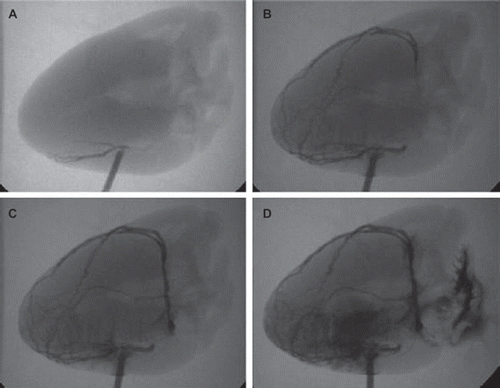
Six milliliters of blood sample were collected from aortic catheter before ligation and 240 min after the ligation of PDA. Thereafter, 10 mL ink was injected into the vein graft on the beating heart at a steady speed in SRCVB groups. And then, the animals were sacrificed with an overdose of potassium chloride and the hearts were harvested for in vitro angiography and histological analysis. In vitro angiography was performed using a C-Arm Cardiac Angiography System (Siemens AG, Healthcare Sector, Erlangen, Germany), with imaging agent injected into the vein graft.
Serum nitric oxide (NO) generation was analysed by the Griess reaction. Serum levels of endothelin (ET) were measured using a commercially available ELISA-Kit (Immundiagnostik, Bensheim, Germany) as previously described [Citation5].
The heart samples were immersed into 10% formaldehyde in phosphate-buffered saline of PH 7.4 at 4°C for 24 hours. After fixation, the samples were embedded in paraffin and sectioned to 5-μm-thick slices. Routine staining was performed with hematoxylin-eosin.
Statistical analysis
All offline measurements were made by investigators blinded to the treatment. Quantitative data were presented as mean ± SD. SPSS 13.0 software package (SPSS Inc, Chicago, USA) was used for statistical analysis. Experimental measurements in each group were compared by one-way analysis of variance (ANOVA) with Bonferroni post hoc correction. A value of P < 0.05 was regarded as statistically significant.
Results
It was detected in control group, but not in SRCVB groups, that the corresponding myocardium became purple color and ST segment was significantly elevated in lead II of the electrocardiogram monitor.
Changes of NO and ET levels
As shown in , no significant differences in the plasma levels of NO and ET were found among the three groups before PDA ligation (P > 0.05). Compared with control group, the plasma concentration of NO and ET respectively increased and decreased in both SRCVB groups 240 min after treatment (P < 0.001). Importantly, there was a more significant improvement in SRCVB 60 mmHg group than in the 90 mmHg (P < 0.001).
Table I. Changes of endothelial factors in each group.
Histological analysis
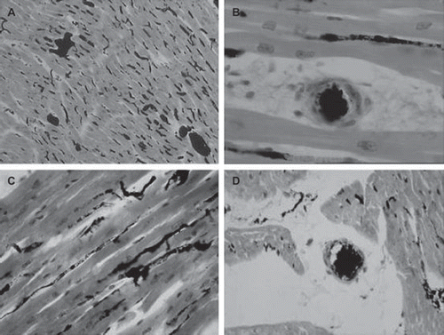
In SRCVB 60 mmHg group, it was revealed by HE staining myocardium sections that the ink distributed in myocardial sinuses (), capillaries and even arterioles (), without angiorrhexis or ink efflux. Although there were similar results in SRCVB 90 mmHg group, significant myocardial tissue edema was detected in this group at the same time ( and ).
Figure 1. Pathological analysis (A) The ink was distributed in myocardial sinuses and no angiorrhexis in SRCVB 60mmHg group; (B) the ink was distributed in capillaries and no angiorrhexis or ink efflux in SRCVB 60 mmHg group; (C) and (D) though ink could be distributed to PDA region, significant myocardial tissue edema was observed in SRCVB 90 mmHg group.

Angiography
The angiography showed that imaging agent could be perfused into the PDA region, and ultimately flowed back to right atrium through contralateral coronary vein ().
Discussion
Our study has demonstrated that SRCVB with low perfusion pressure brought little pressure related cardiac injury, protected endothelial cells against ischemic injury, and provided effective perfusion for ischemic myocardium, at least for the acute period.
As an effective method of myocardial preservation, coronary venous retroperfusion has been widely used for delivering cardioplegic solutions during cardiac surgery [Citation6], which can nourish the myocardium through myocardial sinusoids. In addition, it has been widely accepted that coronary venous system is barely affected by atherosclerosis [Citation7], which is an alternative pathway for myocardium perfusion when the coronary arteries are impaired with diffuse atherosclerosis. Therefore, the coronary veins-derived revascularization is expected to become an effective therapeutic strategy for patients with advanced diffuse lesion in coronary arteries [Citation8].
It has been reported by many investigators that SRCVB could rescue ischemic myocardium, restore myocardial contractile force, and improve cardiac function after myocardial ischemia [Citation3, Citation4, Citation9]. And as shown by angiography and histological analysis in our experiment, blood was perfused into the PDA region through the venous graft and MCV (the ink was distributed in myocardial sinuses, capillaries and even arterioles, without angiorrhexis), and ultimately flowed back to right atrium through contralateral coronary vein, indicating the feasibility of SRCVB again.
NO/ET, a biochemical maker indicating the degree of endothelial injury [Citation10, Citation11], was used in this study to assess the effect of SRCVB on endothelial protection. Compared with control group, there were significant increase in plasma levels of NO and decrease in ET in both SRCVB groups, indicating that this revascularization therapy may alleviate myocardial ischemia and endothelial cell injury. It has been demonstrated that both NO and ET are involved in the pathological process of myocardial ischemia [Citation12–14]. The underlined mechanism may be that myocardial ischemia induces endothelial cells to synthesize and release ET, whilst reduce NO level, which breaks the balance of NO/ET [Citation11]. The disbalance leads to further contraction of peripheral vessels, resulting in severer myocardial ischemia and hypoxia.
Fitzgerald et al. [Citation15] found that blood pressure higher than 60 mmHg could lead to harmful changes such as insufficient venous drainage, myocardial edema and intramyocardial hemorrhage. Our results have also demonstrated that higher perfusion pressure induced larger endothelial injury and more significant myocardial tissue edema, rather than more effective perfusion, and effective control of perfusion pressure could significantly attenuate pressure related cardiac injury. Therefore, it is suggested that the perfusion pressure, on the premise that adequate tissue perfusion is guaranteed, should be maintained as low as possible after SRCVB operation, which may be a decisive factor for the success in SRCVB therapy, although a certain blood pressure is required after coronary arteriovenous anastomosis to ensure the retrograde perfusion.
Despite encouraging results, there are still questions and considerations that need to be further addressed. For instance, only postoperative 4 hours results were investigated in the study, the long-term effects should be further assessed. In addition, the present experiment was performed on the dog model of acute myocardial ischemia, the differences between human and animal hearts has not been taken into account.
In summary, SRCVB may provide effective perfusion for ischemic myocardium, and prevent acute ischemic endothelial cells injury. It is suggested that the mean perfusion pressure should be maintained at low level, at least for the acute period of SRCVB therapy, to avoid high perfusion pressure-induced cardiac injury. Therefore, this study may provide an effective therapeutic strategy for severe CAD.
Acknowledgement
This work was supported by the Research Project of Education Department of Liaoning Province (No.2004C50) and Project of Technology Department of Liaoning Province (No.2006401013-2). Technical assistance of cardiac surgeons Xue-Tao Bai, Yu-Hai Zhang, and Zhong-Wu Wang is gratefully acknowledged.
Declaration of interest: The authors report no conflicts of interest. The authors alone are responsible for the content and writing of the paper.
References
- Frazier OH. Current status of cardiac transplantation and left ventricular assist devices. Tex Heart Inst J 2010;37: 319–21.
- Deng MC. Orthotopic heart transplantation: highlights and limitations. Surg Clin North Am 2004;84:243–255.
- Kassab GS, Navia JA, March K, Choy JS. Coronary venous retroperfusion: an old concept, a new approach. J Appl Physiol. 2008;104:1266–72.
- Syeda B, Schukro C, Heinze G, Modaressi K, Glogar D, Maurer G, . The salvage potential of coronary sinus interventions: meta-analysis and pathophysiologic consequences. J Thorac Cardiovasc Surg. 2004;127:1703–12.
- Wagner FD, Buz S, Knosalla C, Hetzer R, Hocher B. Modulation of circulating endothelin-1 and big endothelin by nitric oxide inhalation following left ventricular assist device implantation. Circulation. 2003;108 Suppl 1:II278–84.
- Harig F, Hoyer E, Labahn D, Schmidt J, Weyand M, Ensminger SM. Refinement of pig retroperfusion technique: Global retroperfusion with ligation of the azygos connection preserves hemodynamic function in an acute infarction model in pigs (Sus scrofa domestica). Comp Med. 2010;60: 38–44.
- von Lüdinghausen M. Clinical anatomy of cardiac veins, Vv. Cardiacae. Surg Radiol Anat 1987;9:159–168.
- Partington MT, Acar C, Buckberg GD, Julia PL. Studies of retrograde cardioplegia. II. Advantages of antegrade/retrograde cardioplegia to optimize distribution in jeopardized myocardium. J Thorac Cardiovasc Surg 1989;97:613–22.
- Raake P, Hinkel R, Kupatt C, von Brühl ML, Beller S, Andrees M, . Percutaneous approach to a stent-based ventricle to coronary vein bypass (venous VPASS): comparison to catheter-based selective pressure-regulated retro-infusion of the coronary vein. Eur Heart J. 2005;26:1228–34.
- Battistini B, Kingma JG. Changes in plasma levels of ET-1 and its precursor, big ET-1, in the arterial and venous circulation following double myocardial ischemia-reperfusion injury in dogs. J Cardiovasc Pharmacol. 2000;36(5 Suppl 1): S215–20.
- Kurita A, Matsui T, Ishizuka T, Takase B, Satomura K. Significance of plasma nitric oxide/endothelial-1 ratio for prediction of coronary artery disease. Angiology. 2005;56: 259–64.
- Fraccarollo D, Widder JD, Galuppo P, Thum T, Tsikas D, Hoffmann M, . Improvement in left ventricular remodeling by the endothelial nitric oxide synthase enhancer AVE9488 after experimental myocardial infarction. Circulation. 2008;118:818–27.
- Zhu SG, Kukreja RC, Das A, Chen Q, Lesnefsky EJ, Xi L. Dietary nitrate supplementation protects against Doxorubicin-induced cardiomyopathy by improving mitochondrial function. J Am Coll Cardiol. 2011;57:2181–9.
- Reriani M, Raichlin E, Prasad A, Mathew V, Pumper GM, Nelson RE, . Long-term administration of endothelin receptor antagonist improves coronary endothelial function in patients with early atherosclerosis. Circulation. 2010;122:958–66.
- Fitzgerald PJ, Hayase M, Yeung AC, Virmani R, Robbins RC, Burkhoff D, . New Approaches and Conduits: In Situ Venous Arterialization and Coronary Artery Bypass. Curr Interv Cardiol Rep. 1999;1:127–137.

