Abstract
Objectives. Bronchial artery revascularization in lung transplantation is disputed. This study examined the physiological consequences of porcine bronchial transsection and reanastomosis with and without bronchial artery blood supply with relation to lung transplantation. Design. Translational, controlled animal study. Twelve pigs were operated through a left lateral thoracotomy. The left bronchus was transsected and reanastomosed. In the control group (n = 6), the bronchial arteries were preserved and in the study group (n = 6) they were severed. Bronchial mucosa blood flow (BMBF) was measured with laser-Doppler velocimetry and bronchial mucosa haemoglobin saturation and concentration with diffuse reflectance spectrophotometry. Measurements were made preoperatively, postoperatively and after 1 week. Results. In the study group, left postoperative BMBF was significantly lower than preoperatively (115 vs. 210 PU/s, p = 0.0001) and lower than in the control group (115 vs. 205 PU/s, p = 0.002). Repeated measurement ANOVA showed a significant treatment effect depending on time (p = 0.0034). The left mucosal haemoglobin saturation in the study group was significantly reduced postoperatively, 92% versus 61%, with a treatment effect depending on time (p = 0.0080). The reduction in left/right ratio of the mucosal haemoglobin concentration 1 week postoperatively in the study group was insignificant. Conclusion. Bronchial transsection and reanastomosis without bronchial artery blood supply was followed by significant decrease in mucosal blood flow and saturation postoperatively, and also in tissue haemoglobin concentration at section, and provides a physiologic explanation of histological changes.
Introduction
The bronchial artery provides systemic arterial blood supply to the lower airways and the lung parenchyma. It affects a number of airway properties, including airway resistance (Citation1), infection resistance (Citation2), and it may participate in gas exchange under certain circumstances (Citation3,Citation4). Its significance in human lung transplantation is disputed, especially if its absence and ischaemia plays a role obliterative bronchiolitis (Citation5–9) or not (Citation10,Citation11). Most centres do not restore bronchial vascularity. The fact that a transplanted lung does survive contradicts older investigations showing that obliteration of the bronchial artery was lethal (Citation12). Many studies have examined the blood flow of the bronchi with a number of methods (Citation13,Citation14), only few have done that non-invasively with the possibility of repetition over time (Citation15–17), and to our knowledge only one have measured the haemoglobin saturation or the haemoglobin concentration of bronchial mucosa in vivo (Citation5). In previous studies, we reported on the importance of the bronchial artery from a morphological view, when the bronchi is transsected and anastomosed (Citation19,Citation20). In the present study, the relationship between physiological properties (i.e. blood flow, haemoglobin saturation and haemoglobin concentration in the bronchial mucosa) and bronchial transsection and reanastomosis with and without systemic arterial blood supply was assessed with laser-Doppler velocimetry (LDV) and diffuse reflectance spectrophotometry (DRS) with visible light. The purpose was to investigate if these examinations would disclose insufficient blood and oxygen supply and relate these to morphological changes and to discuss possible implications for OB. The study was the physiological aspect of a previously published study in which severe morphological changes were demonstrated after bronchial devascularization (Citation19).
Material and methods
Animals
Twelve female pigs from a production herd (Danish domestic/Yorkshire, crossbreed DDY), weighing between 36 and 44 kg, were used. Pre- and postoperatively they were kept in the laboratory yard with artificial daylight and fed twice daily. Six pigs were in a control group and six pigs were in a study group. Perioperative care included intravenous injection of 1.5 g cefuroxime (Zinacef; Glaxo-Wellcome) before operation, and postoperative intramuscular injection of 0.6 mg buprenorphine (Anorphin; GEA) twice daily for 2 days.
Anatomical considerations
The porcine bronchial blood supply originates from the broncho-oesophageal artery that divides into several bronchial branches and a branch for the upper thoracic oesophagus. The principal porcine bronchi are each followed by two major bronchial arterial branches that divide into minor branches. The bronchial arteries have frequent and considerable anastomoses with the lower oesophageal artery, which supplies the lower oesophagus directly from the aorta, and with the coronary arteries (Citation21). A relatively high proportion of the systemic flow to the bronchi is provided by these anastomoses (Citation22). The implication of these anastomoses to the surgical procedure of the present study is described below.
Surgical, bronchoscopic and angiographic procedures
The pigs were operated in general anaesthesia induced by thiopental 60 mg/kg (Pentothal; Abbott/Denmark), and maintained with NO2, with pentobarbital 50 mg/mL (Mebumal; Nycomed DAK, Sweden) and fentanyl 50 μg/mL (Haldid; Janssen, Belgium) infusions, and with pancuron bolus injections 2 mg/mL (Pavulon; Organon Teknika, The Netherlands). This procedure was also applied at sectioning, except for pancuron. A bronchoscopy with LDV and DRS measurements in both the left and right principal bronchus was performed peripheral to the first ramification of the main bronchi. Then, a left lateral thoracotomy was performed and the left main bronchus, the broncho-oesophageal artery and the pulmonary artery were exposed with no vascular damage. The pigs were heparinized (1000 IU of heparin). The left principal bronchus of both groups was subsequently transsected between the carina and the first ramification. It was reanastomosed with a continuous, non-absorbable stitch (Prolene 4 - 0, Johnson & Johnson, USA). In the study group, the lateral and medial bronchial branches were divided using hemoclips and efforts were made to severe all vessels between the lungs on one hand and the oesophagus and the pericardium on the other. The latter was done to ensure interruption of all possible anastomoses with the oesophageal and coronary arteries. In the control group, efforts were made not to harm the bronchial vascularity during transsection and anastomosis. The chest was closed without tube drainage, but full lung expansion was ensured clinically and radiographically. To remove blood from the mucosa surface, a bronchial lavage was made bronchoscopically in the left main bronchus. Postoperative LDV and DRS measurements were made at the same left and right measuring sites as before, which at the left site was peripheral to the anastomosis. These measurements were repeated before section 1 week later. After a sternal split, the heart-lung block was perfused with saline until death occurred, removed with the entire mediastinum and cannulated for contrast injection in the bronco-oesophageal, oesophageal and coronary arteries, and the patency of the bronchial arterial branches was investigated angiographically in the inflated lung. After angiography, specimens for histology and scanning electron microscopy were taken from lungs and bronchi peripheral to LDV and DRS measuring points, as reported earlier (Citation19,Citation20).
Laser-Doppler velocimetry
A laser-Doppler unit (PF3, Perimed, Sweden) was employed together with a flexible optic fibre probe and a bronchoscope for LDV measurements. The laser-Doppler was calibrated before each operation, sampling frequency was set to 12 kHz. It measures in arbitrary ‘Perfusion Units/s’ (PU/s), which refers to a calibration standard and corresponds to – but is not a direct measure of – flow in mL/s. Measuring sites were chosen as described above. We aimed at 6–8 measurements in each location. The mucosa was touched as gently as possible with the probe, and to avoid movement artefacts the respirator was stopped for a few seconds while measuring. The pigs were then ventilated for some minutes before another measurement was made. Measuring direction was across a secondary carina and towards the lumen of the principal bronchus. This was done to avoid measuring into the large pulmonary vessels, since LDV has a spatial resolution up to 6 mm (Citation23). For calculations, we chose the highest, stable part of the curve of each measurement, and beside the direct LDV flow on both sides, a left/right ratio was calculated. The latter was done because cardiac output and thereby tissue circulation would probably change secondary to operative stress. The left/right ratio was calculated for preoperative, postoperative and section comparison. Validation of the laser-Doppler technique has been done by a number of authors (Citation13,Citation24,Citation25) and it is known that accuracy is improved with the number of measurements.
Diffuse reflectance spectrophotometry
For DRS, we used a mini-spectrophotometer (S2000, Ocean Optics Inc., USA) with 2048 diodes arranged in a CCD-array with a spectral resolution of 0.35 nm, together with a 7 W halogen light source and a flexible probe for bronchoscopic measurements. The probe had an arrangement of six 200 μm light emitting optical fibres in a circular arrangement around one central light absorbing fibre within a probe diameter of 2.5 mm. The spectra were recorded with the original software, OOI-Base®, through a digital analogue converter (DAQ 700®, National Instruments, USA) interface on a laptop computer (TS 2130, Toshiba, Japan). To obtain a white reference the probe was inserted into a barium-sulphate solution of 1 g per ml. The software was set to average 10 spectra, so what is referred to as one measurement in the following comprises the average of 10 spectra, obtained with a frequency of 50 spectra per second.
For saturation calculation, we choose a method (Citation26), which we in an earlier study found superior to other spectrophotometrical methods with visible light spectrophotometry (VIS), and from that study we used a modification of the Knoefel method based on our own extinction coefficients for calculation of tissue haemoglobin concentration (Citation27). Measuring locations were the same as above, 6–8 measurements were aimed at, and the probe touched the mucosa as gently as possible. For the same reason as above, a left/right ratio for preoperative, postoperative and sectioning comparison was also calculated. The method was not as sensitive to movements as the LDV-method and the respirator was not stopped while measuring.
Morphological examinations
Morphological examinations consisted of conventional histology of lung tissue and major airways and scanning electron microscopy of the latter. Preparation details have been given earlier (Citation19,Citation20).
Statistics
Weight was analysed with a t-test. The physiological data were analysed using a multivariate repeated measurement ANOVA with treatment and time as factors. The larger variation postoperatively and at section in the treatment group compared to the control group was taken into account by allowing for different standard deviations and correlations in the two groups. Data were analysed using Stata 11.2. A p-value of 0.05 was chosen as level of significance.
Ethics
The operated pigs received humane care in accordance with the national Danish regulations on experimental animal welfare. The investigation was approved by the national Danish Inspection on Animal Welfare.
Results
Clinical course and angiography
None of the 12 pigs had surgical complications, such as bleeding or infection, and none had bronchial necrosis or dehiscence. A relative bronchial stenosis (passable with bronchoscope) occurred in 2 study pigs and 1 control. Angiography showed complete bronchial artery interruption in 5 of 6 study pigs and partial interruption in 1. In the control group, 2 of 6 pigs had partial interruption of the bronchial vascularity. There was a weight gain in the control group and a small weight loss in the study group, but the difference was not significant.
Laser-Doppler velocimetry
Following operation, left BMBF was reduced from 210 (95%-CI: 170–251) to 115 (95%-CI: 89–141) PU/s in the study group and increased from 161 (95%-CI: 118–203) to 205 (95%-CI: 154–256) in the control group (). Repeated measurements ANOVA showed a significant treatment effect depending on time (p = 0.0034). Preoperative and at section, the two treatment groups did not differ significantly (p = 0.10 and p = 0.63), but postoperatively the blood flow was significantly lower in the intervention group compared to the control group (90 PU/s, 95%-CI: 33–147, p = 0.002). Also, the change in blood flow from baseline to after operation was significantly higher in the study group than in the control group (140 PU/s, 95%-CI: 74–206, p < 0.0001). To compensate for possible changes caused by operation as such or the anaesthesia, the left/right ratio was calculated as described earlier. There was a significant treatment effect in the ratio depending on time (p = 0.0023). The ratio showed no significant difference between the two treatment groups preoperatively or at section (p = 0.06 and p = 0.30, respectively), but postoperatively the ratio was significantly lower in the study group compared to the control group (0.61, 95%-CI: 0.17–1.06, p = 0.007). The change in ratio from preoperative to postoperative was significantly higher in the study group than in the control group (0.95, 95%-CI: 0.42–1.47, p < 0.0001). The change in ratio did not differ between the groups from postoperative to section (p = 0.42). gives an overview of the results.
Figure 1. Bronchial mucosa blood flow (BMBF) with laser-Doppler velocimetry. Left panel: Left BMBF in study and control group. Right panel: Left/right BMBF ratio in study and control group. Filled circle: study group. Open circle: control group.
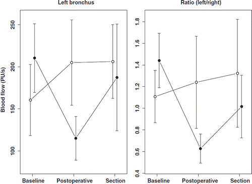
Table I. Overview of results.
Diffuse reflectance spectrophotometry
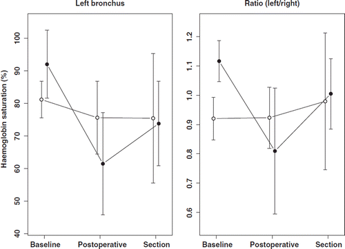
Haemoglobin saturation measurements. The postoperative left bronchial mucosal haemoglobin saturation was not significantly different from the preoperative in the control group, 81% (95%-CI: 76–87) and 76% (95%-CI: 64–87), but reduced from 92% (95%-CI: 82–102) preoperatively to 61% (95%-CI: 46–77) postoperatively in the study group. Repeated measurement ANOVA showed a significant treatment effect depending on time (p = 0.0080). At no specific time-point did the two treatment groups differ significantly (preoperative p = 0.07, postoperative p = 0.15 and section p = 0.89). However, the drop in saturation in the study group from the pre- to postoperative situation was significantly larger compared to that in the control group (25.0%, 95%-CI: 11–39.0%, p < 0.0001) (see ). The left/right ratio showed no significant treatment effect depending on time (p = 0.0644). Postoperatively and at section, the groups did not differ significantly (p = 0.35 and p = 0.85, respectively), but preoperatively the ratio was significantly higher in the study group compared to the control group (0.20, 95%-CI: 0.10–0.30, p < 0.0001). Again, also the change in ratio from the preoperative to the postoperative time-point was significantly higher in the study group than in the control group (0.31, 95%-CI: 0.07–0.55, p = 0.010) (see ). The change in ratio did not differ between the groups from postoperative to section (p = 0.33). gives an overview of the results.
Figure 2. Bronchial mucosa haemoglobin saturation with DRS. Left panel: Bronchial mucosa saturation of left bronchus in study and control group. Right panel: Left/right ratio of bronchial mucosa saturation in study and control group. Filled circle: study group. Open circle: control group.
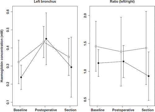
Tissue haemoglobin concentration measurements. The treatment effect on the left bronchus was not significant (p = 0.26), whereas there was a significant effect of time (p = 0.0120). The postoperative haemoglobin tissue concentration was significantly higher than the preoperative (p < 0.0001) and also higher than the haemoglobin level at section (p = 0.05). This also applied to the right bronchus (p = 0.001 and p = 0.010, respectively). The preoperative haemoglobin concentration was not significantly different from that at section (p = 0.17). The left/right ratio of haemoglobin concentration did not change significantly over time (p = 0.92), but there was a significant treatment effect independent of time (p = 0.037). The haemoglobin ratio was lower in the study group compared to the control group (0.32, 95%-CI: 0.02–0.63). The difference at section alone, however, was insignificant (0.50, 95%-CI: − 0.28–1.29, p = 0.21). The study group’s lower ratio in haemoglobin concentration at section mostly depended on an increase in haemoglobin concentration of the right mucosa (0.12, 95%-CI: − 0.02–0.26, p = 0.10) (see and ). This was insignificantly higher than the control group (p = 0.46). The change in ratio (preoperative to section) was not significantly different in the two groups (p = 0.62).
Figure 3. Bronchial mucosa haemoglobin concentration with DRS. Left panel: Bronchial mucosa con-centration of left bronchus in study and control group. Right panel: Left/right ratio of bronchial mucosa concentration in study and control group. Filled circle: study group. Open circle: control group.
Morphological examinations
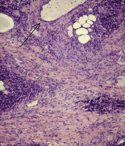
Morphological examinations have been described in detail earlier [19,20], but in short scanning electron microscopy of the main bronchi showed wrinkled surface and loss of cilia and conventional histological examination showed oedema, slight fibrosis and inflammation not only of main bronchi but also in lung parenchyma. Especially remarkable was the finding of oedematous lung septa with inflammation and beginning fibrosis ().
Figure 4. Section of left lung with major bronchi (H&E, × 100), showing severe broadening of lung septae with inflammation, oedema and slight fibrosis. Specimen was taken peripheral to both anastomosis and measuring site. Arrow: Small bronchus.
Discussion
The most important factor hampering long term survival in lung transplantation is obliterative bronchiolitis. The role of ischaemia in the occurrence of obliterative bronchiolitis is controversial (Citation5,Citation6,Citation28).
Despite the dual blood supply of the lung, many experimental studies have shown that interruption of the bronchial arterial circulation causes a number of severe changes to the bronchi and the lung parenchyma in the first week after interruption of the bronchial vascularity (Citation12,Citation19,Citation20,Citation29–31). These studies on bronchial vascularity mostly employed morphologic examinations. However, anatomical and physiological studies have shown that the bronchial mucosa is also supplied from the pulmonary artery via precapillary anastomoses with up to 50% (Citation31,Citation32). The blood flow of the bronchial vascularity has been studied with LDV (Citation33), and microsphere injection (Citation22,Citation32) in human and animal studies, and with arteriography and perfusion scintigraphy in humans (Citation17,Citation18), but few studies address the central issue, tissue oxygenation: two invasively (Citation34,Citation35) and one non-invasively (Citation5). The aim of the present study was to apply both LDV and DRS measurements to the porcine bronchial mucosa and investigate their ability to disclose insufficient blood and oxygen supply. To our knowledge, a study with this combination of non-invasive measurement of blood flow, haemoglobin saturation and haemoglobin concentration in the major airways has not been undertaken before.
The results showed a significant reduction of blood flow and saturation in the left bronchus of the study group from preoperative to postoperative conditions, but no such reduction in the control group. We have chosen to pay special attention to changes in the left/right ratio of the parameters in question, because this to some extent would eliminate changes caused by the operation as such, for example, a lower arterial oxygenation or a higher heart rate, which may represent a confounder. For example, higher heart rate would probably result in higher BMBF. The left/right saturation ratio was much lower in the study group postoperatively, and although this difference was insignificant in itself, ANOVA showed that the change was significantly different. One reason may be the overlapping of groups commented on below. Altogether, it is concluded that the severe morphological changes previously reported (Citation19) were preceded by significant changes in flow and saturation at the time of operation, but these physiological changes were normalised after 1 week. The latter is in accordance with our previous findings (Citation20).
The reduction of blood flow has been reported by others. In a porcine study, Korpela et al. reported a twothird reduction with the bronchial artery clamped and almost no flow with both the pulmonary and the bronchial artery clamped (Citation33). In a canine study with bronchial transsection, blood flow was reduced to 59% of baseline value and recovered after 2 weeks (Citation15) and in a study with radioactive microspheres the pulmonary circulation was estimated to contribute 50% to the blood flow of the main bronchi of dogs (Citation32). The reduction rate of the present study was in accordance with these two studies, since the systemic collateral flow was eliminated. Tissue oxygenation of the bronchi is difficult to measure. Herold et al. (Citation34) and Kamler et al. (Citation35) used a Licox probe (which must be placed within the tissue) to show that the oxygen tension of peribronchial tissue (lymph nodes) was severely decreased in porcine allotransplantation, but pO2 of the bronchial wall or the mucosa was not measured. DRS offers an attractive option, which is non-invasive as far as the tissue is concerned, but we have found only one report employing this method to examine the oxygenation of the bronchi (Citation5). We measured both haemoglobin saturation and haemoglobin concentration in the bronchial mucosa.
Higher postoperative haemoglobin concentration would be expected due to vaporization, and the oedema at 1 week reported earlier (Citation19), would be expected to cause lower tissue haemoglobin concentration in the left bronchus of the study group. Although lower, the latter difference was insignificant. The drop in ratio at section in the study group () is interesting, especially because it mainly reflects a higher haemoglobin concentration (although insignificant) in the right bronchus, suggesting that the blood haemoglobin concentration was higher. Unfortunately, we did not measure this in venous blood, neither preoperatively nor postoperatively. Furthermore, the haemoglobin concentration of the mucosa varied very much, not only between groups and individuals, but also within the same individual and at the same time. For example, the measured preoperative haemoglobin concentration varied in the most extreme case from 0.24 mM to 0.43 mM, from one measurement to the next. We have developed this method for the present study and found that variation was much less in a tissue phantom (Citation27). The huge variations of the present study may be due to high sensibility to probe pressure against the mucosa, where blood possibly is squeezed out of the capillaries. Altogether, the measurement of tissue haemoglobin concentration does not seem to be a reliable method at present, but needs further development. It has, however, the potential to reveal if reduced flow or saturation is due to either arterial stenosis or venous stasis/congestion, and if tissues are oedematous.
Two other weaknesses of the study should be mentioned. First, of course, is the considerable, though insignificant, preoperative difference in left BMBF between the groups, and the significantly different saturation ratio, for which we have no exact explanation. The pigs were operated according to a planned schedule, but not randomised. Second, since complete devascularisation was not obtained in 1 study pig, and partial devascularisation happened in 2 control pigs, these pigs were devascularised to approximately the same extend. Thus, differences may have been more marked with 2 more “clean” groups.
The strength of the study, on the other hand, is also double: First, of course, the use of a control group, which ensures that the effect is due to the severing of the bronchial artery and not the operation itself. Second, talking of lung transplantation, the study shows the pure effect of ischaemia without alloimmunity as a confounder.
The findings may be of relevance for human lung transplantation, where bronchial obliterans syndrome is still a significant problem. Hence, our findings of inflammation, oedema and fibrosis in lung parenchyma have some similarity with obliterative bronchiolitis, although fibrosis is less pronounced in our study. Together with the present physiological findings, it poses the hypothesis that enhanced connective tissue formation of the bronchioles is due to initial hypoxaemia. Long term experimental studies would be needed to clear this up. To our knowledge, no such studies have yet been undertaken.
Acknowledgements
This work was supported by grants from the following institutions and foundations: The Danish Medical Association Research Foundation, The Danish National Association against Lung Diseases, The Danish Hospital Foundation for Medical Research, The Beckett Fund, The Leo Research Foundation, Felo Aps, The Ib Henriksen Fund, The Dagmar Marshall Fund and the Research Fund at the Aalborg Hospital (University Hospital of Århus). Laboratory technician Karin Jensen is thanked for valuable assistance throughout the study, medical students Bettina Breitowicz and Heidi Jacobsen are thanked for operative assistance and Ph.D. Peter Snoer Jensen, Department of Optics and Fluid Dynamics, Risø, Denmark, is specially thanked for the development of programs for data acquisition.
Declaration of interest: The authors report no conflicts of interest. The authors alone are responsible for the content and writing of the paper.
References
- Wagner EM, Brown RH. Blood flow distribution within the airway wall. J Appl Physiol 2002;92:1964–9.
- Charan NB, Turk GM, Dhand R. The role of bronchial circulation in lung abscess. Am Rev Respir Dis 1985;131:121–4.
- Charan NB, Carvalho P. Angiogenesis in bronchial circulatory system after unilateral pulmonary artery obstruction. J Appl Physiol 1997;82:284–91.
- Henig NR, Glenny RW, Aitken ML. A hypertrophied bronchial circulatory system may participate in gas exchange. Lancet 1998;351:113.
- Dhillon GS, Zamora MR, Roos JE, Sheahan D, Sista RR, Van der Starre P, . Lung transplant airway hypoxia: a diathesis to fibrosis? Am J Respir Crit Care Med 2010;182:230–6.
- Norgaard MA, Andersen CB, Pettersson G. Does bronchial artery revascularization influence results concerning bronchiolitis obliterans syndrome and/or obliterative bronchiolitis after lung transplantation? Eur J Cardiothorac Surg 1998;14:311–8.
- Nowak K, Kamler M, Bock M, Motsch J, Hagl S, Jakob H, . Bronchial artery revascularization affects graft recovery after lung transplantation. Am J Respir Crit Care Med 2002;165:216–20.
- Babu AN, Murakawa T, Thurman JM, Miller EJ, Henson PM, Zamora RM, . Microvascular destruction identifies murine allografts that cannot be rescued from airway fibrosis. J Clin Invest 2007;117: 3774–85.
- Akindipe O, Fernandez-Bussy S, Jantz M, Lu L, Deem A, Swafford W, . Obliterative bronchiolitis in lung allografts removed at retransplant for intractable airway problems. Respirology 2009;14:601–5.
- Nicolls MR, Zamora MR. Bronchial blood supply after lung transplantation without bronchial artery revascularization. Curr Opin Organ Transplant 2010:15:563–7.
- Verleden GM, Vos R, van Raemdonck D, Vanaudenaerde B. Pulmonary infection defense after lung transplantation: does airway ischemia play a role? Curr Opin Organ Transplant 2010;15:568–71.
- Ellis FH, Grindlay JH, Edwards JE. The bronchial arteries: experimental occlusion. Surgery 1951;30:810–6.
- Kvietys PR, Shepherd AP, Granger DN. Laser-Doppler, H2 clearance, and microsphere estimates of mucosal blood flow. Am J Physiol 1985;49:G221–7.
- Baile EM, Nelems J, Schulzer M, Paré P. Measurement of regional bronchial arterial blood flow and bronchovascular resistance in dogs. J Appl Physiol 1988;53:1044–9.
- Fujino S, Inoue S, Yamashita N, Mori A. An experimental study on direct revascularization of bronchial circulation by microvascular anastomosis. J Thorac Cardiovasc Surg 1992;104:1067–74.
- Sundset A, Hansen G, Haanaes OC, Line PD, Kvernebo K. Human bronchial perfusion evaluated with endoscopic laser Doppler flowmetry. Int J Microcirc Clin Exp 1993;13: 233–45.
- Norgaard MA, Gadsboll N, Efsen F, Hesse B, Rabol A, Pettersson G. Bronchial artery perfusion scintigraphy to assess bronchial artery blood flow after lung transplantation. J Nucl Med 1999;40:290–5.
- Norgaard MA, Hove JD, Efsen F, Saunamaki K, Hesse B, Pettersson G. Human bronchial artery blood flow after lung Tx with direct bronchial artery revascularization. J Appl Physiol 1999;87:1234–9.
- Gade J, Qvortrup K, Andersen CB, Olsen PS. Bronchial transsection and reanastomosis in pigs with and without bronchial arterial circulation. Ann Thorac Surg 2001;71:332–6.
- Gade J, Quortrup K, Andersen CB, Thorsen S, Svendsen UG, Olsen PS. Bronchial transsection and reanastomosis in pigs with and without bronchial arterial circulation. An experimental study in pigs. Scand Cardiovasc J 2001;35: 212–20.
- Gade J, Nørgaard M, Andersen CB, Breitowizc B, Jakobsen H, Svendsen UG, . The porcine bronchial artery. Anastomoses with coronary, oesophageal and intercostal arteries. J Anat 1999;195:65–3.
- Ashley K, Herndorn D, Traber L, Traber D, Deubel-Ashley K, Stothert JR, . Systemic blood flow to sheep lung: comparison of flow probes and microspheres. J Appl Physiol 1992;73:1996–2003.
- Johansson K, Ahn H, Lindhagen J, Lundgren O. Tissue penetration and measuring depth of laser Doppler flowmetry in the gastrointestinal application. Scand J Gastroenterol 1987;22:1081–8.
- Ahn H, Lindhagen J, Nilsson GE, Salerud EG, Jodal M, Lundgren O. Evaluation of laser Doppler flowmetry in the assessment of intestinal blood flow in cat. Gastroenterology 1985;88:951–7.
- Shepherd AP, Riedel GL, Kiel JW, Haumschild DJ, Maxwell LC. Evaluation of an infrared laser-Doppler blood flowmeter. Am J Physiol 1987;252:G832–9.
- Knoefel WT, Kollias N, Rattner DW, Nishioka NS, Warshaw AL. Reflectance spectroscopy of pancreatic microcirculation. J Appl Physiol 1996;80:116–23.
- Gade J, Palmquist D, Plomgård P, Greisen G. Diffuse reflectance spectrophotometry with visible light. Comparison of 4 different methods in a tissue phantom. Phys Med Biol 2006;51:121–36.
- Huang HJ, Yusen RD, Meyers BF, Walter MJ, Mohanakumar T, Patterson GA, . Late primary graft dysfunction after lung transplantation and bronchiolitis obliterans syndrome. Am J Transplant 2008;8:2454–62.
- Mills NL, Boyd AD, Gheranpong C. The significance of bronchial circulation in lung transplantation. J Thorac Cardiovasc Surg 1970:60:866–74.
- Ventemiglia RA, Braverman B, DiMauro J, Castro R, Blair W, Spigos D, . The ischemic lung: Role of the bronchial arteries in lung function. Cardiovasc Dis 1981;8: 480–98.
- Korpela A, Aarnio P, Taskinen E, Ikonen T, Harjula A. Healing of bronchial allografts in pigs. Scand Cardiovasc J 2000;34:433–7.
- Barman SA, Ardell JL, Parker JC, Perry ML, Taylor AE. Pulmonary and systemic blood flow contributions to upper airways in canine lung. Am J Physiol 1988;255: H1130–5.
- Korpela A, Aarnio P, Harjula A. Evaluation of bronchial mucosal blood flow by laser Doppler flowmeter. Int J Angiology 1995;4:110–2.
- Herold U, Jakob H, Kamler M, Thiele R, Tochtermann U, Weinmann J, . Interruption of bronchial circulation leads to a severe decrease in peribronchial oxygen tension in standard lung transplantation technique. Eur J Cardiothorac Surg 1999;13:176–83.
- Kamler K, Nowak K, Bock M, Herold U, Motsch J, Hagl S, . Bronchial artery revascularization restores peribronchial tissue oxygenation after lung transplantation. J Heart Lung Transplant 2004;23:763–6.