Abstract
Two novel proton transfer compounds were prepared between 2,4-dichloro-5-sulphamoylbenzoic acid (lasamide) (Hsba) and ethylenediamine (en), namely ethane-1,2-diaminium 2,4-dichloro-5-sulphamoylbenzoate (1), and also between Hsba and 2-amino-3-methylpyridine (2-amino-3-picoline) (amp), namely 2-amino-3-methylpyridinium 2,4-dichloro-5-sulphamoylbenzoate (2). All these were characterised by elemental, spectral (IR and UV-vis), thermal analyses, and single crystal X-ray diffraction studies. Compounds 1 and 2 crystallised in the P-1 and P21/c space groups, respectively. Intermolecular non-covalent interactions, such as ion pairing, hydrogen bonding, and π-π stacking were observed for these ionic compounds. The free ligands Hsba, en and amp, the products 1 and 2, and acetazolamide (AAZ) as the control compound, were also evaluated for their in vitro inhibitor effects on the human carbonic anhydrase isoenzymes (hCA I and hCA II) purified from erythrocyte cells by affinity chromatography for their hydratase and esterase activities. The half maximal inhibitory concentration (IC50) values for products 1 and 2 with respect to hydratase activity are 0.15 and 0.32 µM for hCA I and 0.06 and 0.15 µM for hCA II, respectively. The IC50 values of the same inhibitors for esterase activity are 0.13 and 0.8 µM for hCA I and 0.14 and 0.1 µM for hCA II, respectively. In relation to esterase activities, the inhibition equilibrium constants (Ki) were also determined and found to be 0.137 and 0.99 µM on hCA I and 0.157 and 0.075 µM on hCA II for 1 and 2, respectively. The comparison of the inhibition studies of the newly synthesised compounds 1 and 2 to the parent compounds Hsba and amp and also to AAZ indicated that 1 and 2 have an effective inhibitory activity on hCA I and II, and might be used as potential inhibitors.
Introduction
The sulphonamides constitute an important class of drugs, with several types of pharmacological actions including antibacterial, anti-glaucoma, diuretic, hypoglycemic, antithyroid, protease inhibitory and anticancer activities [Citation1–4]. Diversification of the sulphonylamide structure has led to the development of improved antibiotics, insulin-releasing hypoglycaemic drugs, and antihypertensive drugs. With sulphonamides as lead structures, different classes of pharmacological agents have been obtained, such as the antiglaucoma sulphonamides that can inhibit the zinc enzyme carbonic anhydrase (CA; EC 4.2.1.1), in which the zinc ion is the prosthetic group and is coordinated by histidine side chains in the active site. The CA is able to catalyse the reversible hydration of carbon dioxide to bicarbonate and protons, a very simple but critically important physiological reaction for organisms [Citation5–10]. This enzyme has 16 different isoenzymes currently known. Several of these isoenzymes (hCA II and hCA IV) are present in human eyes [Citation11–14] causing glaucoma, which is a group of diseases characterised by a gradual loss of the visual field due to an elevation in intraocular pressure (IOP), and being the second leading cause of blindness worldwide [Citation14,Citation15]. Since CA inhibitors have been shown to reduce intraocular pressure exclusively by lowering the aqueous humour flow and these compounds have been used for the treatment of glaucoma for years [Citation16,Citation17].
In recent years the different aspects of proton transfer systems have been studied by chemists [Citation18–22]. There have been several attempts at employing proton transfer from carboxylic acids to amines [Citation23–27] and at preparing their complexes with different metal ions [Citation28–33]. The first proton transfer compound obtained between Hsba (carboxylic acid including the sulphonylamide group) and amp, with its metal complex have been prepared in our laboratories [Citation34].
In this study, we have prepared proton transfer compounds between 2,4-dichloro-5-sulphamoylbenzoic acid (lasamide) (Hsba) and ethylenediamine (en), namely ethane-1,2-diaminium 2,4-dichloro-5-sulphamoylbenzoate (1), and between Hsba and 2-amino-3-methylpyridine (2-amino-3-picoline) (amp), namely 2-amino-3-methylpyridinium 2,4-dichloro-5-sulphamoylbenzoate (2). They have been characterised by elemental, spectral (IR and UV-vis) and thermal analyses, as well as structural studies by single X-ray diffractions. Furthermore, we have investigated the potential use of these compounds as new inhibitors of hCA isoenzymes in the treatment of glaucoma.
Materials and methods
All chemicals and reagents were of analytical grade and used as received from a commercial source (Sigma-Aldrich, Munich, Germany). Elemental analyses for C, H, N and S were performed on an Elementar Vario III EL (Hanau, Germany). FT-IR spectra were recorded in the 4000–400 cm−1 region with aBruker Optics, Vertex 70 FT-IR spectrometer (Ettlingen, Germany) using ATR techniques. Thermal analyses were performed on the SII Exstar 6000 TG/DTA 6300 model (Shimadzu Co, Japan) using a platinum crucible with 10 mg samples. TG/DTA measurements were taken in static air, within a 20–700°C temperature range. The UV–Vis spectra were obtained for aqueous solution and DMSO solution of the compounds 1 and 2 (10−3 M) and the free ligands, amp and Hsba (10−3 M), with a SHIMADZU UV-2550 spectrometer (Shimadzu Co, Japan) in the range of 900–200 nm.
Synthesis of proton transfer compounds 1 and 2
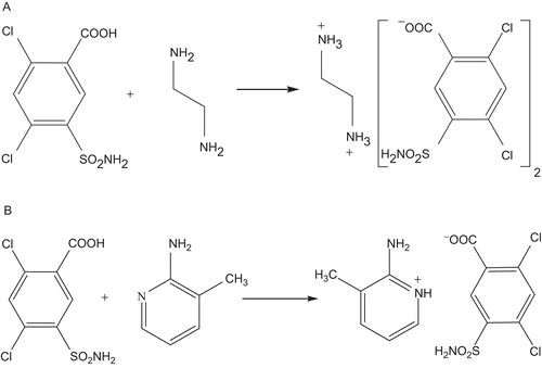
Solutions of amines (0.3 g, 5 mmol en for compound 1 and 0.54 g, 5 mmol amp for compound 2) in 10 mL distilled water were added to the solutions of Hsba (1.35 g, 5 mmol) in 10 ml water separately. The mixtures were refluxed for 3 h, and then were cooled to room temperature. The reaction mixtures were kept at room temperature for 2 weeks to give colourless needle crystals for both studies (1.95 g, 65 % yield for 1, 1.51 g, 80.1% yield for 2). The single crystals suitable for X-ray analyses were obtained by slow evaporation of water solutions for both compounds ().
Figure 1. Syntheses of compounds; (A) for 1, and (B) for 2.
Anal. Calcd. for 1 (C8H9Cl2N2O4S): C, 32.01; H, 3.02; N, 9.33; S, 10.68. Found: C, 32.03; H, 3; N, 9.32; S, 10.7; for 2 (C13H13Cl2N3O4S): C, 41.28; H, 3.46; N, 11.11; S, 8.48. Found: C, 41.29; H, 3.45; N, 11.10; S, 8.46.
Crystal structure determinations of 1 (C8H9Cl2N2O4S) and 2 (C13H13Cl2N3O4S)
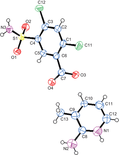
The crystal and instrumental parameters used in the unit-cell determination and data collection are summarised in for compounds 1 and 2. Measurements of 1 and 2 were made on a Stoe IPDS II CCD X-ray diffractometer (STOE & Cie GmbH, Darmstadt, Germany) employing plane graphite monochromatised with Mo Kα radiation (λ = 0.71073 Å), using the ω - 2θ scan mode. The unit cell parameters were refined from the setting angles of the 25 centred reflections in the range of 1.69 ≤ θ ≤ 27.54 for 1 and 1.46 ≤ θ ≤ 26.19 for 2. The structures were solved by the direct methods using SHELXS-97 and refined by full-matrix least-squares techniques on F2 with SHELXL-97 [Citation35]. The empirical absorption corrections were applied by the integration method via X-RED software [Citation36]. All non-hydrogen atoms were refined with the anisotropic displacement parameters and the hydrogen atoms were included in their idealised positions and refined isotropically, except for the N1 (in compound 1) and N3 (in compound 2). The hydrogen atoms of N1 for compound 1 and of N1 and N3 for compound 2 were located from the difference maps and refined with the isotropic thermal parameters. The crystal data and structure refinement details for compounds 1 and 2 are given in . The ORTEP drawings [Citation37] of the molecules with a 40% probability displacement thermal ellipsoids and atom-labelling schemes are shown in for 1 and for 2.
Table 1. Crystal data and structure refinement details for compounds 1 and 2.
Figure 2. The molecular structure and atomic labeling scheme of 1. Displacement ellipsoids are drawn at the 40% probability level.
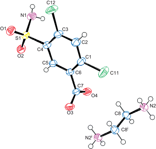
Figure 3. The molecular structure and atomic labeling scheme of 2. Displacement ellipsoids are drawn at the 40% probability level.
Purification of isoenzymes hCA I and II from human erythrocytes
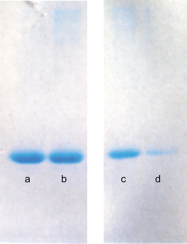
In order to purify the hCA I and II isoenzymes, human blood was centrifuged at 1500 rpm for 20 min, and after the removal of the plasma, the erythrocytes were washed with an isotonic solution (0.9% NaCl). After that, the erythrocytes were lysed with 1.5 volume of ice-cold water. The lysate was centrifuged at 20 000 rpm for 30 min to remove any cell membranes and non-lysed cells. The pH of the supernatant was adjusted to 8.7 with tris and was then loaded onto an affinity column containing Sepharose-4B-L-tyrosine-p-aminobenzene sulphonamide as the binding group. After extensive washing with 25 mM tris–HCl/22mM Na2SO4 (pH 8.7), the hCA I and II isoenzymes were eluted with 1 M NaCl/25 mM Na2HPO4 (pH 6.3) and 0.1 M CH3COONa/0.5 M NaClO4 (pH 5.6) [Citation38,Citation39]. The amount of purified protein was estimated by the Bradford method [Citation40] and SDS–PAGE was carried out to determine whether the elute contained the enzyme () [Citation41].
Figure 4. SDS-PAGE analyses of CA isoenzymes: (a) Standard hCA I, (b) isolated hCA I, (c) Standard hCA II and (d) isolated hCA II.
Hydratase activity assay
The carbonic anhydrase activity was assayed by following the hydration of CO2 according to the method described by Wilbur and Anderson [Citation42]. The CO2-hydratase activity as an enzyme unit (EU) was calculated by using the equation ((t0-tc)/tc) where t0 and tc are the times for pH change of the nonenzymatic and the enzymatic reactions, respectively. The concentration of inhibitor producing a 50% inhibition of CA activity (IC50) values were obtained in vitro for the free ligands en, amp and Hsba, and the synthesised compounds 1 and 2, with acetazolamide (AAZ) as the control compound.
Esterase activity assay
The carbonic anhydrase activity was assayed by following the change in absorbance at 348 nm of the 4-nitrophenylacetate (NPA) to the 4-nitrophenylate ion over a period of 3 min at 25°C using a spectrophotometer (CHEBIOS UV–VIS, Rome, Italy) according to the method described in the literature [Citation43,Citation44]. The enzymatic reaction, in a total volume of 3 mL, contained 1.4 mL of 0.05 M tris–SO4 buffer (pH 7.4), 1 mL of 3 mM 4-nitrophenylacetate, 0.5 mL H2O and 0.1 mL enzyme solution. A reference measurement was obtained by preparing the same cuvette without enzyme solution. The IC50 values have been obtained as in vitro for the free ligands en, amp and Hsba, and the synthesised compounds 1 and 2, with AAZ as the control compound.
Determination of Ki values
The method for determination of the Ki values is described elsewhere [Citation45–49]. In the first part of this study, the IC50 values have been obtained as in vitro. The IC50 of the inhibitors (free ligands en, amp and Hsba, and the synthesised compounds 1 and 2, with AAZ as the control compound) were assayed by the hydrolysis of p-nitrophenylacetate on the esterase activities of the CA isoenzymes in the presence of various inhibitor concentrations. The absorbance was determined at 348 nm after 3 min [Citation45]. Regression analysis graphs were drawn by plotting inhibitor concentrations versus enzyme activity using Microsoft Excel [Citation50].
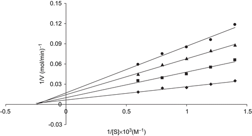
In the second part of the study, the enzyme activity was measured in the presence of five different substrate concentrations. At each of these inhibitor concentrations (30%, 50%, and 70%) the data were linearised using the Lineweaver-Burk plot in order to obtain the Ki values as shown in .
Figure 5. Ki graph obtained from in vitro studies for compound 1 on hCA II.
Results and discussion
Descriptions of the structures of 1 (C8H9Cl2N2O4S) and 2 (C13H13Cl2N3O4S)
The molecular structures of compounds 1 and 2 with theiratomic numbering schemes are shown in and , respectively, whereas the selected bond lengths and angles are presented in . Compounds 1 and 2 crystallised in the P-1 and P 21/c space groups, respectively. The asymmetric unit of 1 was found to contain one sba anion and half of the ethylenediammonium cation. All bond lengths and angles for both compounds are consistent with those found in related compounds [Citation51–53]. In the symmetric units of both compounds, the S1 and C7 atoms deviate from the mean plane of the sba anion with −0.3316(8) Å and −0.0923(10) Å for compound 1 and −0.3095(14) Å and −0.0238(17) Å for compound 2. The plane of amp is out of the plane of sba by 51.04(4)° in compound 2.
Table 2. Selected bond distances (Å) and angles (°) for compounds 1 and 2.
The non-covalent interactions between neighbouring molecules, such as ion pairing, hydrogen bonding, and π-π stacking, were observed in these ionic compounds as they have been considered as an effective factor on the stability of biological and chemical systems [Citation51,Citation54–57].
FTIR Measurements
The FTIR spectra of all the compounds studied are given in . In the high frequency region, weak bands between 3098 and 3072 and between 3005 and 2849 cm−1 are attributed to the stretching vibrations υ(C–H)aromatic and υ(C–H)aliphatic, respectively.
Figure 6. FTIR spectra of en, Hsba, amp, compounds 1 and 2 (KBr).
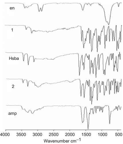
The NH2 vibrations at 3357 and 3281 cm−1 in en were shifted to lower frequencies in compound 1 as 2794, 2676, 2547 cm−1 due to the proton transfer to the amine group [Citation58]. The absorption bands at 3317 and 3175 cm−1 of the NH2 group of amp were slightly shifted from those found for compound 2 (3327 and 3162 cm−1) due to the weak intermolecular interactions. The relatively weak and broad band at 2500 cm−1 can be attributed to the ν(N-H)pyridinium vibration of the compound 2 due to proton transfer to the pyridine nitrogen [Citation59].
The NH2 vibrations of the sulphonamides in free Hsba (3425, 3278 cm−1), for compounds 1 (3326, 3156 cm−1) and 2 (3473, 3328 cm−1) were observed with a similar pattern. The absorption bands for compound 1 has shifted almost 100 cm−1 to the lower frequencies while compound 2 has shifted almost 50 cm−1 to the upper frequencies due to molecular interaction in the solid state. The strong absorption bands for the SO2 groups in Hsba for compounds 1 and 2 were observed at the region of 1400–1170 cm−1 with similar profiles and almost similar vibrations [Citation60].
The relatively weak and broad band at 2900 cm−1 can be attributed to the COOH group of Hsba. The strong C=O vibration at 1684 cm−1 of Hsba is shifted to 1634 and 1670 cm−1 for compounds 1 and 2, respectively, indicating the difference between the carboxylic acid group of the free Hsba and proton transfer compounds 1 and 2 [Citation61]. The ν(C-O) vibration of Hsba also shifted from 1169 cm−1 to 1073 and 1074 cm−1 in 1 and 2 [Citation62,Citation63].
The strong absorption bands at the region of 1400–1580 cm−1 are attributed to the ν(C=N) and ν(C=C) vibrations in free ligands, amp and Hsba, and compounds 1 and 2 [Citation64].
The ring wagging vibrations of the pyridine groups are also observed at 768 and 701 cm−1 for amp and 743 and 683 cm−1 for compound 2.
Thermal Analyses of 1 (C8H9Cl2N2O4S) and 2 (C13H13Cl2N3O4S)
and show the TG-DTG and DTA curves of compounds 1 and 2, respectively. The endothermic first stage (DTGmax = 240 and 260°C), between 25 and 266°C, corresponds to the loss of the CH5N from half of the ethylenediammonium group in the asymmetric unit of 1 (found 10.5, calcd 10.33). One mole of sulphonamide (SO2NH2) and one mole of the carboxylate (COO) groups of Hsba are decomposed in the second stage between 266 and 362°C with a DTGmax at 307 and 318°C (found 41.4, calcd 41.34). The residue of sba, C6H2Cl2, was decomposed between 362 and 590°C with DTGmax at 548 and 580°C (found 48.1, calcd 48.3).
Figure 7. The TG-DTG and DTA curves of 1.
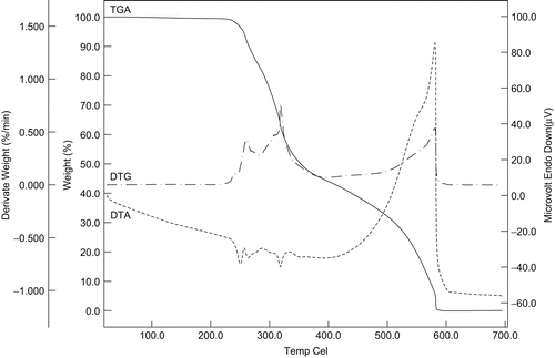
Figure 8. The TG-DTG and DTA curves of 2.
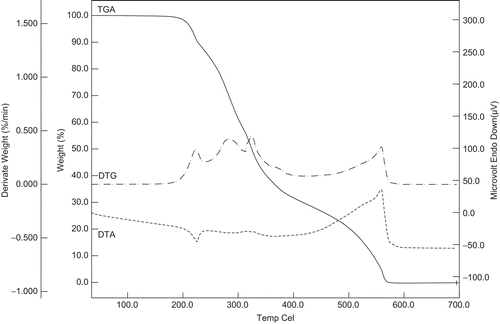
For compound 2, the first stage, an endothermic peak (DTGmax = 222°C) between 30 and 232°C, corresponded to the loss of the CH3N2 from the amine group of 2 (found 11.4, calcd 11.4). The second stage, an endothermic peak (DTGmax = 282°C) between 232 and 282°C, corresponded to the loss of C5H6 from the amine group of 2 (found 17.5, calcd 17.5). The sulphonamide (SO2NH2) and carboxylate (COO) groups of Hsba were decomposed in the third stage between 282 and 360°C with a DTGmax at 325 and 364°C (found 32.7, calcd 32.7). The final stage was the decomposition of the residue of sba, C6H2Cl2, between 360 and 575°C with a DTGmax at 559°C (found 38.4, calcd 38.5).
UV–Vis Spectra
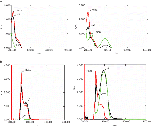
The electronic spectra of compounds 1 and 2, and the free ligands, amp and Hsba were recorded in solution at a 1 × 10⁻³ M concentration at room temperature ( and ). The characteristic π- π* transitions are in the spectra of 1 (207 nm) and of 2 (209 and 291 nm) in a water solution with the same profiles as the free ligands, Hsba (209 nm), amp (226 and 291 nm). Two sets of characteristic π- π* transitions were observed for all compounds in the DMSO solutions, which were located around 255 and 290 nm. The more energetic π-π* transition bands in water around 210 nm for all compounds were red shifted to around 255 nm in the DMSO solution spectra. The other π- π* absorption bands at 291 nm for both amp and compound 2 in water were also observed in almost the same region (296 and 294 nm respectively) in the spectra recorded in the DMSO. All the data which we obtained from the electronic spectra of the free ligands did not show any marked differences from those of the proton transfer compounds for both solutions
Table 3. Optical properties of compounds 1 and 2, and the free ligands amp and Hsba, in water and DMSO.
Figure 9. UV-Vis spectra of en, Hsba, amp, compounds 1 and 2; (A) in water, (B) in DMSO.
In vitro inhibition studies
The inhibition effects of the parent compounds (amp and Hsba), newly synthesised proton transfer compounds (1) and (2) together with AAZ as the control compound on the hCA I and hCA II enzymes were studied by both hydratase and esterase activity methods and then the Ki values were determined for each compound ().
Table 4. IC50 and Ki values of hCA-I and hCA-II isoenzyme hydratase and esterase activities after inhibition experiments.
According to the in vitro studies, the IC50 values for the hydratase activities of the newly synthesised compounds 1 and 2 (0.15 and 0.32 µM for hCA I and 0.06 and 0.15 µM for hCA II, respectively) were lower than the IC50 values for Hsba (0.45 and 0.4 µM for hCA I and II) and of AAZ (3.6 and 2.8 µM for hCA I and II). The IC50 values obtained from the esterase activities of all the compounds studied have been observed in a similar trend, and are 0.13 and 0.8 µM for hCA I and 0.14 and 0.1 µM for hCA II for 1 and 2, respectively. The Hsba and AAZ have esterase activities as 3.2 and 6.1 for hCA I and 18 and 4.5 µM for hCA II, respectively. The other parent compounds, amp and en, did not present any hydratase and esterase activities.
In relation to esterase activities, the inhibition equilibrium constants (Ki) were also determined. The novel compounds 1 and 2 had significant Ki values which are, respectively 0.137 and 0.157 µM for hCA I and 0.99 and 0.075 µM for hCA II compared to Hsba (7.7 and 232 µM for hCA I and II) and AAZ (5.2 and 3.8 µM for hCA I and II).
The parent compound Hsba showed inhibitory effects on hCA I and II as expected since it is a sulphonamide compound [Citation1–4]. Nevertheless, the other parent compounds, amp and en, did not show any inhibitory effects on these isoenzymes due to a lack of -SO2NH2 groups in their structures. The novel compounds 1 and 2 bear sulphonamide functional groups like Hsba and AAZ, and bear additionally carboxylate and protonated nitrogen functional groups unlike the parent compounds () and AAZ. These novel compounds (1 and 2) had higher inhibitory effects than the parent compounds and AAZ, which might be due to the interactions between the functional groups (sulphonamide, carboxylate and protonated nitrogen) in their structures and the zinc ion and histidine residue in the active site of CA [Citation65].
As the novel proton transfer compounds (1 and 2) have a higher potential inhibitory effects than the parent inhibitors (Hsba, amp and en) and also than AAZ as in our previous study [Citation34], they could be seen as candidates for the treatment of glaucoma.
Conclusion
In the present work, two novel proton transfer compounds (1 and 2) were prepared for the first time. The compounds 1 and 2 crystallised in the P-1 and P21/c space groups, respectively. The non-covalent interactions between neighbouring molecules, such as ion pairing, hydrogen bonding, and π-π stacking seemed to be effective in the stabilisation of the crystal structures. Elemental analyses and all measurements showed good agreement with the structures. These two novel proton transfer compounds possessed a significant inhibition effect on hCA I and on hCA II, and thus might be considered as possible drugs for glaucoma. These results suggest that further studies on such compounds would be worthwhile to obtain a correlation of the inhibitory effects on hCA I and hCA II and the derivatives of such compounds should be the subject to further inhibition in vivo tests.
Supplementary data
Crystallographic data (excluding structure factors) for the structures in this paper have been deposited with the Cambridge Crystallographic Data Centre as supplementary publication Nos CCDC 739346 for 1 and CCDC 739347 for 2. Copies of the data can be obtained, free of charge, on application to CCDC, 12 Union Road, Cambridge CB2 1EZ, UK, (fax: +44 (0)1223-336033 or e-mail: [email protected]).
Declaration of interest
The authors acknowledge the support provided by Dumlupınar University Research Fund (grant No. 2007/2). In addition, the authors would like to thank the Faculty of Arts and Sciences, Ondokuz Mayıs University, Turkey, for use of the Stoe IPDS-2 diffractometer purchased under grant F.279 of the University Research Fund.
References
- Supuran CT, Casini A, Mastrolorenzo A, Scozzafava A. Carbonic anhydrase inhibitors. Synthesis and inhibition of cytosolic/tumor-associated carbonic anhydrase isozymes I, II, and IX with boron-containing sulfonamides, sulfamides, and sulfamates: Toward agents for boron neutron capture therapy of hypoxic tumors. Mini Rev Med Chem 2004;4:625–632
- Owa T, Nagasu T. Novel sulphonamide derivatives for the treatment of cancer. Expert Opinion Therapeutic Patents 2000;10:1725–1740.
- Scozzafava A, Owa T, Mastrolorenzo A, Supuran CT. Anticancer and Antiviral Sulfonamides. Curr Med Chem 2003;10:925–953
- Choan ZH, Hassan M, Khan MK, Supuran CT. In-vitro antibacterial, antifungal and cytotoxic properties of sulfonamide-derived Schiff’s bases and their metal complexes. J Enzyme Inhib Med Chem 2005;20:183–188.
- Supuran CT. Carbonic anhydrases: novel therapeutic applications for inhibitors and activators. Nat Rev Drug Disc 2008;7:168–181.
- Bülbül M, Saracoğlu N, Küfrevioğlu Öİ, Çiftci M. Bile acid derivatives of 5-amino-1,3,4-thiadiazole-2-sulfonamide as new carbonic anhydrase inhibitors: Synthesis and investigation of inhibition effects. Bioorg Med Chem 2002;10:2561–2567.
- Bülbül M, Kasımoğulları R, Küfrevioğlu Öİ. Amide derivatives with pyrazole carboxylic acids of 5-amino-1,3, 4-thiadiazole 2-sulfonamide as new carbonic anhydrase inhibitors: Synthesis and investigation of inhibitory effects. J Enzyme Inhib Med Chem 2008;23:895–900.
- Nuti E, Orlandini E, Nencetti S, Rossello A, Innocenti A, Scozzafava A, Supuran CT. Carbonic anhydrase and matrix metalloproteinase inhibitors. Inhibition of human tumor-associated isozymes IX and cytosolic isozyme I and II with sulfonylated hydroxamates. Bioorg Med Chem 2007;15:2298–2311.
- Özensoy Ö,Koçkar F, Arslan O, Işık S, Supuran CT. An evaluation of cytosolic erythrocyte carbonic anhydrase and catalase in carcinoma patients: An elevation of carbonic anhydrase activity. Clin Biochem 2007;40:835.
- Supuran CT, Scozzafava A. Carbonic anhydrases as targets for medicinal chemistry. Bioorg Med Chem 2007:15:4336–4350.
- Di Fiore A, Scozzafava A, Winum JY, Montero JL, Pedone C, Supuran CT, Simone GD. Carbonic anhydrase inhibitors: Binding of an antiglaucoma glycosyl-sulfanilamide derivative to human isoform II and its consequences for the drug design of enzyme inhibitors incorporating sugar moieties. Bioorg Med Chem Lett 2007;17:1726–1731.
- Innocenti A, Vullo D, Pastorek J, Scozzafava A, Pastorekova S, Nishimori I, Supuran CT. Carbonic anhydrase inhibitors. Inhibition of transmembrane isozymes XII (cancer-associated) and XIV with anions. Bioorg Med Chem Lett 2007;17:1532–1537.
- Winum JY, Thiry A, Cheikh KE, Dogne JM, Montero JL, Vullo D, Scozzafava A, Masereel B, Supuran CT. Carbonic anhydrase inhibitors. Inhibition of isoforms I, II, IV, VA, VII, IX, and XIV with sulfonamides incorporating fructopyranose-thioureido tails. Bioorg Med Chem Lett 2007;17:2685–2691.
- Scozzafava A, Banciu MD, Popescu A, Supuran CT. Carbonic anhydrase inhibitors: Inhibition of isozymes I, II and IV by sulfamide and sulfamic acid derivatives. J Enzyme Inhib Med Chem 2000;15:443–553.
- Schuman JS. Antiglaucoma medications: A review of safety and tolerability issues related to their use. Clin Ther 2000;22:167–208.
- Wilkinson BL, Bornaghi LF, Houston TA, Innocenti A, Vullo D, Supuran CT, Poulsen SA. Inhibition of membrane-associated carbonic anhydrase isozymes IX, XII and XIV with a library of glycoconjugate benzenesulfonamides. Bioorg Med Chem Lett 2007;17:987–992.
- Santos MA, Marques S, Vullo D, Innocenti A, Scozzafava A, Supuran CT. Carbonic anhydrase inhibitors: Inhibition of cytosolic/tumor-associated isoforms I, II, and IX with iminodiacetic carboxylates/hydroxamates also incorporating benzene sulfonamide moieties. Bioorg Med Chem Lett 2007;17:1538–1543.
- Glaser R, Lewis M. Single- and double-proton-transfer in the aggregate between cytosine and guaninediazonium ion. Org Lett 1999;1:273–276.
- Semo N, Osheroff MA, Koskl WS. Proton-transfer from the formyl and isoformyl positive-ions to acetylene. J Phys Chem 1983;87:2302–2303.
- He F, Ramirez J, Lebrilla CB. Evidence for an intermolecular proton-transfer reaction induced by collision in gas-phase noncovalently bound complexes. J Am Chem Soc 1999;121:4726–4727.
- Tarkka RM, Zhang X, Jenekhe SA. Electrically generated intramolecular proton transfer: Electroluminescence and stimulated emission from polymers. J Am Chem Soc 1996;118:9438–9439.
- Nogami M, Daiko V, Akai V, Kasuga V. Dynamics of proton transfer in the sol-gel-derived P2O5-SiO2 glasses. J Phys Chem 2001;105:4653–4656.
- Sharif MA, Aghabozorg A, Shokrollahi A, Kickelbick G, Moghimi A, Shamsipur M. Novel proton transfer compounds containing 2,6-pyridinedicarboxylic acid and melamine and their Pb-II complex: Synthesis, characterization, crystal structure and solution studies. Polish J Chem 2006;80:847–863.
- Moghimi A, Moosavi SM, Kordestani D, Maddah B, Shamsipur M, Aghabozorg H, Ramezanipour F, Kickelbick G. Pyridine-2,6-bis(monothiocarboxylic) acid and 2-aminopyridine as building blocks of a novel proton transfer compound: Solution and X-ray crystal structural studies. J Mol Struct 2007;828:38–45.
- Zhang J, Zhu LG. Different supramolecular assemblies in two 1:1 proton-transfer compounds of sulfobenzoic acids with aromatic amines. Acta Cryst 2007;C63:o484–o486.
- Franklin S, Balasubramanian T. Salts of maleic and fumaric acids with oxine: the role of isomeric acids in hydrogen-bonding patterns. Acta Cryst 2009;C65:o58–o61.
- Soleimannejad J, Aghabozorg H, Morsali A, Hemmati F, Manteghi F. 4,4′-Bipyridinium bis(2-carboxypyridine-3-carboxylate). Acta Cryst 2009;E65:O153–U2591.
- Yenikaya C, Poyraz M, Sarı M, Demirci F, İlkimen H, Büyükgüngör O. Synthesis, characterization and biological evaluation of a novel Cu(II) complex with the mixed ligands 2,6-pyridinedicarboxylic acid and 2-aminopyridine. Polyhedron 2009;28:3526–3532
- Moghimi A, Alizadeh R, Aghabozorgb H, Shockravib A, Aragonic MC, Demartind F, Isaiac F, Lippolisc V, Harrisone A, Shokrollahif A, Shamsipurf M. Ion pairing, H-bonding, and pi-pi interactions in copper(II) complex-organo-networks derived from a proton-transfer compound of the 1,10-phenanthroline-2,9-dicarboxylic acid. J Mol Struct 2005;750:166–173.
- Ramezanipour F, Aghabozorg H, Shokrollahi A, Shamsipur M, Stoeckli-Evans H, Soleimannejad J, Sheshmani S. Different complexation behavior of a proton transfer compound obtained from 1,10-phenanthroline and pyridine-2,6-dicarboxylic acid with In-III and Ce-III: Synthesis, crystal structures and solution studies. J Mol Struct 2005;779:77–86.
- Aghabozorg H, Derikvand Z, Olmstead MM, Gharamaleki JA. Hydroxonium hydrate tris(2,4,6-triamino-1,3,5-triazin-1-ium)bis[bis(pyridine-2,6-dicarboxylato)manganate (II/III)] hydroxide pyridine-2,6-dicarboxylic acid solvate pentahydrate. Acta Cryst 2008;C64:m372–m374.
- Aghabozorg H, Motyeian E, Soleimannejad J, Ghadermazi M, Gharamaleki JA. Aqua(4-hydroxypyridine-2,6-dicarboxylato)(1,10-phenanothroline)copper(II)4.5-hydrate. Acta Cryst 2008;E64:m252–u2372.
- Aghabozorg H, Derikvand Z, Nemati A, Bahrami Z, Gharamaleki JA. Poly[piperazinediium [aquabis(mu-pyridine-2,5-dicarboxylato)zincate] dihydrate] Acta Cryst 2008;E64:m111–u1098.
- Yenikaya C, Sarı M, Bülbül M, İlkimen H, Çelik H, Büyükgüngör O. Synthesis, characterization and antiglaucoma activity of a novel proton transfer compound and a mixed-ligand Zn(II) complex. Bioorg Med Chem 2010;18:930–938.
- Sheldrick GM. SHELXS97 and SHEXL97. Program for Crystal Structure Solution and Refinement. University of Göttingen, Germany, 1997.
- Stoe & Cie, X-AREA (Version 1.18) and X-RED32 (Version 1.04). Stoe & Cie, Darmstadt, Germany, 2002.
- Farrugia LJ. ORTEP-3 for Windows - a version of ORTEP-III with a Graphical User Interface (GUI) J Appl Cryst 1997;30:565.
- Arslan O, Nalbantoğlu B, Demir N, Özdemir H, Küfrevioğlu Öİ. A new method for the purification of carbonic anhydrase isozymes by affinity chromatography. Trop J Med Sci 1996;26:163–166.
- Rickli EE, Ghazanfar SA, Gibbson BH, Edsall JT. Carbonic Anhydrases from Human Erythrocytes: Preparation and properties of two enzymes. J Biol Chem 1964;239:1065–1068.
- Bradford MM. A rapid and sensitive method for the quantitation of microgram quantities of protein utilizing the principle of protein-dye binding. Anal Biochem 1976;72:248–254.
- Laemmli UK. Cleavage of Structural Proteins during the Assembly of the Head of Bacteriophage T4. Nature 1970;227:680–685.
- Wilbur KM, Anderson NG. Electrometric and colorimetric determination of carbonic anhydrase. J Biol Chem 1948;176:147–154.
- Verpoorte JA, Mehta S, Edsall JT. Esterase activities of human carbonic anhydrases B and C. J Biol Chem. 1967;242:4221–4229.
- Innocenti A, Scozzafava A, Parkkila S, Pucceti L, De Simone G, Supuran CT. Investigations of the esterase, phosphatase, and sulfatase activities of the cytosolic mammalian carbonic anhydrase isoforms I, II, and XIII with 4-nitrophenyl esters as substrates. Bioorg Med Chem Lett 2008;18:2267–2271.
- Landolfi C, Marchetti M, Ciocci G, Milanese C. Development and pharmacological characterization of a modified procedure for the measurement of carbonic anhydrase activity. J Pharmacol Toxicol Methods 1998;38:169–172.
- Bülbül M, Hisar O, Beydemir S, Ciftci M, Küfrevioğlu Öİ. The in vitro and in vivo inhibitory effects of some sulfonamide derivatives on rainbow trout (Oncorhynchus mykiss) erythrocyte carbonic anhydrase activity. J Enzyme Inhib Med Chem 2003:18;371–375.
- Ciftci M, Bülbül M, Gül M, Gümüştekin K, Dane Ş, Süleyman H. Effects of nicotine and Vitamin E on carbonic anhydrase activity in some rat tissues in vivo and in vitro. J Enzyme Inhib Med Chem 2005:20;103–109.
- Hisar O, Beydemir Ş, Bülbül M, Yanık T. Kinetic properties of carbonic anhydrase purified from gills of rainbow trout (Oncorhynchus mykiss). J Appl Anim Res 2006;30:185–188.
- Winum JY, Cecchi A, Montero JL, Innocenti A, Scozzafava A, Supuran CT. Carbonic anhydrase inhibitors. Synthesis and inhibition of cytosolic/tumor-associated carbonic anhydrase isozymes I, II, and IX with boron-containing sulfonamides, sulfamides, and sulfamates: Toward agents for boron neutron capture therapy of hypoxic tumors. Bioorg Med Chem Lett 2005;15:3302–3306.
- Lineweaver H, Burk D. The determination of enzyme dissociation contants. J Am Chem Soc 1934;56:658–666.
- Siddiqui WA, Ahmad S, Siddiqui HL, Tariq MI, Parvez M. o-sulfamoylbenzoic acid. Acta Cryst 2007;E63:o4117–u3678.
- Yeşilel OZ, Büyükgüngör O. The novel monomeric bis(orotate-N,O) supramolecular complex of manganese(II): Synthesis, spectrothermal properties and crystal structures of (enH)2[Mn(HOr)2(H2O)2].2.5H2O. Solid State Science 2007;9:485–490.
- Aghabozorg H, Saei AA, Ramezanipour F. 2,6-Diaminopyridinium pyridinium-2,6-dicarboxylate: a redetermination. Acta Cryst 2005;E61:o3242–o3244.
- Nolan SJ, Shiels JC, Tuite JB, Cecere KL, Baranger AM. Recognition of an essential adenine at a protein-RNA interface: Comparison of the contributions of hydrogen bonds and a stacking interaction. J Am Chem Soc 1999;121:8951–8952.
- Madalan AM, Kravtsov VC, Simonov YA, Voronkova V, Korobchenko L, Avarvari N, Andruh M. A unique diamondoid network resulting from the convolution of pi-pi stacking and lipophilic interactions. Cryst Growth Des 2005;5:45–47.
- Bergman SD, Reshef D, Frish L, Cohen Y, Goldberg I, Kol M. From eilatin to isoeilatin: A skeletal rearrangement strongly influences pi-stacking of Ru(II) complex. Inorg Chem 2004;43;3792–3794.
- Rathore R, Abdelwahed SH, Guzei IA. Synthesis, structure, and evaluation of the effect of multiple stacking on the electron-donor properties of pi-stacked polyfluorenes. J Am Chem Soc 2003;125:8712–8713.
- Nakamoto K. Infrared And Raman Spectra of Inorganic and Coordination Compounds, 5th Edition, Interscience-Wiley, NewYork, 1997:16.
- Cooks, D. Vibrational spectra of pyridinium salts. Canadian J Chem 1961;39(10):2009–2024.
- Gowda BT, Jyothi K, Souza JDD. Infrared and NMR spectra of arylsulphonamides, 4-X-C6H4SO2NH2 and i-X, j-YC6H3SO2NH2 (X = H; CH3; C2H5; F; Cl; Br; I or NO2 and i-X, j-Y = 2,3-(CH3)(2); 2,4-(CH3)(2); 2,5(CH3)(2); 2-CH3, 4-Cl; 2-CH3, 5-Cl; 3-CH3, 4-Cl; 2,4-Cl-2 or 3,4-Cl-2). Z Naturforsch 2002;57:967–973.
- Nakamoto K. Infrared and Raman Spectra of Inorganic and Coordination Compounds. 5th Edition, NewYork: Interscience-Wiley, 1997:59.
- Mao L, Wang Y, Qi Y, Cao M, Hu C. A novel three-dimensional supramolecular framework with one-dimensional channels: synthesis and crystal structure of [Cu(DPC)(H2O)3] (H2DPC=Pyridine-2,6-dicarboxylic acid). J Mol Struct 2004;688:197–201.
- Uçar İ,Karabulut B, Bulut A, Büyükgüngör O. Synthesis, structure, spectroscopic and electrochemical properties of (2-amino-4-methylpyrimidine)-(pyridine-2,6-dicarboxylato) copper(II) monohydrate. J Mol Struct 2007;834:336–344.
- Kirillova MV, Da Silva MFCG, Kirillov AM, Da Silva JJRF, Pombeiro AJL. 3D hydrogen bonded heteronuclear Co-II, Ni-II, Cu-II and Zn-II aqua complexes derived from dipicolinic acid. Inorg Chim Acta 2007;360:506–512.
- Lindskog S. Structure and mechanism of carbonic anhydrase. Pharmacol Ther 1997;74:1–20.