Abstract
A new carbonic anhydrase (CA, EC 4.2.1.1) from the thermophilic bacterium Sulfurihydrogenibium yellowstonense YO3AOP1 was identified and characterized. The bacterial carbonic anhydrase gene was expressed in Escherichia coli yielding an active enzyme, which was purified in large amounts. The recombinant protein (SspCA) was found to belong to the α-CA class and displays esterase activity. The kinetic parameters were determined by using CO2 and p-nitrophenylacetate (p-NpA) as substrates. The bacterial enzyme presented specific activity comparable to that of bovine carbonic anhydrase (bCA II) but it showed biochemical properties never observed for the mammalian enzyme. The thermophilic enzyme, in fact, was endowed with high thermostability and with unaltered residual activity after prolonged exposure to heat up to 100°C. SspCA and the bovine carbonic anhydrase (bCA II) were immobilized within a polyurethane (PU) foam. The immobilized bacterial enzyme was found to be active and stable at 100°C up to 50 h.
Introduction
Carbonic anhydrases (CAs, EC 4.2.1.1) are zinc-containing enzymes that catalyze the reversible conversion of carbon dioxide to bicarbonate and protons (CO2 + H2O ↔ HCO3− + H+) with very high efficiency, and are among the most efficient enzymes in the cell. CAs play roles in carbon metabolism, human health, and the global carbon cycle and are found in most eukaryotic and many microbial organismsCitation1,Citation2. In eukaryotes, these enzymes participate in various physiological functions including respiration, pH homeostasis, ion transports and photosynthetic CO2 fixationCitation1,Citation3.
CAs are divided into five evolutionary distinct classes, the α, β and γ classes and the recently discovered classes, δ and ζ. The human CA II (hCA II) and IX (hCA IX), two of the human isoenzymes existing, are catalytically the fastest members of the α-class of the CA family, exhibiting near-diffusion-limited kineticsCitation4. The catalytic mechanism and inhibition of α-CAs has been intensively investigatedCitation5. These enzymes contain an active site zinc ion (Zn2+) coordinated by three histidine residues and a water molecule/hydroxide ion. Some of the catalytically active α-CAs can also catalyze the hydrolysis of esters, for example 4-nitrophenyl acetate (4-NpA) and other hydrolytic reactions. Known α-CA inhibitors (CAIs) include various anions, imidazole, phenol, hydroxyurea, carboxylates, organic phosphates and phosphonates, and various sulfonamide compounds (R-SO2NH2)Citation6.
Members of three enzyme classes have been described for prokaryotic organismsCitation7, i.e. α-, β- and γ-CAs, with the β-class enzymes being the most diffuseCitation8. Within the Bacteria domain, the enzymes purified from Neisseria gonorrhoeae, Neisseria sicca and Helicobacter pylori belong to the α classCitation9–12. The N. gonorrhoeae CA had a molecular mass of 28 kDa, being quite homologous to mammalian CAs and showed a high CO2 hydratase activity, similarly to the human isoforms hCA II and an esterase activity for the hydrolysis of p-NpACitation10. Instead, the H. pylori CA displayed an activity similar to that of the human isoform hCA I for the CO2 hydration reactionCitation11. CAs belonging to the β-class were cloned, purified, and characterized from Escherichia coli, Salmonella enterica, Helicobacter pylori, Haemophilus influenza and from other pathogensCitation8. The tri-dimensional structures of these enzymes are similar to that of the Salmonella enterica (stCA 1)Citation13. However, many of them displayed excellent activity for the CO2 hydration but lack esterase activityCitation14,Citation15. A β-CA from the thermophilic bacterium Methanobacterium thermoautotrophicum has been also identified. Interestingly, the enzyme from M. thermoautotrophicum was stable at temperatures up to 75°CCitation16. In the Archaea domain, a γ-CA from Methanosarcina thermophila was discovered by Ferry’s groupCitation7.
Here, we report the characterization of an innovative carbonic anhydrase (SspCA) identified by translated genome inspection of a novel thermophilic bacterium, the species YO3AOP1 of Sulfurihydrogenibium, isolated in the Yellowstone National Park, USACitation17. This investigation aimed to characterize the CA from Sulfurihydrogenibium yellowstonense YO3AOP1 (Sulfurihydrogenibium sp.), to study its biochemical properties and to provide additional insights in the field of biotechnology and carbon dioxide sequestration. The encoding gene was cloned and expressed in Escherichia coli and the recombinant protein purified to homogeneity. This enzyme (SspCA) belongs to the α class of the carbonic anhydrase family. Thermoactivity and thermostability studies showed that SspCA is active in the temperature range from 0 to 100°C and retains full activity after 2 h incubation at 100°C. SspCA was immobilized within a polyurethane (PU) foam and was found to be unalterably active and stable up to 50 h at 100°C.
Materials and methods
Gene identification, construct preparation, protein expression and purification
The search of CAs was performed with the “FASTA” program and sequences selected for significant similarity scores. The nucleotide sequence encoding for the carbonic anhydrase of Methanosarcina thermophila (accession number: U08885.1), a moderate thermophile archeon, was used as reference sequence. Among others, the CA sequence from Sulfurihydrogenibium sp. YO3AOP1 (SspCA) was screened. The Mr. Gene Company, specialized in gene synthesis, designed the synthetic Sulfurihydrogenibium sp. gene encoding for the α carbonic anhydrase lacking the peptide signal (first 20 amino acid of the peptide sequence)Citation18 and containing a NdeI and XhoI site at the 5′ and 3′ end of the SspCa gene, respectively. The resulting plasmid was amplified into E. coli DH5 α cells. The Sulfurihydrogenibium sp. DNA fragments were separated on 1% agarose gel. The recovered Sulfurihydrogenibium sp. gene and the linearized expression vector (pET15-b) were ligated by T4 DNA ligase to form the expression vector pET15-b/Ssp. In order to confirm the integrity of the Sulfurihydrogenibium sp. gene and that no errors had taken place at the ligation sites, the vector containing the fragment was sequenced. Competent E. coli BL21 (DE3) cells were transformed with pET15-b/Ssp, grown at 37°C and induced with 1 mM IPTG. After additional growth for 5 h, the cells were harvested and disrupted by sonication at 4°C. Following centrifugation, the cell extract was heated at 90°C for 30 min and centrifuged. The supernatant was loaded into His-select HF Nickel affinity gel and the protein was eluted with 250 mM imidazole. At this stage of purification the enzyme was at least 95% pure and the obtained recovery was of 12 mg of the recombinant bacterial carbonic anhydrase.
SDS-PAGE
Sodium dodecyl sulfate (SDS)-polyacrylamide gel electrophoresis (PAGE) was performed as described previouslyCitation19, using 12% gels.
Assay for carbonic anhydrase with CO2 as substrate
CA activity assay was a modification of the procedure described by ChiricaCitation10. The assay was based on the monitoring of pH variation due to the catalyzed conversion of CO2 to bicarbonate. Bromothymol blue was used as the indicator of pH variation. The assay was performed at 0°C adding 1.0 mL ice-cold CO2-saturated water to 1.0 mL mixtures of 25 mM Tris-SO4 buffer containing different amounts of the enzyme. The CO2-satured solution was prepared by bubbling CO2 into 100 mL distilled water for approximately 3 h. The CO2 solution was chilled in an ice-water bath. To test the activity of carbonic anhydrase, 1 mL of 25 mM Tris, pH 8.3, containing bromothymol blue as a dye (to give a distinct and visible blue color) was added to two test tubes chilled in an ice bath. Ten to 50 µL of the enzyme solution (e.g. cell extract or purified enzyme) were added to one tube, and an equivalent amount of buffer was added to the second tube as control. One milliliter of CO2 solution was added very quickly and simultaneously a stopwatch was started. The time required for the solution to change from blue to yellow was recorded (transition point of bromothymol blue is pH 6–7.6). The production of hydrogen ions during the CO2 hydration reaction lowers the pH of the solution until the color transition point of the dye is reached. The time required for the color change is inversely related to the quantity of carbonic anhydrase present in the sample. Detecting the color change is somewhat subjective but the error for triple measurements was in the range of 0–1 s difference for the catalyzed reaction. Wilbur-Anderson units were calculated according to the following definition: One Wilbur-Anderson unit (WAU) of activity is defined as (T0 – T)/T, where T0 (uncatalyzed reaction) and T (catalyzed reaction) are recorded as the time (in seconds) required for the pH to drop from 8.3 to the transition point of the dye in a control buffer and in the presence of enzyme, respectively.
Esterase activity
Activity for p-nitrophenylacetate (p-NpA) hydrolysis was determined at 0°C using a modification of the method proposed by Armstrong et al. (1996). The reaction mixture contained 0.3 mL of freshly prepared 3 mM p-NpA and 0.7 mL of 15 mM Tris sulphate buffer, pH 7.6. Ten µL of enzyme solution were added, and the catalyzed reaction was monitored reading the increase in A348 nm for 5 min. The catalyzed reactions were corrected for the non-enzymatic reaction. One enzyme unit was defined as the amount capable of producing an OD348 nm = 0.03 in 5 min.
Determination of kinetic constants using CO2 as substrate
Kinetic measures with CO2 as the substrate were performed at 0°C. In order to determine Michaelis–Menten constants, increasing concentrations of CO2 were used. An ice-cold CO2-saturated water (70 mM) was prepared and concentrations from 12 to 62.5 mM were made using ice-cold CO2 non-saturated water as diluent. To measure the activity of carbonic anhydrase, 0.1 mL of 250 mM Tris, pH 8.3, containing bromothymol blue was added to 0.9 mL of CO2 at different concentration. Reactions were started by adding 30 ng of the CA preparation. The time required for the solution to change from blue to yellow was recorded as described in the section “assay for carbonic anhydrase.” All the reactions were performed in triplicate and in parallel with bovine carbonic anhydrase.
Determination of kinetic constants using p-NpA as substrate
Enzyme activity at 10 different p-NpA concentration (0.125–5 mM) was determined at 0°C by measuring the increase of absorbance at 348 nm. The Michaelis–Menten constants (Km and Vmax) were calculated at pH 7.6. The enzyme concentration in the assay was 300 ng. All the reactions were performed in triplicate and in parallel with bovine carbonic anhydrase.
Temperature studies
Effect of temperature on activity
The temperature dependence activity of SspCA was measured using the p-NpA as substrate. Protein concentration used in the assay was 300 ng. The activity was measured in the temperature range from 25 to 100°C. All the reactions were performed in triplicate.
Effect of temperature on the SspCA and bCA II stability
To compare the stability of SspCA and bCA II at different temperatures, enzymes at the concentration of 3 µg/mL in 10 mM Tris/HCl, pH 8.3 were incubated at 40, 50, 60, 70, 80, 90 and 100°C for different time (30, 60, 120 and 180 min). Enzyme aliquots (30 ng) were withdrawn at appropriate times and the residual activity was measured at 0°C using CO2 as substrate.
Immobilization of carbonic anhydrases within PU foam
Ten mg of CA enzyme were dissolved in 3 mL of distilled water and poured onto about 3 g of viscous HYPOL2060 prepolymer in a 50 mL falcon tubeCitation20. After completion of enzyme immobilization, CA activity was determined on a piece of CA-immobilized foam (3 mg of foam containing 10 µg of enzyme) which was cut from the middle of the whole product. The reaction was started by addition of the foam to the substrate-containing mixture under stirring. All the experiments were carried out in parallel with the mammalian enzyme. For studies on stability, foam slices containing the immobilized SspCA or bCA II were incubated at 100°C. The immobilized enzyme activity was determined every 12 h over a total of 48 h. The assay was carried out using SspCA or bCA II and CO2 as substrate.
Results
Enzyme purification
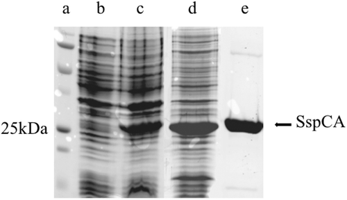
The recombinant SspCA was isolated and purified to homogeneity at room temperature from Escherichia coli (DE3) cell extract (). Most of the carbonic anhydrase activity was recovered in the soluble fraction of cell extract after sonication and centrifugation as described in Materials and methods section. shows that the heterologously expressed SspCA enzyme was purified 3-fold with the thermoprecipitation step. Using the affinity column (His-select HF Nickel affinity gel), SspCA was purified 16-fold to apparent homogeneity, as indicated by a single protein band after SDS-PAGE (, lane e). shows the SDS-PAGE of the recombinant SspCA at various steps during purification from E. coli.
Table 1. Purification of recombinant SspCA produced in E. coli.
Figure 1. SDS-PAGE of the recombinant SspCA purified from E. coli. Lane a, molecular markers, M.W. starting from the top: 250 kDa, 150 kDa, 100 kDa, 75 kDa, 50 kDa, 37 kDa, 25 kDa, 20 kDa; Lane b, cell extract protein from E. coli before induction with IPTG; Lane c, cell extract protein after induction with IPTG; Lane d, proteins after thermoprecipitation at 90°C and centrifugation; Lane e, purified SspCA from His-tag affinity column.
Biochemical characterization
Determination of molecular weight
The molecular weight estimated by SDS-PAGE was 26.0 kDA. A subunit molecular mass of 26.4 kDa was calculated on the basis of the amino acid sequence translated from the gene.
Hydratase and esterase activities
Using CO2 as substrate, the specific activities for SspCA and bCA II were determined. As indicated in , the specific activity of SspCA was 1.4 times greater than that of the mammalian enzyme. Carbonic anhydrases (α-type) from mammalian sources catalyze the reversible hydrolysis of esters. With p-nitrophenyl acetate as substrate, the presence of esterase activity was investigated in the bacterial enzyme. SspCA showed a specific activity of 322 U/mg of protein that is 4 times smaller than that of the commercially available bovine bCA II ().
Table 2. Comparison of the hydratase and esterase activity for SspCA and bCA II.
Kinetic analysis
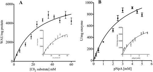
Kinetic parameters were determined for the purified recombinant SspCA and the commercially available bCA II (). The kinetic constants (Km and Vmax) were calculated for either CO2 () or p-NpA as substrates () using the program GraphPad Prism 5.0 software (GraphPad Software, San Diego, CA). Using CO2 as substrate, the Km of SspCA was approximately 2.7 times lower than Km calculated for the bovine enzyme suggesting a higher affinity of the bacterial enzyme for the substrate. The values for the reaction rates (Vmax) of the bacterial and bovine enzymes differed of about 1.5-fold. Opposite to this, the esterase activity of the two enzymes showed approximately the same Km. Nevertheless, the Vmax calculated for the bovine carbonic anhydrase was about 3.6 times higher than the Vmax relative to the bacterial enzyme ().
Table 3. Comparison of the kinetic constants for SspCA and bCA II using CO2 or p-NpA as substrate.
Figure 2. Michaelis–Menten curves for the determination of the apparent Km and Vmax calculated using CO2 (A) or p-NpA (B) as substrate. The kinetic constant values are reported in . Inserts represent the Michaelis–Menten curve calculated for the mammalian enzyme (bCA II). Each point is the mean ±SEM of 3 independent determinations. Data have been analyzed by means of GraphPad Prism 5.0 software (GraphPad Software, San Diego, CA).
Stability studies
Effect of temperature on activity
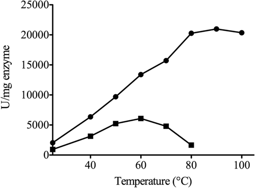
The temperature dependent activity of SspCA was assayed using the p-NpA as substrate, and measurements were performed in parallel with bCA II using 300 ng of each enzyme (). The optimum temperature range for SspCA and bCA II were determined by incubating enzymes at 25, 40, 50, 60, 70, 80, 90 and 100°C. The reaction was monitored following the absorbance at 348 nm for 5 min. The optimum temperature for bovine enzyme activity was found to be 60°C, while the SspCA (optimum at 95°C) was still active at 100°C ().
Figure 3. Effect of temperature on the activities of SspCA and bCA II. The enzyme activity was measured at the indicated temperatures and using p-NpA as substrate. SspCA, filled circle; bCA II, filled square.
Temperature effect on the stabilities of SspCA and bCA II
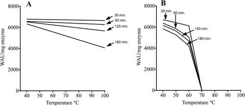
Stabilities of SspCA and bCA II were compared at the temperatures indicated on the X-axis ( and ). After 30, 60, 120 and 180 min of incubation, the activities of SspCA and bCA II were determined using CO2 as substrate. The bovine enzyme was inactivated at temperature higher than 60°C () for all incubation times. Noticeably, the bacterial enzyme retained its activity at temperature of 100°C (). These studies indicate that the bacterial enzyme was more stable at high temperature and retained its activity for a longer time (e.g. 180 min) when compared to the mammalian enzyme.
Figure 4. Thermostability of SspCA and bCA II. The enzymes were incubated for 30, 60, 120 and 180 min at the indicated temperatures and assayed using CO2 as substrate. (A) thermostability of SspCA; (B) thermostability of bCA II.
Immobilization of SspCA and bCA II within the PU foam
Immobilization was performed as described in Materials and methods section. The specific activities of the immobilized enzymes are listed in . The bacterial enzyme was 1.4-fold more active than the bovine counterpart and it maintained its activity after several reaction cycles. The PU foam containing the immobilized SspCA or bCA II was incubated at 100°C for 3 and 48 h. The activity of the immobilized bCA II showed a 50-fold decrease over 48 h, while the immobilized SspCA retained its full activity ().
Table 4. Comparison of the hydratase activity for the immobilized SspCA and bCA II.
Discussion and conclusions
Carbonic anhydrases, isolated from mammals or prokaryotes and other mesophilic sources, are active at physiological temperature (37°C) and are, like many enzymes, quite unstable under operative conditions. Studies on the enzymes from thermophiles, microorganisms living at temperatures from 70°C to 110°C, demonstrated that they are thermostable, thermoactive and generally stable to the common enzymes denaturantsCitation21,Citation22. With the aim of the recombinant DNA technology, a novel carbonic anhydrase, SspCA, identified in the genome of the thermophilic bacterium, Sulfurihydrogenibium sp., was overexpressed in E. coli. The enzyme shared significant similarity with other α-CA class enzymes, previously believed to be present only in mammalian organisms. The dependence of the SspCA activity on temperature was determined in comparison with bCA II. Starting from 25°C, temperature at which the activity of both enzymes was comparable, the SspCA activity increased constantly up to 95°C whereas the bovine enzyme had a maximal activity at 60°C. However, the activity of the bovine enzyme at the optimum temperature was still about one half of the bacterial enzyme activity. The results of our studies on thermostability showed that soluble SspCA was stable for 180 min at 100°C whereas the bovine enzyme was fully inactivated at 70°C. The first documented CA from a thermophile was purified from the thermophilic methanoarcheon M. thermoautotrophicum, but the enzyme was thermostable at temperatures up to 75°C and belonged to the β-CA classCitation7,Citation16. Comparative kinetic studies on SspCA and bCA II revealed that, at 0°C, the bacterial enzyme showed lower values of Km, namely a higher affinity toward both CO2 and p-NpA, whereas the Vmax was lower. One intriguing fact is that the temperature of the CO2 assay, 0°C, was many degrees below the optimal growth temperature of Sulfurihydrogenibium sp., which is between 55 and 78°C. With p-NpA, the optimal temperature for SspCA was 80°C while that of the bCA II was 60°C (see ). The low temperature of the CO2 assay enables appropriate molecular motions required for biological activity of a thermostable protein like SspCA, determining an activity lower than that expected.
In addition to biochemical studies, carbonic anhydrases have received much attention to their potential use in carbon dioxide sequestration. The use of carbonic anhydrases, either free or immobilized, has been reported in several technical approaches for capture of CO2 from combustion or from gases mixtures, for its subsequent utilization or disposal. This utilization of CA requires stable enzymes, even at high temperature, and the possibility of re-using the enzyme in many subsequent cycles for an efficient sequestration process. SspCA was demonstrated to be efficiently immobilized within PU foam at 100°C. The bacterial enzyme maintained 100% activity over 50 h while the mammalian CA was fully inactivated already after 3 h at 100°C. The results obtained in this work strongly suggest that the thermostable SspCA is an almost ideal candidate for hydration of CO2 in biomimetic sequestration.
In the prokaryotes, the existence of genes encoding carbonic anhydrases from all three classes with the β and γ classes predominatingCitation23 suggests that these enzymes may play an important role in prokaryotic physiology. Various roles were proposed for the known carbonic anhydraseCitation7. The most significant are (i) transport of CO2 or HCO3−; (ii) supply of CO2 or HCO3− for the enzymatic reactions; (iii) involvement in cyanate degradation; and (iv) survival of intracellular pathogens within their host. Most of the evidences about the function of carbonic anhydrase in the prokaryotic organisms are related to the fixation of CO2 by cyanobacteria. However, the role of these enzymes is still basically unknown. Our research on the new thermostable carbonic anhydrase from the thermophilic bacterium, Sulfurihydrogenibium sp. might contribute to elucidate novel functions for this ubiquitous prokaryotic enzyme.
Acknowledgments
The authors thank Dr. Claudiu T. Supuran (University of Florence) for helpful discussions concerning this work.
Declaration of interest: This research was financed by a grant of the “Accordo di Programma CNR-MSE.”
References
- Supuran CT. Carbonic anhydrases–an overview. Curr Pharm Des 2008;14:603–614.
- Armstrong JM, Myers DV, Verpoorte JA, Edsall JT. Purification and properties of human erythrocyte carbonic anhydrases. J Biol Chem 1966;241:5137–5149.
- Tashian RE. The carbonic anhydrases: Widening perspectives on their evolution, expression and function. Bioessays 1989;10:186–192.
- Supuran CT. Carbonic anhydrases: Novel therapeutic applications for inhibitors and activators. Nat Rev Drug Discov 2008;7:168–181.
- Neri D, Supuran CT. Interfering with pH regulation in tumours as a therapeutic strategy. Nat Rev Drug Discov 2011;10:767–777.
- Bertucci A, Innocenti A, Scozzafava A, Tambutté S, Zoccola D, Supuran CT. Carbonic anhydrase inhibitors. Inhibition studies with anions and sulfonamides of a new cytosolic enzyme from the scleractinian coral Stylophora pistillata. Bioorg Med Chem Lett 2011;21:710–714.
- Smith KS, Ferry JG. Prokaryotic carbonic anhydrases. FEMS Microbiol Rev 2000;24:335–366.
- Supuran CT. Bacterial carbonic anhydrases as drug targets: Toward novel antibiotics? Front Pharmacol 2011;2:34.
- Adler L, Brundell J, Falkbring SO, Nyman PO. Carbonic anhydrase from Neisseria sicca, strain 6021. I. Bacterial growth and purification of the enzyme. Biochim Biophys Acta 1972;284:298–310.
- Chirica LC, Elleby B, Jonsson BH, Lindskog S. The complete sequence, expression in Escherichia coli, purification and some properties of carbonic anhydrase from Neisseria gonorrhoeae. Eur J Biochem 1997;244:755–760.
- Chirica LC, Petersson C, Hurtig M, Jonsson BH, Borén T, Lindskog S. Expression and localization of α- and β-carbonic anhydrase in Helicobacter pylori. Biochim Biophys Acta 2002;1601:192–199.
- Guilloton MB, Korte JJ, Lamblin AF, Fuchs JA, Anderson PM. Carbonic anhydrase in Escherichia coli. A product of the cyn operon. J Biol Chem 1992;267:3731–3734.
- Vullo D, Nishimori I, Minakuchi T, Scozzafava A, Supuran CT. Inhibition studies with anions and small molecules of two novel ß-carbonic anhydrases from the bacterial pathogen Salmonella enterica serovar Typhimurium. Bioorg Med Chem Lett 2011;21:3591–3595.
- Supuran CT. Carbonic anhydrase inhibitors. Bioorg Med Chem Lett 2010;20:3467–3474.
- Supuran CT. Carbonic anhydrase inhibition/activation: Trip of a scientist around the world in the search of novel chemotypes and drug targets. Curr Pharm Des 2010;16:3233–3245.
- Smith KS, Ferry JG. A plant-type (β-class) carbonic anhydrase in the thermophilic methanoarchaeon Methanobacterium thermoautotrophicum. J Bacteriol 1999;181:6247–6253.
- Nakagawa S, Shtaih Z, Banta A, Beveridge TJ, Sako Y, Reysenbach AL. Sulfurihydrogenibium yellowstonense sp. nov., an extremely thermophilic, facultatively heterotrophic, sulfur-oxidizing bacterium from Yellowstone National Park, and emended descriptions of the genus Sulfurihydrogenibium, Sulfurihydrogenibium subterraneum and Sulfurihydrogenibium azorense. Int J Syst Evol Microbiol 2005;55:2263–2268.
- Chirica LC, Elleby B, Lindskog S. Cloning, expression and some properties of α-carbonic anhydrase from Helicobacter pylori. Biochim Biophys Acta 2001;1544:55–63.
- Laemmli UK. Cleavage of structural proteins during the assembly of the head of bacteriophage T4. Nature 1970;227:680–685.
- Kanbar B, Ozdemir E. Thermal stability of carbonic anhydrase immobilized within polyurethane foam. Biotechnol Prog 2010;26:1474–1480.
- Rossi M. (1995). Proteins and enzymes from extremophiles: Academical and industrial prospects. New York: Plenum Press.
- Pouwels J, Moracci M, Cobucci-Ponzano B, Perugino G, van der Oost J, Kaper T et al. Activity and stability of hyperthermophilic enzymes: A comparative study on two archaeal β-glycosidases. Extremophiles 2000;4:157–164.
- Smith KS, Jakubzick C, Whittam TS, Ferry JG. Carbonic anhydrase is an ancient enzyme widespread in prokaryotes. Proc Natl Acad Sci USA 1999;96:15184–15189.