Abstract
Background: On spirometry the FEV1/FEV6 ratio has been advocated as a surrogate for the FEV1/FVC. The significance of isolated reductions in either the FEV1/FEV6 or FEV1/FVC is not known. Methods: First-time adult spirograms (n = 22,837), with concomitant lung volumes (n = 12,040), diffusion (n = 14,154), and inspiratory capacity (n = 12,480) were studied. Four groups were compared. 1) Only FEV1/FEV6 reduced (n = 302). 2) Only FEV1/FVC reduced (n = 1158). 3) Both ratios reduced (n = 6593). 4) Both ratios normal (n = 14,784). Results: In patients with obstructed spirometry (either a reduced FEV1/FVC and/or FEV1/FEV6), 3.8% only had a reduced FEV1/FEV6, while 14.4% only had a reduced FEV1/FVC. The mean FEV1 was lower when both ratios were reduced. The group with only a reduced FEV1/FEV6, compared to only the FEV1/FVC reduced, had a lower FEV1, FVC, BMI, Expiratory Time, and IC (p values < 0.0001). DLCO was also lower (p = 0.005), and the FEV1/FVC and RV/TLC were higher (p values < 0.0001). When the patients with only a reduced FEV1/FEV6 had a subsequent spirogram, 60% had a reduced FEV1/FVC when their mean expiratory times were 3.5 seconds longer. Ninety percent of this group had strong clinical evidence of airways obstruction. Conclusions: The FEV1/FEV6 is not as sensitive as the FEV1/FVC for diagnosing airways obstruction, but in the presence of a normal FEV1/FVC, subjects have greater physiologic abnormalities than when only the FEV1/FVC is reduced. The FEV1/FEV6 ratio should not replace the FEV1/FVC as the standard for airways obstruction, but there is benefit including this measurement to identify individuals with greater air trapping and diffusion abnormalities.
| Abbreviations | ||
| ATS | = | American Thoracic Society |
| BMI | = | Body Mass Index |
| CI | = | Confidence Interval |
| COPD | = | Chronic Obstructive Lung Disease |
| DLCO | = | Diffusing Capacity |
| ERS | = | European Respiratory Society |
| FEV1 | = | Forced Expiratory Volume in 1 second |
| FEV6 | = | Forced Expiratory Volume in 6 seconds |
| FVC | = | Forced Vital Capacity |
| GOLD | = | Global Initiative for Chronic Obstructive Lung Disease |
| IC | = | Inspiratory Capacity |
| LLN | = | Lower Limit of Normal |
| OR | = | Odds Ratio |
| RV | = | Residual Volume |
| SVC | = | Slow Vital Capacity |
| TLC | = | Total Lung Capacity. |
Introduction
Since the publication of the National Health and Nutrition Examination Survey (NHANES) III data in 1999, creating the first reliable predicted equations for the FEV1/FEV6 ratio, with a defined lower limit of normal (LLN) for multiple races, there have been a number of studies arguing the importance of this value for diagnosing airways obstruction (Citation1). The National Lung Health Education Program consensus statement in 2000 advocated the use of the FEV6 and FEV1/FEV6 as a replacement for the FVC and FEV1/FVC for the diagnoses of airways obstruction (Citation2).
The reasoning was that measuring the volume at 6 seconds is a more consistent endpoint across a broad patient population, is easier to perform, and is associated with less patient discomfort. Although a number of studies promote the use of the FEV1/FEV6 as an alternative or substitute for detecting airways obstruction (Citation3–9), there also have been voices of caution regarding its sensitivity compared to the FEV1/FVC (Citation10–14).
A systematic review of the literature (Citation11 studies) by Jing et al. looked at the relationship of the FEV1/FEV6 to the FEV1/FVC. In spite of reporting a wide range in the sensitivity of the FEV1/FEV6 ratio, varying from 73% to 97%, and specificity ranging from 70% to 100%, they supported using this ratio as valid alternative for the FEV1/FVC (Citation3). The purpose of our study was to establish the prevalence of discordant abnormalities of these two ratios in our large institutional pulmonary function lab, and determine if there may be a physiological explanation for these differences using additional lung volume measurements and diffusing capacity.
Methods
This study was performed with the permission of the Institutional Review Board for Henry Ford Hospital, Detroit, Michigan. The authors had no conflicts of interests. Data were collected from the pulmonary division database of pulmonary function tests performed over 8 years. Only Vmax equipment and software was used for testing, though different versions were used. Spirometry, lung volumes, and diffusion measurements were performed using Legacy, Spectra, and Encore systems (from CareFusion). The tests were performed by a core group of pulmonary function technicians with experience testing almost 50,000 patients during the study period.
Senior staff pulmonologists, on a daily basis would evaluate each test for quality control issues, i.e., examining flow volume loops, volume time curves, expiratory times, consistency of efforts, and achieving zero airflow. Testing protocols adhered to guidelines for calibration and testing recommended by the ATS, and most recently updated by the ATS and ERS (Citation15–19). Spirometers were calibrated daily using a 3 L syringe. Maximum efforts were made to achieve reproducibility of 3% between the two best test efforts, zero flow, and maximum expiratory effort and times. In the real world setting, patients with respiratory diseases experience discomfort, and may have trouble achieving these goals, especially on first time testing, and could improve with training after given bronchodilators or on future tests. The spirogram with the best FEV1+FVC effort was reported and it was on this trial that the FEV6 was also selected.
Plethysmography was performed using variable pressure technique calibrating daily according to manufacturer's guidelines and monthly using biological controls, with equipment meeting published standards recommended by the ATS/ERS. Manufacturer frequency response was verified. Patients were seated comfortably and allowed time for thermal drift. Patients were coached to achieve panting frequencies between 0.5 and 1.0 Hz while holding hands against cheeks. A minimum of 3 efforts were obtained. The order of ERV and VC maneuvers may have been adjusted based on the severity of underlying lung disease and degree of dyspnea the patient was experiencing. Nitrogen was also calibration daily according to manufacturer's guidelines, and monthly using biological controls achieving an N2 concentration <1.5% for at least 3 successive breaths while closely examining for air leaks.
Diffusion calibration was performed internally prior to each patient test. Manufacturer's guidelines were again followed closely as well as using frequent biological controls. Using a single breath technique, a minimum of 2 acceptable efforts was collected with averaging of results. The goal was to achieve a breath hold of >10 sec, and VC capacity within 85% of the best FVC maneuver in <4 seconds. At least 4 minutes elapsed between each effort, up to 10 minutes in more severely impaired patients.
Patients younger than the age of 20 were excluded to confine results to adults with reliable predicted values. A very small number of tests with expiratory times less than 6 seconds were also excluded since the purpose of this study was to examine the clinical significance of abnormalities related to the FEV6. Only Caucasians and African-Americans were included because of the small number of subjects in the other racial groups and the lack of well defined lower confidence limits of normal. Patients self-selected their race from an institutional approved list of accepted ethnic groups. Though post-bronchodilator spirometry is often advocated to evaluate, diagnose, and classify COPD, we reviewed an institutional database in which testing was performed for all possible diagnoses. Often post-bronchodilator studies were not ordered with initial testing. Exclusively examining only post-bronchodilator studies would have underestimated the number of our patients with airways obstruction due to asthma if they had reversibility after bronchodilators, and we wanted to examine all patients with airway obstruction.
Lung volume measurements were performed primarily by plethysmography, but N2 washout values were used of if plethysmography could not be performed. The volumes studied were inspiratory capacity (IC), total lung capacity (TLC), residual volume (RV), and RV/TLC. If the patient did not have a recent hemoglobin value within the previous month, or had recent significant changes in their medical status, a finger stick hemoglobin was obtained whenever possible. In only a small percentage of cases was a non-hemoglobin corrected diffusing capacity (DLCO) used for analysis.
The use of tobacco was self-reported by the patient during the entering of demographic data by the lab technician.
Patients were categorized into 4 groups () using strict NHANES III 95% lower confidence limits of normal for the FEV1/FEV6 and the FEV1/FVC:
Table 1. Database Characteristics of the 4 Groups Studied
A reduced FEV1/FEV6 with normal FEV1/FVC.
A reduced FEV1/FVC with normal FEV1/FEV6.
Both ratios reduced.
Both ratios normal.
We then identified the patients who had simultaneous lung volumes and DLCO to look for physiologic characteristics unique to each of these groups. To adjust for demographic differences in race, sex, age, and height when making comparisons between groups, percent predicted values were compared. Crapo predicted volumes (TLC, RV, RV/TLC, IC) were used for Caucasians, and corrected for African-Americans according to ATS/ERS guidelines (TLC × 0.88, RV × 0.93, and RV/TLC × 1.05) (Citation20). The Miller et al. non-smoking equations were used for diffusion-predicted values and corrected by 0.93 for African Americans (Citation21).
The patients in our group that only had an isolated reduction of the FEV1/FEV6 ratio, were further evaluated by searching for those subjects who had spirometry at a future date. The clinical diagnosis of these 302 patients was determined by carefully reviewing their medical records for diagnoses of airways obstruction, conditions for which they were being treated, and also to more accurately obtain their smoking history.
The open source, R-statistical package (r-project.org) version 2.8.0, was utilized for data analysis with two-sample t-test and chi-square test used for significance testing, with 95% CI of the group differences reported for comparisons of continuous and dichotomous data, respectively.
Results
shows the breakdown of the numbers of the individual pulmonary functions tests performed during the study period. Of the 43,630 patient tests analyzed, 22,837 were first-time spirograms with 12,040 having concomitant lung volumes, and 14,154 having simultaneous DLCO. 12,480 had IC measured as either part of a slow vital capacity (SVC) maneuver during lung volume measurements or SVC ordered as a separate test. The greater number of subjects who had DLCO compared to lung volumes is due to ordering staff only requesting diffusion with spirometry, and not volumes.
Caucasians were more likely to have obstruction when both ratios were reduced (OR = 1.15, 95% CI 1.08, 1.22, p < 0.05) and when only the FEV1/FEV6 was reduced (OR = 1.68, 95% CI 1.30, 2.18, p < 0.05). In addition, males were more likely to have both ratios reduced (OR = 1.29, 95% CI 1.22, 1.36, p < 0.05). Tobacco use was more likely if both ratios were reduced (OR = 1.83, 95% CI 1.73, 1.95, p < 0.05). The groups with only a reduced FEV1/FEV6 or FEV1/FVC had similar reported tobacco use.
shows the mean values of the variables examined in the 4 groups studied, along with their standard deviations. There appeared to be differences in almost all the mean variables analyzed between the 4 study groups. Using the mean values in , we performed pair-wise comparisons of the 3 groups with a reduced FEV1/FEV6 and/or a reduced FEV1/FVC. shows the differences between the mean values in for the 3 groups in which the FEV1/FVC and/or FEV1/FEV6 ratios were reduced below their 95% lower limit of normal, along with their p-values. There were significant differences between all groups for almost all of the variables analyzed.
Table 2. Mean of Variables of 4 Study Groups ± Standard Deviation
Table 3. Differences Between Mean Values Studied
The ages of the 3 groups with reduced ratios were similar. The group with only a reduced FEV1/FEV6 had a lower BMI than if only the FEV1/FVC was reduced (28.7 ± 8.7 vs. 30.6 ± 6.9, p < 0.0001), but similar to when both ratios were reduced (28.5 ± 7.4).
Spirometry revealed significant differences in the mean FEV1 and FEV1/FVC between the 3 groups with reduced ratios (p < 0.0001). The lowest values occurred when both ratios were reduced indicating a greater degree of airway obstruction (FEV1 = 56.2% predicted, FEV1/FVC = 53.6%).
Comparing the 2 milder groups of obstruction (only the FEV1/FEV6 or only the FEV1/FVC reduced), when only the FEV1/FEV6 was reduced, the FEV1 was significantly lower (71.0% vs. 76.3%, p < 0.0001), as well as the FVC (79.6% vs. 90.3%, p < 0.0001), but the FVC was comparable to the more impaired group in which both ratios were reduced (79.6% vs. 79.7%, p = .958).
Not unexpectedly, the TLC and RV/TLC, measures of hyperinflation and air trapping, were highest when both ratios were reduced. But when only the FEV1/FEV6 was reduced, the RV/TLC was significantly higher compared to the group in which only the FEV1/FVC was reduced (122.4% vs. 113.8%, p < 0.0001), and significantly lower than when both ratios were reduced (134.8%, p < 0.0001).
The IC, also useful in monitoring air trapping, corresponded to the above findings. In the group in which only the FEV1/FEV6 was reduced, the IC was lower than if only the FEV1/FVC was reduced (p < 0.0001), and similar to when both ratios were reduced (p = 0.7522). The differences in DLCO were also highly significant. When only the FEV1/FEV6 was reduced, the DLCO was lower than if only the FEV1/FVC was reduced (72.1% vs. 76.6%, p < 0.005), but higher than if both ratios were reduced (65.2%, p < 0.0001).
One of the most striking findings between all of our obstructed groups was the differences in mean expiratory times. The group in which only the FEV1/FEV6 was reduced had a significantly shorter mean expiratory time (8.1 sec) than all the other groups (p < 0.0001), with the greatest difference occurring between the groups in which only the FEV1/FEV6 or only the FEV1/FVC was reduced (). For this reason, we searched the database to find how many of the patients in this group had future repeat spirometry. Of 100 patients identified (), 60% demonstrated a reduced FEV1/FVC on subsequent testing. This group of 60 patients showed a significantly longer mean expiratory time increase (test2 minus test1) than the 40 patients in whom the FEV1/FVC remained within the confidence limits of normal (3.5 seconds longer vs. 1 second, p < 0.0001).
Figure 1. Expiratory Times if Only FEV1/FEV6 or FEV1/FVC is Obstructed.
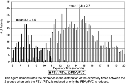
Table 4. Of the 302 patients with only a reduced FEV1FVC6, 100 of them (below) had future repeat spirograms
Reviewing the medical records of the 302 patient with only a reduction in the FEV1/FEV6 (), 45.4% had a clinical diagnosis of COPD, 29.8% asthma, and 2.7% other obstructive diseases (tracheal stenosis, bronchiectasis) and were being treated for these conditions. Though another 11.9% did not have an obstructive diagnosis listed, they had significant smoking histories (>15 pack-years) and almost all of them were on bronchodilator therapy and/or were being managed for advanced stages of lung cancer. It is noted that only 4% had a diagnosis consistent with a restrictive process with 2.7% having diagnosis of both obstructive and restrictive disease.
Table 5. Diagnoses of subjects with only a reduced FEV1/FEV6 ratio (302 subjects)
Discussion
The literature reports a wide range in sensitivity and specificity regarding the usefulness of substituting the FEV1/FEV6 for the FEV1/FVC. Half favor doing so, while half urge caution. This study highlights that though both these ratios are measurements of airways obstruction, there appears to be physiologic reasons for their discordance.
Our results indicated that if only the FEV1/FEV6 is reduced, when compared to the group with only a reduced FEV1/FVC ratio, this group had a higher TLC and RV/TLC, with a lower IC, DLCO, FEV1, and FVC. In addition, the FVC, BMI and IC in this group were similar to the group that had both ratios reduced suggesting a greater degree of air trapping with the relatively shorter expiratory times masking a reduction in the FEV1/FVC. This finding is further supported by looking at future tests on these subjects and finding that with a longer expiratory time, their FEV1/FVC ratios decreased below the confidence limit of normal ().
Because 90% of the subjects with only a reduced FEV1/FEV6 () had strong clinical evidence for having airways disease (reviewing their medical records), this supports that these subjects are not the 5% of the normal population that falls outside the 95% lower limit of normal. A chart review of the 1,158 patients with only a reduced FEV1/FVC was not performed since this value is considered the standard for defining airways obstruction on spirometry.
shows that if we substituted the FEV1/FEV6 ratio for the FEV1/FVC, as some have recommended, out of our total patients with a reduced ratio (airways obstruction), based on spirometry alone we would ultimately have misclassified 1158 subjects (14.4% of 8053 subjects). In contrast, using both ratios together, we would have included another 4% of patients who had greater physiologic abnormalities than when only the FEV1/FVC was reduced. Our results were not too dissimilar from a recent review.
Table 6. Breakdown of patients with one or both ratios reduced.
Five of those studies used the GOLD guidelines of an FEV1/FVC < 70% and FEV1/FEV6 < 70% to define obstruction (Citation4,5,Citation8,9,Citation10,Citation22), even though the GOLD document has no guidelines for interpreting the FEV1/FEV6 ratio. Furthermore, using the criteria of an FEV1/FVC ratio < 70% as defining obstruction is increasingly discouraged. Hansen et al. have pointed out the problem using the GOLD consensus opinion of creating a cutoff of 70% for the lower limit of normal for an FEV1/FVC ratio. We know that predicted values and lower limits of normal decline with age, qualifying the GOLD conclusion that obstruction worsens with age (Citation23).
Our study looked at all patients with airway obstruction, not just patients at risk for COPD. Recent COPD studies advocate using only post-bronchodilator spirometry (Citation5,Citation12,Citation13), which could lead to erroneous conclusions if one is also evaluating patients for possible asthma, by normalizing airway obstruction after bronchodilator therapy. Hanson et al, stated that the significance of an isolated reduction in the FEV1/FEV6 is not known (Citation14).
It appears too simplistic to try to replace the FEV1/FVC ratio with the FEV1/FEV6, attempting to find a substitute, especially if screening patients who have a milder degree of airways obstruction. Though using only the FEV1/FVC ratio will be more inclusive for finding airways obstruction, it can miss identifying a small number of patients as having normal airflows that have more pronounced abnormalities in lung volumes and diffusion. shows how these 2 milder subgroups separate out from each other based on their expiratory times, supporting our premise that these groups may differ by greater air trapping in the group with only reduced FEV1/FEV6. Although we did not selectively examine the flow volume curves of these subjects, we may have found that this subgroup had a higher number of subjects that did not actually achieve zero flow on spirometry even with expiratory times meeting ATS criteria.
The authors believe that the FEV1/FEV6 ratio should not be used as a substitute for the FEV1/FVC ratio. We recognize that including this value on spirometry could potentially increase the occurrence of a false positive result (the 5% of the normal population that falls outside the normal 95% LLN). However, since 90% of this group had strong clinical support for having an obstructive disease that would have been missed only using the FEV1/FVC, the benefit may outweigh the risk. This abnormality should make one more carefully scrutinize a clinically symptomatic patient with the additional measurements of lung volumes and diffusion. And in particular, it could identify patients for repeat future spirometry with concentrated efforts on achieving a more prolonged expiratory time to >11 seconds.
Conclusion
When this FEV1/FEV6 is reduced, with a normal FEV1/FVC ratio, it may identify a group with a greater degree of physiologic abnormalities than if only the FEV1/FVC is reduced. And substituting the FEV1/FEV6 for the FEV1/FVC in a large pulmonary population can result in a significant reduction in the diagnosis of airways obstruction on spirometry in patients with milder obstruction.
Declaration of Interests
Zachary Q. Morris, MD was responsible for the design of the study, creation of the database, writing of programs to extract data, and writing of the manuscript. Najia Huda, MD was responsible for research and assisted in manuscript preparation. Robert R. Burke, MD was responsible for statistical analysis and assisted in manuscript preparation. The authors have no conflicts of interests to disclose.
References
- Hankinson JL, Odencrantz JR, Fedan KB. Spirometric reference values from a sample of the general US population. Am Rev Respir Crit Care Med 1999; 159:179–187.
- Ferguson GT, Enright PL, Buist AS, Office spirometry for lung health assessment in adults: A consensus statement from the National Lung Health Education Program. Chest 2000; 117:1146–1161.
- Jing J, Huang T, Cui W, Should FEV1/FEV6 replace FEV1/FVC ratio to detect airway obstruction? Chest 2009; 135: 991–998.
- Vandevoorde J, Verbanck S, Schuemans DW, FEV1/FEV6 and FEV6 as an alternative for FEV1/FVC and FVC in the spirometric detection of airway obstruction and restriction. Chest 2005; 127:1560–1564.
- Lundgren F, Cabral M, Climaco D, Determination of the efficacy of FEV6 as a surrogate for FVC in the diagnostic screening from chronic obstructive pulmonary disease through the comparison of FEV1/FVC and FEV1/FEV6 ratios. J Bras Pneumol 2007; 33(2):148–151.
- Swanney MP, Jensen RL, Crichton DA, FEV6 is an acceptable surrogate for FVC in the spirometric diagnosis of airway obstruction and restriction. Am J Respir Crit Care Med 2000; 162:917–919.
- Jensen RL, Crapo, RO, Enright P. A statistical rationale for the use of forced expired volume in 6 s. Chest 2006; 130;1650–1656.
- Melbye H, Medbo A, Crockett A. The FEV1/FEV6 ratio is a good substitute for the FEV1/FVC ratio in the elderly. Prim Care Respir J 2006; 15:294–298.
- Rosa FW, Perez-Padilla R, Camelier A, Efficacy of the FEV1/FEV6 ratio compared to the FEV1/FVC ratio for the diagnosis of airway obstruction in subjects aged 40 years or over. Braz J Med Biol Res 2007; 40:1615–1621.
- Demir T, Ikitimur HD, Koc N, The role of FEV6 in the detection of airway obstruction. Respir Med 2005; 99:103–106.
- Gleeson S, Mitchell B, Pasquarella C, Comparison of FEV6 and FVC for detection of airway obstruction in a community hospital pulmonary function laboratory. Respir Med 2006; 100:1397–1401.
- Akpinar-Elci M, Fedan KB, Enright PL. FEV6 as a surrogate for FVC in detecting airways obstruction and restriction in the workplace. Eur Respir J 2006; 27:374–377.
- Lamprecht B, Schirnhofer L, Tiefenbacher F, Six-second spirometry for detection of airway obstruction. Am J Respir Crit Care Med 2007; 176:460–464.
- Hansen JE, Sun XG, Wasserman K. Should forced expiratory volume in six seconds replace forced vital capacity to detect airway obstruction? Eur Respir J 2006; 27;1244–1250.
- Miller, MR, Crapo R, Hankinson J, ATS/ERS Task Force: Standardisation of lung function testing. General considerations for lung function testing. Eur Respir J 2005; 26:153–161.
- Miller MR, Hankinson J, Brusasco V, ATS/ERS Task Force: Standardisation of lung function testing. Standardisation of spirometry. Eur Respir J 2005; 26:319–338.
- Wanger J, Clausen JL, Coates A, Pedersen OF, ATS/ERS Task Force: Standardisation of lung function testing. Standardisation of the measurement of lung volumes. Eur Respir J 2005; 26:511–522.
- MacIntyre N, Crapo RO, Viegi G, ATS/ERS Task Force: Standardisation of lung function testing. Standardisation of the single-breath determination of carbon monoxide uptake in the lung. Eur Respir J 2005; 26:720–735.
- Pellegrino R, Viegi G, Brusasco V, ATS/ERS Task Force: Standardisation of lung function testing. Interpretive strategies for lung function tests. Eur Respir J 2005; 26:948–968.
- Crapo RO, Morris AH, Clayton PD, Lung volumes in healthy nonsmoking adults. Bull Europ Physiopathol Respir 1982; 18:419–425.
- Miller A, Thomton JC, Raphel W, Single breath diffusing capacity in a representative sample of the population of Michigan, a large industrial state. Am Rev Respir Dis 1983; 127:270–277.
- Lung and Blood Institute. Global Initiative for Chronic Obstructive Lung Disease: Global Strategy for the Diagnosis, Management, and Prevention of Chronic Obstructive Pulmonary Disease. Date last updated: January 2008. www.goldcopd.org. Date last accessed: May 7, 2011.
- Hansen JE, Sun XG, Wasserman K. Spirometric criteria for airway obstruction. Chest 2007; 131:349–355.