
Abstract
Although numerous investigations have demonstrated a direct effect of cadmium (Cd) on peripheral blood mononuclear cell (PBMC) activity in humans, there is virtually no data concerning the in vivo impact of this metal on circulatory mononuclear cells. In this study, the effects of a sub-lethal Cd (1 mg/kg) dose were examined in rats 48 h following a single intraperitoneal injection. Cd treatment resulted in increased total peripheral blood leukocyte levels; however, decreases in PBMC numbers were seen. These changes coincided with an accumulation of mononuclear cells in the lungs and an increase in mononuclear cells expressing CD11b. A lack of effect of Cd on spontaneous nitric oxide (NO) production and on iNOS mRNA levels in the PBMC was also noted. Differential effects of Cd on PBMC inflammatory cytokine (IL-1β, TNFα, IL-6, IFNγ, and IL-17) gene expression and production were also seen. Specifically, except for IL-1β (levels increased), there were decreases (relative to controls) in mRNA levels for all the other cytokines examined. While there were no Cd treatment-related changes in spontaneous production of the cytokines assessed, there seemed to be a trend (p = 0.06) toward a decrease in spontaneous IL-6 release. When these harvested cells were stimulated ex vivo, there was no effect from Cd exposure on LPS-stimulated IL-1β and TNFα or on ConA-stimulated IFNγ or IL-17 production, but a decrease in IL-6 production in response to LPS was, again, noted. A preliminary study with a lower Cd dose (0.5 mg/kg) revealed some of the same outcomes noted here (mononuclear cell infiltration into lungs, increases in PBMC IL-1β mRNA levels), but differential (increased IL-17 mRNA levels) or newly detected outcomes (increased levels of IL-1α mRNA) as well. The described effects of the single in vivo exposure to Cd on PBMC might contribute to a better overall understanding of the immunomodulatory potential of this environmental contaminant.
Introduction
Cadmium is an environmental pollutant responsible for toxicity in humans and animals. It adversely affects a number of organs and tissues, including kidneys, liver, lungs, testes, bone, brain, and blood (US Department of Health and Human Services, Citation1997; WHO, Citation1992). While toxicity of this metal is expressed primarily in liver and kidneys (Kayama et al., Citation1995a, Citationb), other distal organs, including lungs and brain, are affected as well (Manca et al., Citation1991). The data obtained from the livers implies that oxidative stress and inflammation were likely mechanisms responsible for toxicity in this tissue (Rikans & Yamano, Citation2000). It was shown by our group and by other investigators that these processes were present in the lungs as well (Kataranovski et al., Citation2009a; Manca et al., Citation1994). An infiltration of leukocytes into affected tissues during the acute phase is a common histopathologic finding in mice and rats administered Cd (Kayama et al., Citation1995a,Citationb; Kataranovski et al., Citation2009a). Local tissue activation of circulating inflammatory cells, neutrophils and monocytes, has also been suggested as a mechanism of indirect tissue damage from the metal (Rikans & Yamano, Citation2000).
Administration of Cd affects circulating inflammatory cells (neutrophils and monocytes), causing changes that might be important for their migration and activity in peripheral tissues. In this respect, the presence of neutrophils as well as their activation was observed in lungs within 24 h of rats being administered Cd (Kataranovski et al., Citation1998). Later investigations showed that Cd administration to rats resulted in peripheral blood granulocyte activation (i.e. propensity to form aggregates in vivo) and priming (i.e. potentiation of functional responses induced by prior exposure to stimuli) for adhesion and respiratory burst (Kataranovski et al., Citation2009b). Lower relative numbers of circulating mononuclear cells were observed in the blood of these animals as well; this implies that there were effects of Cd on these cells too. As far as is known, to date, there are no studies that have reported on the in vivo effects of Cd on peripheral blood mononuclear cells (PBMC).
In vitro investigations wherein human PBMC were used demonstrated that Cd exerted influence on activity of these cells. Effects on cytokine production were investigated mainly, but there is paucity of data concerning effects on other activities of these cells (Boscolo et al., Citation2005). Generally, inhibition of production of inflammatory cytokines was observed with regard to interleukin (IL)-1β (Marth et al., Citation2001; Theocharis et al., Citation1994), tumor necrosis factor (TNF)-α (Boscolo et al., Citation2005; Marth et al., Citation2001), and interferon (IFN)-γ (Boscolo et al., Citation2005) within 2–24 h of exposure to Cd at levels ≥10−5 M. However, differential effects of Cd on PBMC production of some of these cytokines (i.e. lack of effect on IFNγ and an increase in TNFα) were also observed (Villanueva et al., Citation2000). In addition, an increase in production of other cytokines (including IL-8 and IL-6) was observed following ex vivo exposure of PBMC to Cd (Horiguchi et al., Citation1993; Villanueva et al., Citation2000). Taking these findings together, this suggested that Cd could differently affect PBMC production of various cytokines.
In view of the above cited findings that showed that Cd exerted influence on peripheral blood leukocytes in vivo and the knowledge that this metal can affect PBMC in vitro, the aim of the present study was to investigate effects of in vivo Cd administration on circulating mononuclear cells and their activities in rats. Included among the measured effects here were changes in PBMC number and in cell expression of CD11b (a key molecule needed for tissue infiltration by the cells), production of inflammatory mediators including nitric oxide (NO), and select inflammatory cytokines that are most often measured in studies with human PBMC (i.e. IL-1β, TNFα, IL-6, IFNγ), as well as the formation of IL-17, an inflammatory cytokine that stimulates leukocyte effector activities (Curtis & Way, Citation2009).
Materials and methods
Chemicals
Cadmium chloride (CdCl2; Serva Feinbiochemica, Heidelberg, Germany) was prepared in sterile pyrogen-free saline. Lipopolysaccharide (LPS; type 0111:B4 from Escherichia coli), Concanavalin A (ConA), 3-(4,5-dimethyl-thiazol-2-yl)-2,5-diphenyltetrazolium bromide (MTT), naphtylenediamine dihydrochloride, and sulphanilamide were all purchased from Sigma (St. Louis, MO). For use in experiments, LPS and ConA were dissolved in RPMI-1640 medium (PAA Laboratories, Pasching, Austria); MTT was dissolved in phosphate-buffered saline (PBS, pH 7.2). All solutions for cell culture experiments were prepared under sterile conditions or were sterile-filtered (Minisart, 0.20-µm pore; Sartorius Stedim Biotech, Goettingen, Germany) before use. Dextran T 500 (Sigma) was prepared as a 6% solution in apyrogenic saline and autoclaved at 110 °C for 25 min. RPMI 1640 supplemented with 20 μg gentamicin/ml (Galenika AD, Zemun, Serbia), 2 mM glutamine, and 5% (v/v) heat-inactivated fetal calf serum (PAA Labs) were used in all cultures. Monoclonal antibody OX-42 (mouse anti-rat CD11b/CD11c) and fluoroisothiocyanate (FITC)-conjugated F(ab′)2 goat anti-mouse IgG were purchased from Serotec Ltd. (Oxford, UK). Sodium heparin was from Galenika AD; buffer used for red blood cell lysis was purchased from eBioscience (San Diego, CA). OptiPrep (Nycomed AS, Oslo, Norway) was used for isolation of mononuclear cells from peripheral blood.
Animals
Dark Agouti (DA) rats (male, 12–16-weeks-old, 200–240 g) raised in the Institute for Biological Research “Sinisa Stankovic” (IBISS, Belgrade, Serbia) were used in this study. As great inter-individual variability was observed in peripheral blood mononuclear cell (PBMC) cytokine responses in humans (Marth et al., Citation2001), the experiments here had to be conducted in inbred rats. The DA rat strain in particular was used as these rats have been shown to respond vigorously to chemical insult (Mijatovic et al., 1997). Further, other investigators have used this strain to establish systemic (peripheral blood) inflammation in response to several chemicals (Popov et al., Citation2013a, Citationb), including cadmium (Cd) (Djokic et al., Citation2014).
All rats were housed conventionally in pathogen-free facilities that were maintained at 24 °C with a 60% relative humidity and with a 12-h light:dark cycle. All rats had ad libitum access to standard rodent chow and filtered water throughout the study; Cd levels in the chow and water were well below permissible limits according to the manufacturer (Veterinary Institute Subotica, Subotica, Serbia) and the Institute for Public Health (Belgrade, Serbia). For the studies, rats were weighed and then randomly allocated into the experimental groups (4–6 rats per group/experiment, for use in each of at least two independent experiments). Rats were not pre-assigned for the studies of Cd content or post-exposure histologic analyses; Cd content and histology were each determined in four randomly chosen animals from each group (in each of two independent experiments; total n = 8/group for the given end-point assessed). In contrast, all measurements of PBMC activity were assessed using cells isolated from the blood of every individual in each group (either control or treated) prior to euthanization. All procedures used herein complied with the European Communities Council Directive (86/609/EEC) and were approved by the Ethical Committee for the Use of Laboratory Animals.
Cadmium treatment
The CdCl2 was dissolved in pyrogen-free saline, filtered, and then administered by intra-peritoneal injection (IP) in a 0.5 ml dosing volume so the rats received 1 mg Cd/kg body weight (BW). Saline was administered to other rats as the control. All treatments were repeated in two-to-three independent experiments. In a preliminary experiment, rats (n = 4) received a dose of 0.5 mg Cd/kg body weight (BW) or saline (n = 4).
At 48 h post-exposure, following blood withdrawal, all rats were euthanized by over-dose (40 mg/kg, IP) of sodium thiopental (Rotexmedica, Tritau, Germany). To assess effects at an intermediate timepoint, one sub-set of animals (n = 4) was euthanized 24 h after the Cd injection. At necropsy, selected tissues/organs – including bone marrow (femur) contents (see below) – were collected for use in experiments outlined below. The 48-h post-exposure point was selected as the key timepoint based on preliminary experiments that showed this was the earliest wherein significant effects of Cd on PBMC were documented. Specifically, there was a decrease in levels/percentages of mononuclear cell numbers; only a slightly lower percentage of cells was seen at 24 h post-treatment. In addition, a pronounced accumulation of mono-nuclear cells in the lungs of rats was also seen at 48 h in earlier studies (Kataranovski et al., Citation2009a; Stosic et al., Citation2010).
Cadmium determinations
The Cd content in peripheral blood, lungs, spleen, kidneys, and liver was determined by atomic absorption spectrometry (AAS; SpectrAA-50, Varian, Inc., Palo Alto, CA). After the samples were harvested, each was weighed and ashed at 550 °C. The ash was then dissolved in 1 M HCl and diluted with metal-free ultrapure water for use in the analyses. All concentrations were ultimately expressed as μmol Cd/kg tissue (wet weight) or per kg blood.
Clinical biochemistry
Serum was obtained from the whole blood samples collected at necropsy and assessed for levels of aspartate aminotransferase (AST) and alanine aminotransferase (ALT) activity using an autoanalyzer (Ciba Corning Express, Oberlin, OH) and commercially available reagents.
Histology
Lung and liver samples collected at necropsy from rats not used for Cd content analyses were fixed in 4% formaldehyde (pH 6.9) and processed for embedding in paraffin wax for subsequent sectioning at 5-μm. Hematoxylin and eosin-stained histology slides were then prepared and analyzed (in a blinded manner) by a certified pathologist using a Coolscope digital light microscope (Nikon, Tokyo, Japan).
Preparation of bone marrow cells
Femurs collected at necropsy were liberated from muscle tissue and bone peaks cut. For cell number determination, bone marrow cell suspensions were prepared by flushing the femoral cavity of one femur with RPMI 1640 culture medium using a syringe fitted with an 18-G needle and then repeated up-down pipetting. Cells from the other femur were extruded by air pressure and used for preparation of bone marrow smears.
Peripheral blood and bone marrow leukocyte counts
Total peripheral blood leukocyte and bone marrow cell counts were determined using an improved Neubauer hemocytometer (Neubauer, Wertheim, Germany). Differential cell counts were determined by examining at least 300 cells in air-dried whole-blood smears or at least 1000 cells from air-dried bone marrow smears that had been stained using a standard May Grünwald-Giemsa (MGG) protocol.
Flow cytometry
Measurements of CD11b expression were achieved by flow cytometric analysis of leukocytes in the whole blood samples following removal of erythrocytes with lysing buffer. The erythrocyte-free peripheral blood cells (106) were incubated on ice for 30 min with mouse anti-rat CD11b OX-42 antibody (IgG2a; 1 mg/ml). Control sets of samples were processed in parallel using non-specific isotype control antibody. Thereafter, the cells were washed twice with PBS and then stained on ice with FITC-conjugated F(ab′)2 goat anti-mouse IgG (1 mg/ml) for a further 30 min. After washing, the cells were fixed with 1% paraformaldehyde (Serva, Heidelberg, Germany) and fluorescence assayed in a CyFLOW SPACE system (Partec, Munich, Germany). Side- and forward-scatter dot-plots of the leukocytes obtained from the whole blood (after erythrocyte lysis) were used to identify the isolated population of mononuclear cells. The dot-plots were used to first differentiate expression of CD11b by granulated or mononuclear cells, with only CD11b+ cells then being gated. Cells in each histogram (at positions characteristic for mononuclear cells) were analyzed using FlowMax software (Partec). A minimum of 10,000 events/sample was acquired each time.
Peripheral blood mononuclear cell isolation
For use in functional analyses (NO and cytokine production/mRNA expression), PBMC were isolated from the blood samples by dextran sedimentation followed by centrifugation of the leukocyte-enriched suspension over OptiPrep density separation medium (700 × g, 20 min, 20 °C). Mononuclear cells were then harvested from the band formed at the interface of plasma and OptiPrep. Cell purity, determined morphologically after MGG staining, was ≈100%.
MTT assay for PBMC leukocyte viability and survival
A quantitative colorimetric assay (Denizot & Lang, Citation1986) wherein MTT is reduced to formazan by mitochondrial dehydrogenases was used as a measure of viability of the isolated PBMC. In brief, isolated cells were added to a 96-well plate (at 0.25 × 10⁶ cells/well) and incubated with 500 μg MTT/ml (added immediately or after 24 h in culture) for 3 h at 37 °C. Formazan produced in the cells was dissolved by overnight incubation of the cells in 10% SDS-0.01 N HCl solution that had been added to the wells. Absorbance in each well was then measured at 540/650 nm in an ELISA 96-well plate reader (GDV EC, Rome, Italy). The 0 and 24 h (in culture) timepoints were selected to reflect any changes in cell viability that had, respectively, occurred in situ (as a direct result of the host treatment) or due to any induced effect from “damaged” cells due to the actual culture process. The latter has been previously observed in our laboratory, i.e. there was a drop in MTT reduction capacity (both by lymphocytes and granulocytes) after 24 h in culture (Kataranovski et al., Citation2003).
Nitric oxide (NO) production by PBMC
The concentration of nitrite (as an indicator of NO formation) in the medium of 48-h cultures of PBMC was assessed using a Griess reaction (Hibbs et al., Citation1988). Cells were cultured at 5 × 10⁵ cells/well in medium alone (spontaneous production) or in the presence of 100 ng LPS/ml (LPS-stimulated production) for the entire period. For the measurements, aliquots (50 μl) of PBMC supernatant were mixed with an equal volume of Griess reagent (0.1% naphtylenedi-amine dihydrochloride [in water] + 1% sulphanilamide [in 5% phosphoric acid]) and incubated for 10 min at room temperature. After measuring the absorbances at 540 and 670 nm in a 96-well ELISA plate reader (GRD, Rome, Italy) the amount of nitrite present was calculated by extrapolation from a standard curve constructed in parallel with known sodium nitrite solutions.
Cytokine determinations
Cytokine concentrations were determined in the supernatant of cells (5 × 10⁵ cells/well) cultured in 96-well plates for 48 h in medium only (spontaneous production) or in the presence of LPS (100 ng/ml) or of ConA (1 µg/ml) [LPS- or ConA-stimulated production]. Enzyme-linked immunosorbent assay kits (ELISA) for rat TNFα (eBioscience), rat IL-1β, IL-6, and IFNγ (R&D Systems, Minneapolis, MN), and for mouse IL-17 [cross-reactive with rat IL-17] (eBioscience), were used according to manufacturer instructions. Cytokine titers were calculated by extrapolations from standard curves generated in parallel with fixed amounts of kit-provided recombinant cytokines. Kit limits of detection were: 21 pg IL-6/ml, 10 pg IL-1β/ml, 20 pg IFNγ/ml, 16 pg IL-17/ml, and 16 pg TNFα/ml.
Reverse transcription—real time polymerase chain reaction (RT-PCR)
Total RNA was isolated from the PBMC immediately after isolation with an RNA Isolator (Metabion, Martinsried, Germany) according to manufacturer instructions. Reverse transcription of isolated RNA was conducted using random hexamer primers and MMLV (Moloney Murine Leukemia Virus) reverse transcriptase, following manufacturer instructions (Fermentas, Vilnius, Lithuania). Amplification of prepared cDNA was done in a total volume of 20 μl using Power SYBR® Green PCR Master Mix (Applied Biosystems [AB], Foster City, CA) in an ABI PRISM 7000 Sequence Detection System (AB). Thermocycler conditions were: an initial step at 50 °C for 5 min, followed by a step at 95 °C for 10 min and subsequent 2-step PCR program at 95 °C for 15 s and 60 °C for 60 s for 40 cycles. PCR primers (forward/reverse) used were: β-actin (housekeeping): 5′-CCCTGGCTCCTAGCACCAT-3′/5′-GAGCCACCAA-TCCACACAGA-3′; IL-6: 5′-GCCCTTCAGGAACAGCTATGA-3′/5′-TGTCAACAACATCA-GTCCCAAGA-3′; IL-1β: 5′-CACCTCTCAAGCAGAGCA-3′/5′-GGGTTCCATGGTGAAGTC-AAC-3′; IFNγ: 5′-AACAGTAAAGCAAAAAAGGATGCA-3′/5′-TGTGCTGGATCTGTGGGT-TGT-3′; IL-17: 5′-CTACCTCAACCGTTCCACTTCAC-3′/5′-CCTCCCAGATCACAGAAG-GATATC-3′; TNFα: 5′-TCGAGTGACAAGCCCGTAGC-3′/5′-CTCAGCCACTCCAGCTG-CTC-3′; IL-1α: 5′-AAGACAAGCCTGTGTTGCTGAAGG-3′/5′-TCCCAGAAGAAAATG-AGGTCGGTC-3′; and iNOS: 5′-TTCCCATCGCTCCGCTG-3′/5′-CCGGAGCTGTAGCACT-GCA-3′. PCR products were detected in real-time and results analyzed with 7500 System Software (AB) and calculated as 2−ΔCt, where ΔCt was the difference between Ct (cycle threshold) values of specific gene and the endogenous control (β-actin).
Data display and statistical analysis
Results were expressed as means ± SD. Statistical analysis was performed using STATISTICA 7.0 (StatSoft Inc., Tulsa, OK). Statistical significance was defined by a Mann-Whitney U test. A p value < 0.05 was considered significant.
Results
Systemic toxicity from Cd exposure
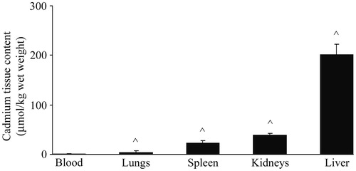
Administration of 1 mg Cd/kg resulted in accumulation of the metal in the isolated organs by 48 h post-injection (). The Cd level was highest in the liver, followed by those in the kidneys, spleen, and lungs. The lowest concentration (1.09 [± 0.25] µmol/kg) was found in the blood; this value was significantly (p < 0.001) lower compared to the Cd content in all other tissues that were examined. Increases (p < 0.001) in serum levels of AST (373 [± 96] versus 203 [± 60] U/ml in controls) and liver ALT (140 [± 36] versus 82 [± 24] U/ml) were observed at 48 h in the rats that received Cd.
Figure 1. Cadmium content in blood, lungs, spleen, kidneys, and liver of rats. Values represent tissue Cd content 48 h after single IP treatment. Data presented as mean (±SD) from at least two independent experiments (n = 4–6 rats/group in each experiment). ^ Significantly differs versus blood value (p < 0.05).
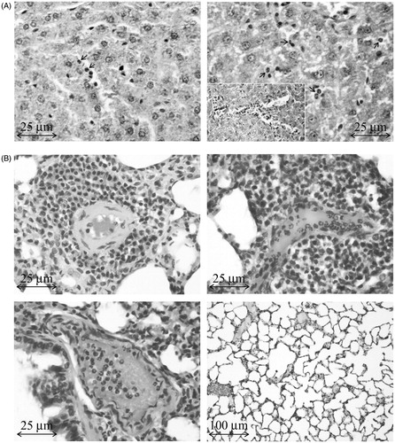
The 1 mg Cd/kg treatment was also associated with neutrophil infiltration into the liver; this effect was scarce at 24 h, but became more pronounced by 48 h post-treatment (). Neutrophil infiltration was also seen in the spleen and lungs; however, only in the lungs, perivascular infiltrates comprised mainly of mononuclear cells were noted (). A measure of mononuclear cell infiltration was observed in the lungs at 24 h after Cd administration, but at this timepoint neutrophils were far more prevalent in any infiltrate ().
Figure 2. Histology of liver and lungs of rats administered Cd. (A) Representative sample of liver from Cd-treated rat. Left: Rare neutrophils (PMN; arrows) in liver 24 h after Cd dosing. Right: PMN migration to sinusoids (arrows) and intravascular accumulation and aggregation of PMN (insert) at 48 h. (B) Representative samples of lung from the same Cd-treated rats. Top left (48 h): Concentric perivascular mononuclear cell infiltrate. Top right (48 h): Perivascular cell infiltrate (predominantly mononuclear with PMN). Intraluminal PMN. Lower left (24 h): Interstitial inflammatory cells, mainly PMN, with lesser numbers of mononuclear cells. Intraluminal PMN. Lower right (control lungs).
Effects of treatment on peripheral blood leukocyte numbers
By 48 h after the treatment with Cd, significant increases in total leukocyte counts were noted (). There was a clear decrease in the percentages of lymphocytes concomitant with an increase in the relative number of neutrophils. However, the analysis of bone marrow smears yielded similar numbers of lymphocytes (43.7 [± 7.8] × 103 and 44.8 [± 7.1] × 103), granulocytes (36.3 [± 8.5] × 103 and 35.4 [± 6.7] × 103), meta-myelocytes (7.9 [± 5.5] × 103 and 7.7 [± 4.2] × 103), and monocytes (2.0 [± 0.5] × 103 and 2.0 [± 0.5] × 103) in samples isolated from Cd-treated and control rats, respectively.
Table 1. Total and differential peripheral blood cell counts.
PBMC CD11b expression
Flow cytometric analysis of blood leukocytes showed that Cd administration caused an increase in the number of mononuclear cells that expressed CD11b (). Cells from this population had a tendency (p = 0.054) toward an increased density of surface expression of the molecules.
Table 2. Peripheral blood mononuclear cell CD11b expression.
Viability and survival of the PBMC
No effect of Cd administration on PBMC metabolic viability was observed. Similar levels of MTT reduction (OD540) by freshly isolated cells from Cd-treated (0.923 [± 0.045]) and control rats (0.949 [± 0.094]) were seen. Furthermore, the same lack of differences was noted following 24 h of culturing (i.e. 0.432 [± 0.040] for Cd-treated versus 0.474 [± 0.043] in controls).
PBMC nitric oxide formation
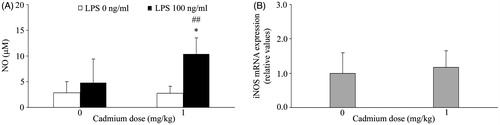
There were no differences between the PBMC from the different host groups with regard to spontaneous NO production. LPS stimulation ex vivo resulted in an increase (compared to spontaneous) NO release by only cells from the rats treated with Cd (). No changes in iNOS mRNA levels in freshly isolated PBMC from Cd-treated animals were noted ().
Figure 3. Effect of Cd on PBMC NO production and iNOS mRNA levels. (A) Spontaneous and LPS stimulated NO production. NO production was analyzed using conditioned medium of mononuclear cells isolated from peripheral blood of control rats (not administered Cd) and rats that received 1 mg Cd/kg. (B) Inducible nitric oxide synthase (iNOS) mRNA expression. Data presented as mean (±SD) from at least two independent experiments (n = 4–6 rats/group in each experiment). mRNA data are expressed as mRNA in PBMC from Cd-treated rats relative to that in cells of control rats. *Value significantly differs from control (p < 0.05). ##Value significantly differs from spontaneous NO production by PBMC from Cd-treated rats (p < 0.01).
PBMC cytokine production
Cadmium administration resulted in differential effects on isolated PBMC inflammatory cytokine production and gene expression ex vivo (). While in general there were no Cd treatment-related changes in spontaneous production of the cytokines assessed, there seemed to be a trend (p = 0.06) toward a decrease in spontaneous IL-6 release. Similarly, there was a general lack of effect from Cd treatment on ex vivo LPS-stimulated IL-1β and TNFα or ConA-stimulated IFNγ and IL-17 production; only mitogen-stimulated IL-6 production appeared to be significantly impacted by the original host treatment with Cd.
Figure 4. Effect of Cd dosing on PBMC inflammatory cytokine production and expression. (A) Spontaneous (no LPS or ConA), LPS-stimulated (100 ng LPS/ml) production of IL-1β, TNFα, and IL-6, and ConA-stimulated (1 µg ConA/ml) production of IFNγ and IL-17, was analyzed using conditioned medium of mononuclear cells isolated from blood of control rats (not administered Cd) and rats that received 1 mg Cd/kg. (B) Inflammatory cytokine mRNA expression. Data presented as mean (±SD) from at least two independent experiments (n = 4–6 rats/group in each experiment). mRNA data are expressed as mRNA in PBMC from Cd-treated rats relative to that in cells of control rats. Value significantly differs from control (0 mg Cd/kg) at *p < 0.05, **p < 0.01, or ***p < 0.001. Value significantly differs from spontaneous cytokine production by PBMC at ##p < 0.01 or ###p < 0.001.
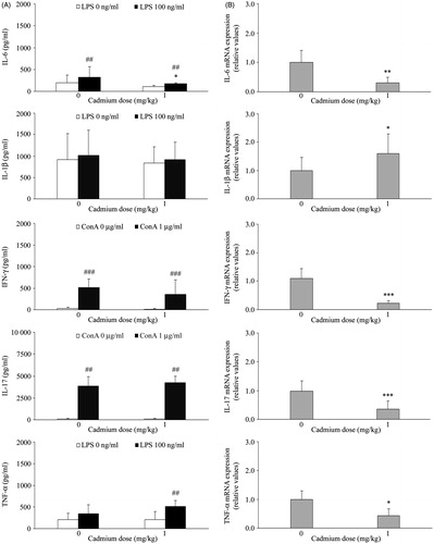
These outcomes were different from the findings regarding mRNA levels of these proteins in the parent PBMC. Specifically, measures of mRNA for each of the assayed cytokines in freshly-harvested PBMC (i.e. that did not undergo any 48 h of culture) revealed there were already increases in levels of IL-1β mRNA in the cells of the Cd-treated hosts, but decreases in message levels for each of the other cytokines to be analyzed.
Effects from a lower Cd dose on some of the measured endpoints
To ascertain if the effects of Cd on PMBC cytokine responses and some of the other endpoints reported here were potentially related to the dose of Cd employed, the impact of administration of a lower Cd dose (0.5 mg/kg) was examined in a preliminary study (with n = 4 individuals per each [control or Cd] treatment). The concentration of Cd in the blood (0.24 [± 0.05] µmol/kg) was significantly lower (p < 0.05) compared to the levels attained following administration of 1 mg Cd/kg (1.09 [± 0.25] µmol/kg), but still significantly (p < 0.05) higher compared to values in control rats (0.045 [± 0.003] µmol/kg). Rarely were neutrophils seen in the liver at 48 h post-treatment, but they were present in the lungs where perivascular mononuclear cells were noted as well. At this Cd dose, no changes (compared to controls) in total or differential leukocyte numbers, percentages of CD11b+cells, levels of membrane (MFI) CD11b expression, or NO production were observed (data not shown).
While the 1 mg Cd/kg dose affected the expression of all pro-inflammatory cytokines analyzed, no effects were seen in mRNA levels with this lower dose, e.g. IL-6 (1.8 [± 1.0] versus 1.0 [± 0.1] in controls), TNFα (0.8 [± 0.3] versus 1.0 [± 0.4] in controls) and IFNγ (1.7 [± 0.9] versus 1.0 [± 0.3] in controls), except for IL-17 and IL-1β, each of which showed a significant (p < 0.05) increase compared to levels in control rat cells (IL-17 = 2.61 [± 0.4] and IL-1β = 1.6 [± 0.3] versus 1.0 [± 0.2] and 1.0 [± 0.1] in respective controls). As IL-1β mRNA levels were found to be up-regulated by both Cd doses, we were intrigued as to whether gene expression of another form of IL-1 (IL-1α) changed following Cd administration as well. Thus, IL-1α mRNA expression was subsequently measured in follow-up analyses with the isolated cells. The effect of the higher Cd dose was examined as well, using mRNA from cells of control or 1 mg Cd/kg-treated rats in the previous experiments. Again, significantly (p < 0.05) higher levels of mRNA for this IL-1 form were noted with both the 1.0 (2.7 [± 0.2]) and 0.5 mg Cd/kg (1.5 [± 0.3]) doses compared to 1.0 [± 0.3] and 1.0 [± 0.3] for the control rat cells from respective experiments.
Discussion
In this study, the acute effect of a single Cd administration on the PBMC of rats was examined by measuring changes in numbers of these leukocytes, their CD11b expression, both their gene expression, and production of several proinflammatory cytokines (e.g. IL-6, IL-1β, IFNγ, IL-17, and TNFα), and their ability to form/release nitric oxide (NO).
While it is clear that exposure to Cd in environmental and occupational settings is often mainly via ingestion or inhalation, and many key studies (see IARC, Citation1993; Waalkes, Citation2000) have used these routes of exposure to assess toxicities induced by Cd, the choice to employ IP exposure here was based on use of this route of administration in numerous rodent studies of toxicity of Cd to various tissues. These included effects on the liver (Casalino et al., Citation2002), lungs (Kataranovski et al., Citation2009a; Kundu et al., Citation2009), spleen and thymus (Pathak & Khandelwal, Citation2007), and gonads/endocrine systems (Ates et al., Citation2004; Hofer et al., Citation2009; Johnson et al., Citation2003; Zhang et al., Citation2008). Our own early studies showed that, when Cd was provided IP, this led to systemic inflammation in rat hosts (Kataranovski et al., Citation1998, Citation2009b).
The highest metal dose used in the current rat studies was 1 mg/kg. Our own preliminary data showed that IP injection with 2 mg Cd/kg was lethal to ≈25% of the test rats, but 1 mg/kg was not lethal and resulted in blood Cd levels close to those noted with the lowest Cd concentrations that imparted an effect on PMBC cytokine production in earlier in vitro studies (Theocharis et al., Citation1994; Villanueva et al., Citation2000). Given that inhibitory effects on IL-1β and TNFα formation/release were observed in vitro using Cd levels of 1–100 µM (Boscolo et al., Citation2005; Theocharis et al., Citation1994) and that differential effects (inhibition versus stimulation) on TNFα production were noted with Cd levels of 3.3–5.0 µM (Theocharis et al., Citation1994; Villanueva et al., Citation2000), it was believed that examination of effects from just a single Cd dose on several different PBMC-derived cytokines was justified and sufficient in the present study. The reader should note that, for the changes noted here, a preliminary study of effects of a lower Cd dose (0.5 mg/kg) was conducted to assess dose-dependency of those outcomes.
The drop in PBMC counts observed in the present study was in line with previous data that showed that Cd affected PBMC levels in rats 24 h after administration (Kataranovski et al., Citation2009b). While this demonstrated a protracted effect of Cd on these types of leukocytes, the decrease in the number of mononuclear cells in the peripheral blood of rats seemed not to be caused by Cd cytotoxicity per se, as similar viabilities (both for freshly isolated or overnight cultured PMBC) were noted among cells from both treated and control rats. This would be in keeping with studies of the susceptibility of human PBMC response to Cd, i.e. direct toxicity of Cd was shown at concentrations >50 μM (Marth et al., Citation2001) – levels far above those measured in the rats’ blood in this study (i.e. 1.09 [± 0.25] μM).
The decrease in relative number of PBMC also seems not to rely upon changes in/effects on hematopoetic processes, as there were no changes noted in bone marrow lymphocyte and monocyte numbers. The lower number of PBMC most likely reflected a re-distribution of these cells from the circulation into peripheral tissues; this was demonstrated histologically in lungs of the rats here. In support of this viewpoint, the increases in the levels of PBMC that expressed CD11b (α subunit of β2 integrin receptors CD11b/CD18) molecules involved in leukocyte blood extravasation and tissue migration (Arnaout, Citation1990) suggested migration of the cells to the lungs (or to other organs) was one potential mechanism to explain the decreases in mononuclear cells in circulation. Interestingly, no changes in either numbers of CD11b+ cells or in the density of membrane expression of CD11b were observed at the 0.5 mg Cd/kg dose (when a presence of mononuclear cells was only rarely seen in the lungs).
In this context, the tendency for the PBMC of rats that received 1 mg Cd/kg to display a higher density of the CD11b marker also suggested to us that these PBMC may have undergone activation in a manner like that seen with other immune cell types that showed this particular pattern of up-regulated expression (Arnaout, Citation1990; Wagner et al., Citation2001). The observed mono-nuclear cell infiltration might also be related to the neutrophil influx that was evident both at 24 and 48 h after dosing with 1 mg Cd/kg. A co-ordinated inter-play between macrophages, neutrophils, and monocytes in the onset – and subsequent resolution – of tissue inflammation has been described by many investigators (Porcheray et al., Citation2005; Schaffer & Barbul, Citation1998; reviewed in Soehnlien and Lindbom, 2010).
The apparent lack of effect of Cd administration on spontaneous NO production and on iNOS mRNA levels in PBMC shown in this study contrasts with findings from in vivo studies of Cd-exposed mice (Ramirez & Gimenez, Citation2003) and in vitro studies with macrophage cell lines (Hassoun and Stohs, 1996), each of which showed up-regulation of iNOS and stimulated NO production. Although there was no change in spontaneous NO production or iNOS mRNA levels, the Cd-treated rats’ cells did display significantly higher LPS-stimulated production of NO. This suggested that the Cd led to a “priming” of the cells toward LPS.
In line with published data that showed that PBMC responded to 1 μM Cd (and higher doses too) with a decrease, increase, or lack of change in production of many cytokines (Villanueva et al., Citation2000), differential effects of Cd on PBMC cytokine production were assessed in this study as well. A lack of effect of Cd on PBMC IFNγ production following stimulation here was in contrast to data from other studies that showed decreases in levels of mitogen-stimulated production (Boscolo et al., Citation2005). Similarly, in contrast to an increase in TNFα production seen by Villanueva et al. (Citation2000), no effect of Cd on spontaneous production of this cytokine was noted in the present study. While decreases in LPS-stimulated TNFα production were reported in two studies (see Boscolo et al., Citation2005; Theocharis et al., Citation1994), in the present study no significant change in LPS-stimulated production was observed. The tendency toward a decrease in spontaneous IL-6 production in the present study also contrasted with the data of Villanueva et al. (Citation2000) wherein a Cd-induced stimulation of IL-6 release was noted. The present study also demonstrated (for the first time) that, at least regarding production, Cd exerted no effect on PBMC IL-17. Along these lines, our observation of a lack of effect from the high-dose Cd treatment on spontaneous TNFα, IFNγ, and IL-17 formation – despite a decrease in corresponding basal mRNA levels for each – reflected the possibility that there was an unintended effect of the cell culturing process, i.e. cell adherence is known to stimulate cytokine mRNA expression during culture (Haskill et al., Citation1988). Having in mind the propensity of monocytes to migrate to peripheral tissues (a process for which adhesion is a prerequisite), a need to examine the effect of Cd on cytokines at both the gene and protein level is recognized.
Host species (rat versus human PBMC) might have accounted for the observed differences in cytokine production at the Cd dose used here and in the above-cited studies. It is also possible that differences in Cd doses used might also be responsible for the differential effects on production of a particular cytokine. For example, the lowest Cd dose tested in the studies of Theocharis et al. (Citation1994) and Boscolo et al. (Citation2005) was 3.3 and 100 μM, respectively; these were all higher than the doses used in the present study. The differences with respect to TNFα mRNA levels compared to that from Theocharis et al. (Citation1994) might be attributable to the fact those authors used human PBMC exposed to Cd in vitro. Similarly, the decreases in IL-6 mRNA levels found in the present study contrasted with the findings of Marth et al. (Citation2001), who reported unchanged levels in human PBMC following exposures in vitro to Cd doses, even up to >50 μM. These variations in outcomes suggest strongly that species differences might exist with respect to the ability of PBMC to form/release certain cytokines following Cd exposure, be it in vivo or in vitro.
Conclusions
In this study, differential effects from a single non-lethal Cd (1 mg/kg) IP dose on rat PMBC biology (including integrin molecule expression, NO production, and the gene expression of select proinflammatory cytokines) ex vivo were shown. Increases in basal gene expression (i.e. immediately at the time of harvest from host) of both forms of IL-1 (and at lower Cd dose as well, when also up-regulation of IL-17 mRNA levels was observed) suggest that there was a potential proinflammatory effect of this metal induced in circulating PBMC. Down-regulation of mRNA levels for other proinflammatory cytokines (not observed, however, at lower Cd dose) suggest immunosuppressive effects at high Cd doses. Other data here suggest Cd exposure could also have modulated PBMC responsivity to LPS, although this effect was not always consistent. Overall, the data obtained here will contribute to a better understanding of the immunomodulatory potential of the dangerous occupational/environmental pollutant cadmium.
Declaration of interest
The authors report no conflicts of interest. The authors alone are responsible for the content and writing of the paper. This study was supported by the Ministry of Education, Science and Technological Development of the Republic of Serbia, Grant #173039.
Acknowledgements
The authors thank Veljko Blagojevic (MSc) for his assistance in the processing of the manuscript.
References
- Arnaout, M. A. 1990. Structure and function of the leukocyte adhesion molecules CD11/CD18. Blood 75:1037–1050
- Ates, I., Suzen, S., Aydin, A., and Karakaya, A. 2004. The oxidative DNA base damage in testes of rats after intraperitoneal cadmium injection. BioMetals 17:371–377
- Boscolo, P., Di Giampaolo, L., Qiao, N., et al. 2005. Inhibitory effects of Cd on peripheral blood mononuclear cell proliferation and cytokine release are reversed by zinc and selenium salts. Ann. Clin. Lab. Sci. 35:115–120
- Casalino, E., Calzaretti, G., Sblano, C., and Landriscina, C. 2002. Molecular inhibitory mechanisms of antioxidant enzymes in rat liver and kidney by cadmium. Toxicology 179:37–50
- Curtis, M. M., and Way, S. S. 2009. IL-17 in host defense against bacterial, mycobacterial, and fungal pathogens. Immunology 126:177–185
- Denizot, F., and Lang, R. 1986. Rapid colorimetric assay for cell growth and survival. Modifications to tetrazolium dye procedure giving improved sensitivity and reliability. J. Immunol. Meth. 89:271–277
- Djokic, J., Ninkov, M., Mirkov, I., et al. 2014. Differential effects on cadmium administration on peripheral blood granulocytes in rats. Environ. Toxicol. Pharmacol. 37:210–219
- Hassoun, E. A., and Stohs, S. J. 1996. Cadmium-induced production of superoxide anion and nitric oxide, DNA single strand breaks, and lactate dehydrogenase leakage in J774 cell culture. Toxicology 1123:219–226
- Haskill, S., Johnson, C., Eierman, D., et al. 1988. Adherence induces selective MRNA expression of monocyte mediators and proto-oncogenes. J. Immunol. 140:1690–1694
- Hibbs, J. B. M. Jr., Taintor, R. R., Vavrin, Z., and Rachlin, E. M. 1988. Nitric oxide: A cytotoxic activated macrophage effector molecule. Biochem. Biophys. Res. Commun. 157:87–94
- Hofer, N., Diel, P., Wittsiepe, J., et al. 2009. Dose- and route-dependent hormonal activity of the metal estrogen cadmium in the rat uterus. Toxicol. Lett. 191:123–131
- Horiguchi, H., Mukaida, N., Okamoto, S., et al. 1993. Cadmium induces IL-8 production in human peripheral blood mononuclear cells with the concomitant generation of superoxide radicals. Lymphokine Cytokine Res. 12:421–428
- IARC (International Agency for Research on Cancer). 1993. Beryllium, cadmium, mercury, and exposures in the glass manufacturing industry. IARC Monogr. Eval. Carcinog. Risk. Humans 58:119–237
- Johnson, M., Kenney, N., Stoica, A., et al. 2003. Cadmium mimics the in vivo effects of estrogen in the uterus and mammary gland. Nat. Med. 9:1081–1083
- Kataranovski, M., Jankovic, S., Kataranovski, D., et al. 2009b. Gender differences in acute cadmium-induced systemic inflammation in rats. Biomed. Environ. Sci. 22:1–7
- Kataranovski, M., Kataranovski, D., Savic, D., et al. 1998. Granulocyte and plasma cytokine activity in acute cadmium intoxication in rats. Physiol. Res. 47:453–461
- Kataranovski, M., Mirkov, I., Belij, S., et al. 2009a. Lungs: Remote inflammatory target of systemic cadmium administration in rats. Environ. Toxicol. Pharmacol. 28:225–231
- Kataranovski, M., Vlaski, M., Kataranovski, D., et al. 2003. Immunotoxicity of epicutaneously applied anti-coagulant rodenticide warfarin: Evaluation by contact hypersensitivity to DNCB in rats. Toxicology 188:83–100
- Kayama, F., Yoshida, T., Elwell, M. R., and Luster, M. I. 1995a. Cadmium-induced renal damage and pro-inflammatory cytokines: Possible role of IL-6 in tubular epithelial cell regeneration. Toxicol. Appl. Pharmacol. 134:26–34
- Kayama, F., Yoshida, T., Elwell, M. R., and Luster, M. I. 1995b. Role of TNFα in cadmium-induced hepatotoxicity. Toxicol. Appl. Pharmacol. 131:224–234
- Kundu, S., Sengupta, S., Chatterjee, S., et al. 2009. Cadmium induces lung inflammation independent of lung cell proliferation: A molecular approach. J. Inflamm. (Lond). 6:19
- Mijatovic, S., Ejdus, L., Pravica, V., et al. 1997. Strain-dependent induction and modulation of autoimmunity by mercuric chloride in two strains of rats. In: Immunoregulation in health and disese. (Lukic, M.L., Colic, M., Mostarica-Stojkovic, M., and Cuperlovic, K., Eds.) San Diego, CA: Academic Press, pp. 181–188
- Manca, D., Ricard, A. C., Tra, H. V., and Chevalier, G. 1994. Relation between lipid peroxidation and inflamation in the pulmonary toxicity of cadmium. Arch. Toxicol. 68:364–369
- Manca, D., Ricard, A. C., Trottier, B., and Chevalier, G. 1991. Studies on lipid peroxidation in rat tissues following administration of low and moderate doses of cadmium chloride. Toxicology 67:303–323
- Marth, E., Jelovcan, S., Kleinhappl, B., et al. 2001. Effect of heavy metals on immune system at low concentrations. Int. J. Occup. Med. Environ. Health 14:375–386
- Pathak, N., and Khandelwal, S. 2007. Role of oxidative stress and apoptosis in cadmium induced thymic atrophy and splenomegaly in mice. Toxicol. Lett. 169:95–108
- Popov, A., Belij, S., Subota, V., et al. 2013a. Oral warfarin affects peripheral blood leukocyte IL-6 and TNFα production in rats. J. Immunotoxicol. 10:17–24
- Popov, A., Mirkov, I., Vasilijic, S., et al. 2013b. Impact of the magnitude of sensitization dose on the incidence and intensity of CHS to dinitrochlorobenzene (DNCB): Insight from ear swelling and challenged-skin draining lymph node response in rats. J. Immunotoxicol. 10:355–360
- Porcheray, E., Viaud, S., Rimaniol, A. C., et al. 2005. Macrophage activation switching: Asset for the resolution of inflammation. Clin. Exp. Immunol. 142:481–489
- Ramirez, D. C., and Gimenez, M. S. 2003. Induction of redox changes, inducible nitric oxide synthase, and cyclo-oxygenase-2 by chronic cadmium exposure in mouse peritoneal macrophages. Toxicol. Lett. 145:121–132
- Rikans, L.E., and Yamano, T. 2000. Mechanisms of cadmium-mediated acute hepatotoxicity. J. Biochem. Mol. Toxicol. 14:110–117
- Schaffer, M., and Barbul, A. 1998. Lymphocyte function in wound healing and following injury. Br. J. Surg. 85:444–460
- Soehnlien, O., and Lindbom, L. 2010. Phagocyte partnership during the onset and resolution of inflammation. Nat. Rev. Immunol. 10:427–439
- Stosic, J., Mirkov, I., Belij, S., et al. 2010. Gender differences in pulmonary inflammation systemic cadmium administration in rats. Biomed. Environ. Sci. 23:293–299
- Theocharis, S. E., Souliotis, V. L., and Panayiotidis, P. G. 1994. Suppression of IL-1β and TNFα biosynthesis by cadmium in in vitro activated human peripheral blood mononuclear cells. Arch. Toxicol. 69:132–136
- U.S. Department of Health and Human Services. 1997. Toxicological Profile for Cadmium. Atlanta, GA: Agency for Toxic Substances and Disease Registry (ATSDR)
- Villanueva, M. B., Koizumi, S., and Jonai, H. 2000. Cytokine production by human peripheral blood mononuclear cells after exposure to heavy metals. J. Health Sci. 46:358–362
- Waalkes, M. P. 2000. Cadmium carcinogenesis in review. J. Inorg. Biochem. 79:241–244
- Wagner, C., Hänsch, G. M., Stegmaier, S., et al. 2001. The complement receptor 3, CR3 (CD11b/CD18), on T-lymphocytes: Activation-dependent up-regulation and regulatory function. Eur. J. Immunol. 31:1173–1180
- WHO (World Health Organization). 1992. Environmental Health Criteria #134: Cadmium. Geneva: World Health Organization
- Zhang, W., Pang, F., Huang, Y., et al. 2008. Cadmium exerts toxic effects on ovarian steroid hormone release in rats. Toxicol. Lett. 182:18–23