
Abstract
First responders (FR) present at Ground Zero in the first 72 h after the World Trade Center (WTC) collapsed have progressively exhibited significant respiratory injuries. The few toxicology studies performed to date evaluated effects from just fine (< 2.5 µm) WTC dusts; none examined health effects/toxicities from atmospheres bearing larger particle sizes, despite the fact the majority (> 96%) of dusts were > 10 µm and most FR likely entrained dusts by mouth breathing. Using a system that generated/delivered supercoarse (10–53 µm) WTC dusts to F344 rats (in a manner that mimicked FR exposures), this study sought to examine potential toxicities in the lungs. In this exploratory study, rats were exposed for 2 h to 100 mg WTC dust/m3 (while under isoflurane [ISO] anesthesia) or an air/ISO mixture; this dose conservatively modeled likely exposures by mouth-breathing FR facing ≈750–1000 mg WTC dust/m3. Lungs were harvested 2 h post-exposure and total RNA extracted for subsequent global gene expression analysis. Among the > 1000 genes affected by WTC dust (under ISO) or ISO alone, 166 were unique to the dust exposure. In many instances, genes maximally-induced by the WTC dust exposure (relative to in naïve rats) were unchanged/inhibited by ISO only; similarly, several genes maximally inhibited in WTC dust rats were largely induced/unchanged in rats that received ISO only. These outcomes reflect likely contrasting effects of ISO and the WTC dust on lung gene expression. Overall, the data show that lungs of rats exposed to WTC dust (under ISO) – after accounting for any impact from ISO alone – displayed increased expression of genes related to lung inflammation, oxidative stress, and cell cycle control, while several involved in anti-oxidant function were inhibited. These changes suggested acute inflammogenic effects and oxidative stress in the lungs of WTC dust-exposed rats. This study, thus, concludes that a single very high exposure to WTC dusts could potentially have adversely affected the respiratory system – in terms of early inflammatory and oxidative stress processes. As these changes were not compared with other types of dusts, the uniqueness of these WTC-mediated effects remains to be confirmed. It also still remains to be determined if these effects might have any relevance to chronic lung pathologies that became evident among FR who encountered the highest dust levels on September 11, 2001 and the 2 days thereafter. Ongoing studies using longer-range post-exposure analyses (up to 1-year or more) will help to determine if effects seen here on genes were acute, reversible, or persistent, and associated with corresponding histopathologic/biochemical changes in situ.
Introduction
The World Trade Center (WTC) collapse on September 11, 2001 not only resulted in an immediate devastating loss of life, but gave rise to health effects that took years to manifest. The collapse resulted in discharge of an expansive dust-laden plume bearing tons of pulverized building materials. ∼Although no explicit measures of air quality immediately after the collapse were taken, extrapolations from airborne PM levels allowed for estimates ranging from > 5000 µg/m3 initially (in collapse cloud) at Ground Zero to < 1500 µg/m3 thereafter (with intermittent spikes or declines dependent on site activity) to be generated (Landrigan et al., Citation2004; Lorber et al., Citation2007). Thus, a huge number of people were potentially exposed to aerosolized WTC dusts, particularly First Responders (FR) (e.g. firefighters, police, rescuers, iron/construction workers).
Of the many people likely to have inhaled WTC dusts at the time of the collapse and during the first 72 h after, FR would seem most at risk to manifest adverse health effects. Based on the Fire Department of New York, 16% of members were present at the collapse and 69% arrived in the next 48 h; exposure status of these FR was deemed moderate-to-high. A majority of those FR at Ground Zero rarely/never wore respiratory protection; Prezant et al. (2002) noted 76–93% non-usage rates and Feldman et al. (Citation2004) rates of 35–74%. A survey of non-firefighter FR noted only ≈20% (in a cohort of 1150) had access to/used respirators 9/11–9/13 (Antao et al., Citation2011; CDC, Citation2004). Thus, FR and others were likely exposed to unprecedented levels of WTC dusts without adequate protection. This, along with continuous re-suspension of settled dusts and increased mouth breathing due to intense physical activity increased the risk that WTC dust particles – particularly those of large aerodynamic diameter (i.e. > 2.5 µm) – could have been entrained in the lungs of FR. It is, thus, not surprising that, in the past decade, upper and lower respiratory diseases/dysfunctions in a large percentage of exposed FR have emerged and continue to increase. Significant associations have been established between the time of arrival at Ground Zero and reductions in pulmonary function and in increased incidence/severity of persistent airway hyperactivity and asthma, sarcoid-like granulomatous pulmonary disease (SLGPD), and cancers (albeit not in lungs) (see Supplemental References).
The airborne WTC dusts were a mix of varying sizes, shapes, densities, and composition derived from an amalgamation of sources from/within the buildings. While 1.5% of the dusts (by mass) collected at/around Ground Zero during 9/11–9/13 were fine (aerodynamic diameter ≤2.5 µm), and ≈0.5% were coarse, the majority (> 96%) were supercoarse (≥10 µm) (see Vaughan et al., 2014). Several WTC dust constituents were believed to pose adverse health effects, including metals, polychlorinated biphenyls, dioxins, asbestos, volatile organics, polycyclic aromatic hydrocarbons, and synthetic vitreous fibers (SVF) (Supplemental References). Despite the fact that the majority of the initial WTC dusts were mostly supercoarse particles, no in vivo studies have been performed to evaluate health effects/mechanisms of toxicity by such particles. The few in vivo studies done to date utilized only the fine fraction (i.e. Gavett et al., Citation2003). As such, attempts to broadly assess WTC dust-related health effects have been limited in scope. We contend that inclusion of the larger diameter particles is essential to accurately assess effects of the entrained WTC dust particles.
To best simulate Ground Zero exposures (in regard to particle size distributions and mode of exposure), we built a system to deliver supercoarse-bearing (i.e. unfractionated part after initial sieving to remove supersize [> 53 µm] debris) WTC dusts to a rat model at representative levels and in a manner to mimic FR mouth breathing (Vaughan et al., 2014). Via this system, rats were exposed to a single dose of WTC dust (from 9/11–13/01 period) that approximated a level that FR would have faced in the 24–72 h period after the buildings collapsed. All exposures were based on a referent period of exposure (i.e. 4-h for average FR at Ground Zero) (Mayor’s WTC Medical Working Group et al., Citation2011). The rats’ lungs were then isolated ≈2 h after exposure and subjected to global gene expression analyses to obtain initial indications of which, if any, genes might have been impacted (up-/down-regulated) by the acute exposure. Of particular interest were genes related to inflammation, oxidative stress, immune function, and cell cycle control, as these could potentially be pertinent to mechanisms underlying various pulmonary pathologies increasingly reported in dust-exposed firefighters and other FR.
As the uniqueness of any WTC-induced changes will still need to be fully established (i.e. by comparing outcomes here to those from other dust types) and because the impact from the WTC dusts were measured here only at 2 h after the exposure, further studies (including use of longer-range post-exposure analyses [i.e. up to 1 year or more] and other coarse/supercoarse ambient dusts) will be needed to firmly establish the relevance of any observed changes in gene expressions to WTC-related long-term diseases in the FR.
Materials and methods
WTC dusts
WTC dusts were collected at representative sites on/around the Main Pile at Ground Zero during September 12–13, 2001. All samples were kept in their original airtight containers in the dark at room temperature to minimize potential light, heat, or ambient gas-induced changes in physicochemical properties that would then affect sample integrity. As large debris (glass/metal shards, stones, carpet fragments) that would never be drawn into the lungs was also present in the samples, parent WTC dusts were sieved to yield all particles of diameters ≤53 µm (i.e. WTC53-) for use in all exposures herein (WTC53-; referred to hereafter as ‘WTC dust’). Details about preparation of these first-sieve WTC53- materials, as well as inherent chemical/physical properties of this and other WTC size fractions were previously reported (Gavett et al., Citation2003; Maciejczyk et al., Citation2006; McGee et al., Citation2003).
WTC dust atmosphere generator
Dust atmospheres were generated via a custom-built Fishing Line system to generate nanoparticle/precious material atmospheres for inhalation studies (Ledbetter et al., 1998; Vaughan et al., 2014). All dilution air required for dust generation was composed of a mixture of filtered air and Isoflurane (ISO; IsoFlo, Abbott Laboratories, North Chicago, IL) in O2 carrier gas (2.5% final concentration after mixing with house air). ISO was selected for use in these exposures in that, unlike other commonly used inhalable (halothane, enthrane, penthrane, etc.) or injectable (pentobarbital, etc.) anesthetics, ISO was not expected to cause changes in body temperature or breathing patterns, or induce generalized inflammation, in rats in the 2-h period (proprietary data; EZ Systems Corp., Palmer, PA). Further confirming the utility of ISO, our data from ongoing studies have shown that ISO had no effect on lung/blood immune cell differentials within the first 7 days post-exposure (Horton and Sisco, Personal Communication).
Temperature/relative humidity (RH) of dilution air was continuously measured by digital probe and maintained to avoid conditions for excessive particle agglomeration. All dilution air was passed through an air-cleansing system prior to combination with ISO or dust. Total flow rates of ≈10 LPM (using air/ISO carrier) were used for all particle generations. All generated dust atmospheres were assessed gravimetrically on Teflon filters (0.2-µm pore Type FG; Millipore, Bedford, MA) and a microbalance (Mettler-Toledo, Hightown, NJ) in an environmentally controlled facility (20–23 °C and 38–42% RH). All samples were obtained by flow-pass collection for fixed periods of time before and after the exposures.
Experimental design
Male F344 rats (8-weeks-old) were purchased from Harlan Labs (Frederick, MD). On arrival, the rats were placed in polycarbonate cages with corncob bedding in a facility maintained at 23 °C with a 30–50% relative humidity and 12-h interval light/dark cycle. Food (Purina lab chow) and tap water were provided ad libitum. All rats were acclimated 1 week prior to use. All animal procedures were conducted under an animal protocol approved by New York University Institutional Animal Care and Use Committee (IACUC).
In this study, sets of rats were exposed once (2-h period) to WTC53- dusts under ISO anesthesia; two controls were also used, i.e. rats exposed to ISO only (2.5% in carrier O2 gas) or naïve hosts. An atmosphere of ≈100 mg WTC dust/m3 was used; this dose was chosen as a conservative estimate to model the rat exposure to correspond to one likely to have occurred in mouth-breathing FR exposed to Ground Zero levels of ≈750–1000 mg WTC dust/m3 (Asgharian, Personal Communication). All rats were exposed to the WTC53- dusts or ISO alone in an intra-tracheal inhalation (ITIH) integrated system (Vaughan et al., 2014) that allowed the WTC particles, including those with da > 2.5 µm, to circumvent the rat nasal region so that they were introduced (and deposited) in the lungs in a manner to mimic mouth-breathing FR.
For the exposure, each rat was anesthetized using 5% ISO (for 3–5 min) generated by an EZ-150C system (EZ Systems). Each rat was then quickly intubated by direct insertion of an 18-G Insyte catheter (Becton Dickinson, Sandy, UT) fitted with a plastic collar (200 µl pipette tip, to assure particles did not escape up trachea) and the catheter was then immediately attached to a system port. Mechanical ventilation was thereafter sustained at 60 breaths/min (0.5 s inhale: 0.5 s exhale). The dust/ISO (ISO now 2.0–2.5% due to dilution) mixture was then delivered at a pressure of 15–20 cm H2O; excess air was vented off via a water column. To maintain body temperature (monitored via rectal probe), each rat was kept on a covered heating pad for the entire exposure. Blood O2 levels were also constantly checked using a Pulse Oximeter (Nonin Medical Inc., Minneapolis, MN).
At 2-h post-exposure, each rat (dust, ISO, or naïve) was euthanized by injection with Sleepaway® (500 mg/kg; Fort Dodge Animal Health, Fort Dodge, IA). The lungs were removed en bloc, separated from the trachea and major bronchi, blot dried, and weighed. The right lung cardiac lobe was removed, frozen, and stored at −80 °C until used for expression analyses. All other lung tissues were processed for histology or for dust/metal burden analyses.
RNA isolation and expression array
Total lung RNA was isolated using the Qiagen RNeasy mini Kit (Valencia, CA). Total RNA integrity was assessed by an RNA 6000 LabChip® kit using a 2100 Bioanalyzer (Agilent Technologies, Palo Alto, CA). Global gene expression changes were evaluated with a bead array gene expression platform (Illumina Inc., San Diego, CA). Representative rat lung tissues were screened for gene expression changes after exposure to air (naïve; n = 3), ISO (n = 3), or ISO plus WTC dust (n = 5). Since a chip contained 12 arrays, and the total number of samples was 11, one RNA sample from naïve rats was run in duplicate. We anticipated that the high-level exposure to WTC dust would induce marked changes in the expression of genes and, therefore, 3–5 replicates/group would provide adequate power for statistical comparison between groups.
Biotin-labeled cRNA was produced from 200 ng total RNA using the Illumina TotalPrep RNA Amplification kit (cat# AMIL1791). Total cRNA was then quantified using an ND-1000 spectrophotometer (NanoDrop Technologies, Wilmington, DE) and evaluated for quality. Amplified RNA size distributions were assessed by an RNA 6000 LabChip® kit using a 2100 Bioanalyzer (Agilent). Samples were hybridized onto Sentrix Rat Ref-12 Expression BeadChips with a multi-sample format allowing 12 samples/chip. After overnight hybridization at 58 °C in an oven (SE-901-1001), bead arrays were washed and stained as recommended by the manufacturer (cat# BD-901-1003). Arrays were then scanned on the iScan module and raw data (.idat files) obtained using illumina iScan control Operating Software (version v3.3.28). All.idat files were analyzed by Illumina GenomeStudio® Data Analysis Software to assess data quality.
Statistics
Lung weight data were analyzed by one-way analysis of variance (ANOVA), with exposure group (naïve, ISO only, or WTC dust/ISO) as the main factor. Prior to performing ANOVA, all data were tested to assure assumptions of normality and homogeneity of variance were met. Data were also screened for outliers via Dixon and Grubbs analyses (Taylor, 1990). Statistical significance in all cases was considered at p < 0.05. Statistical analyses of body weight data were performed using Graphpad Prism software (V5.0, Graphpad Software Inc., San Diego, CA) and R-statistical software package (version 2.15.1; R Development Core Team).
Analysis of microarray data
Normalization and determination of differentially-expressed genes
Illumina bead array data for each sample was normalized using Illumina GenomeStudio, using quantile normalization with no background subtraction. The resulting bead array expression table was downloaded from GenomeStudio into a text file. Statistical contrasts were calculated for ‘naïve’ (untreated) vs ISO only control, ‘naïve’ vs WTC dust (in ISO), and ISO only control vs WTC dust (in ISO). Each contrast was computed on a text file containing all assayed genes as rows and only the contrast samples as columns by a Bayes t-test using R (Baldi & Long, Citation2001). Subsequently, a multiple test correction using the Benjamini-Hochberg method was applied to the Bayes t-test output in Excel with an α of 0.05. To support subsequent analysis, differentially-expressed genes (DEG) from the three contrasts were consolidated into a master DEG expression table.
Functional gene list preparation and hierarchical clustering
Three functional gene lists were generated by NetAffx queries at the Affymetrix website (www.affymetrix.com). The query terms were ‘oxidative stress’, ‘inflammation’, ‘cell cycle’, and ‘cancer’; the latter two term’s lists were combined for analysis. The three lists were exported separately from NetAffx as text files. For each list, gene symbols from each list were used to select the corresponding gene, if present, from the master DEG Illumina expression table to build an expression table that only contains the genes for a given function that were also DEG. These expression tables were then subjected to hierarchical clustering as subsequently described. Hierarchical clustering was computed using Cluster 3.0 (de Hoon et al., Citation2004) and was displayed using Java Treeview (Alok, 2004). The input file was adjusted by median centering of genes and clustering was done on genes with average linkage.
Results
WTC dust generation
Based on filter measurements taken before and immediately after the 2 h exposure (i.e. no concurrent measures due to impact on flow delivery to rats), the rats were exposed to the WTC dusts at an average level of 111.2 mg/m3 (±13.8; SD). The mass median aerodynamic diameter (MMAD) of the WTC dust for this study was confirmed (via horizontal elutriation prior to animal exposures) to be 23 µm (σg = 1.45). This 111.2 mg dust/m3 level here would approximate an ≈850 mg dust/m3 exposure, likely to have been experienced by FR/others at Ground Zero over a period of 4 h during the 72 h period after the buildings’ collapse.
General effects of WTC dust exposure on lungs
F344 rats were exposed to ≈100 mg WTC dust/m3 (dust + 2.5% ISO in O2 carrier gas) for 2 h. Within 2 h of exposure, lung weights of WTC rats had increased ≈0.06 g; compared to those of ISO and naïve rats (net 11% increase). The increase in weight was more than predicted as a result of the dust being deposited during exposure (≈3 mg; Vaughan et al., 2014). We have attributed this to, in part, rapid induction of edema/changes in post-exposure physiology.
Lung gene expression changes
Overall patterns
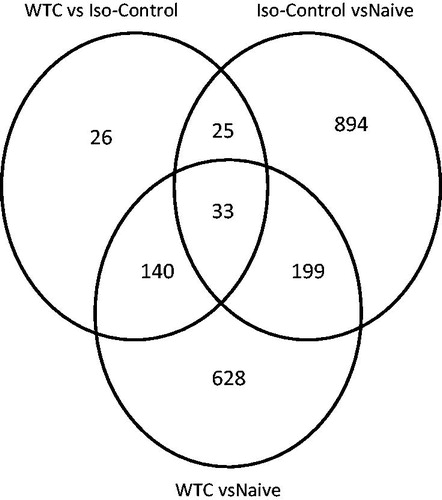
As shown in a Venn diagram, the single acute 2-h exposure of rats to WTC dust (always under ISO anesthesia) resulted in ≈1000 [628 + 140 + 33 + 199] genes that were significantly changed compared to levels in the naïve controls (); of these, 232 [33 + 199] were also noted in samples from rats in the ISO only [vs naïve] group. The use of the ISO anesthetic itself resulted in changes in 1151 [894 + 25 + 33 + 199] genes compared to levels in the naïve rats. When the data for the WTC (+ISO) dust rats was compared with that of ISO alone rats, 224 [140 + 25 + 33 + 26] genes were still found to significantly differ. Of these 224 genes, 58 [33 + 25] were ‘in common’ with the ISO only [vs naïve] hosts, leaving 166 [140 + 26] to be unique due to the WTC (+ISO) dust itself (). Interestingly, because the anesthesia itself actually affected more genes than the WTC dust (1151 vs 1000, respectively), further scrutiny of all gene changes was undertaken (see below).
Figure 1. Venn diagram illustrating number of genes significantly changed by isoflurane (ISO) alone (vs naïve) and by WTC dust exposure (under ISO anesthesia) in rat lungs. ISO by itself changed 1151 genes; when dust was combined with ISO, 1000 genes were changed – of which 232 were in common with ISO, but 166 were due to WTC dust exposure.
To determine the nature of the gene changes, master DEG lists (significant differentially-expressed genes) that included significant genes from all possible contrasts were sorted to derive the 30 maximally-amplified and 30 maximally-inhibited genes (relative to expression in naïve hosts) in rats exposed to WTC dust (under ISO) and those that received ISO only (). In doing this, rather than including a column for fold-change for just one contrast, fold-changes from all contrasts were included. This allowed for determination of relative fold-change and direction of change that might occur in all groups and a determination of how changes induced by WTC dust differed from that by ISO alone.
Table 1. Top 30 genes induced in lungs by WTC dust relative to by isoflurane (ISO) only.
Table 2. Top 30 genes inhibited in lungs by WTC dust relative to by isoflurane (ISO) only.
Table 3. Top 30 genes induced by isoflurane (ISO) only in lungs of rats (relative to in naïve rats).
Table 4. Top 30 genes inhibited by isoflurane (ISO) only in lungs of rats (relative to in naïve rats).
From this analysis, one picture clearly emerged; genes induced (relative to in naive rats) by WTC dust (under ISO) were largely inhibited in rats exposed to just the anesthetic alone, and vice-versa. This suggested that the types of changes induced by WTC dust contrasted with those induced by ISO alone. The genes maximally-induced by WTC dust exposure – relative to that by ISO alone – included several involved in an inflammatory response and maintenance of the proteinase:anti-proteinase balance in the lungs (). Most of these genes remained increased when the WTC group was compared to naïve controls. In contrast, many of these same genes were either unchanged or inhibited in the ISO only group (vs naïve), again emphasizing the likely contrasting effects of ISO and the WTC dust. This remained true as well for genes that were maximally inhibited in the WTC dust rats relative to those in the ISO only regimen, i.e. genes inhibited by the WTC dust were largely induced or unchanged in rats that received ISO only (). Genes inhibited maximally in WTC dust rats – relative to in ISO only rats, included some involved in anti-oxidant function, fibroproliferative responses, and inhibition of energy-generating processes.
The 30 genes maximally-induced by ISO alone (relative to naïve rat levels) were generally inhibited in the WTC dust-exposed rats. When fold-changes (vs naïve hosts) in ISO only-induced genes were compared to those due to the WTC dust regimen, these genes remained induced; this suggested that effects on these genes were specific to ISO and that, in the WTC dust rats, these genes might have been inhibited or unchanged (). Genes induced by ISO alone (vs naïve) included some involved in tissue regeneration and repair processes, including protease inhibitors. In general, those genes inhibited in ISO only rats (vs naïve) generally were also inhibited in the WTC rats (vs naïve); however, as noted above, in direct comparison to in the ISO only hosts, there were many cases wherein the levels of these genes were greater as a result of the WTC dust itself (), once again illustrating likely contrasting effects of ISO and the WTC dust. These included several involved in inflammation and cell signaling.
Genes related to inflammation
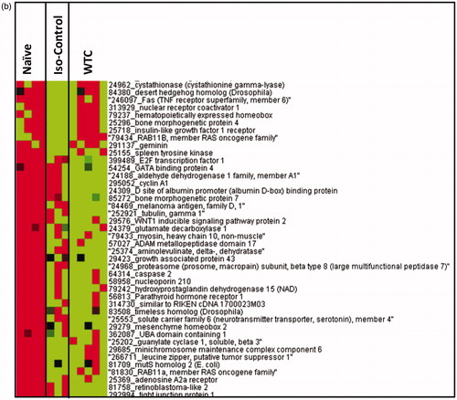
The Master DEGs list was further screened for genes involved in inflammation, oxidative stress, and cell cycle control () based on known associations of each gene in respective processes. presents a heat-map of selected hierarchical clusters of genes within this DEG list that are involved in inflammation. A large cluster of genes that was selectively induced in rats exposed to WTC dust under ISO anesthesia (relative to naïve rat expression), but not changed by ISO alone. These included numerous chemokines and cytokines, as well as genes involved in cell signaling, protease:anti-protease responses, cell adhesion, calcium homeostasis, and cell-mediated immunity (). Interestingly, a cluster of genes that was induced in the ISO only group – but not in WTC dust rats – included suppressors of cytokine signaling and inhibitors of metallopeptidase; this indicated that, in contrast to the WTC dust itself, the anesthetic was likely to produce anti-inflammatory and -proteolytic changes in the lung. Genes inhibited (relative to expression in naïve rats) in WTC dust rats, but not in ISO-only rats, included epoxide hydrolase 2, cytochrome P450 isoform, ICAM2, and mucin 1 ().
Figure 2. Heat-map for relative expression of genes involved in inflammatory response in naïve rats and those exposed to only ISO anesthesia or WTC dust (under ISO anesthesia). Genes were clustered using Eisen’s Cluster Analysis with median centering and average linkage. Resulting clusters were visualized with Treeview. Red = genes with higher expression values than median; green = genes with lower expression values than median; black = median expression. Gene list was truncated to fit in the figure and for clarity.
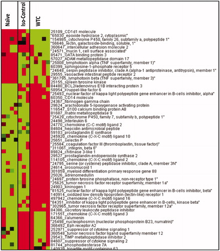
Figure 3. Heat-map for relative expression of genes involved in oxidative stress response in naïve rats and those exposed to only ISO anesthesia or WTC dust (under ISO anesthesia). Genes were clustered using Eisen’s Cluster Analysis with median centering and average linkage. Resulting clusters were visualized with Treeview. Red = genes with higher expression values than median; green = genes with lower expression values than median; black = median expression. Gene list was truncated to fit in the figure and for clarity.
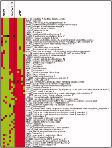
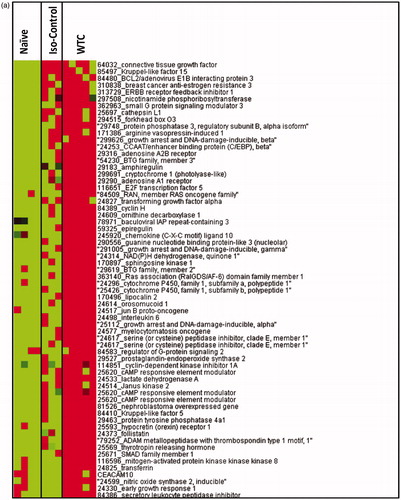
Figure 4. Heat-map for relative expression of genes involved in cell cycle-related processes in naïve rats and those exposed to only ISO anesthesia or WTC dust (under ISO anesthesia). (a) One hierarchical cluster of genes that were induced in rats exposed to WTC dust (+ISO) but not changed in rats exposed to ISO alone (relative to naïve controls). (b) A different hierarchical cluster of genes showing down-regulation in rats exposed to WTC dust (+ISO) and ISO alone (relative to naïve controls). Genes were clustered using Eisen’s Cluster Analysis with median centering and average linkage. Resulting clusters were visualized with Treeview. Red = genes with higher expression values than median; green = genes with lower expression values than median; black = median expression. Gene list was truncated to fit in the figure and for clarity.
Genes related to oxidative stress
Genes implicated in oxidative stress responses were also variably affected by ISO alone and by the WTC dust. A cluster of genes was induced (relative to naïve rats) by ISO alone, but was not changed in the WTC dust rats, suggesting that the presence of WTC dust in the lung inhibited some ISO-induced genes. Those included several genes involved in the processes associated with glutathione metabolism, as well as peroxiredoxin, oxidative stress responsive-1, and glutathione S-transferase. Several other genes involved in tissue repair-related cell signaling, such as mitogen-activated protein kinase (MAPK)-3 and -12, vascular endothelial growth factor (VEGF) A, Bcl2-like-1, transforming growth factor (TGF)-β1, cyclin dependent kinase inhibitor, and Kruppel-like factor-2 were induced in the ISO only rats, but not apparent in the WTC dust rats (). Numerous genes involved in oxidative stress response were induced in the WTC dust exposed rats, but unchanged by the anesthetic alone. These included dual oxidase 2, iNOS, eNOS, glutamate cysteine ligase-C, lipocalin-2, glutathione peroxidase 2, mitochondrial superoxide dismutase, NADPH dehydrogenase, and thioredoxine reductase ().
Genes implicated in cell cycle control
A cluster of genes relevant to cell cycle control that was up-regulated (relative to naïve rats) in WTC dust rats but not changed by ISO alone is shown in the () heat-map. Genes induced only by the anesthetic are not shown in this cluster. It should be emphasized that many genes involved in inflammation and oxidative stress have also been implicated in cell cycle control and, therefore, they can be seen in multiple clusters. Those include several Kruppel-like factors, cAMP responsive element modulators, cyclin-dependent kinase inhibitors, jun B proto-oncogene, Ras associated-RalGDS/AF-6, RAN-Ras oncogene family members, growth arrest and DNA-damage-inducible gene, cyclin H, and several involved in apoptosis (). A cluster of genes that appeared to be inhibited by the ISO itself or by WTC dust is shown in (). This cluster included several genes associated with diverse signaling processes involved in pathologies, including E2F transcription factor-1, insulin-like growth factor receptor-1, bone morphogenic protein-7, ADAM metalloproteases, caspase-2, hydroxyl-prostaglandin dehydrogenase isoforms, RAB11a, and adenosine receptor-A2a ().
Discussion
The few in vivo studies performed to date to examine WTC dust-related health effects did not utilize exposures that accurately reflected conditions in the early days post-9/11. Those studies used fine dust fractions/exposure methods either non-representative (instillation, aspiration) or optimal (nose-only inhalation) for use with large particles. A study doing exposures to total WTC dust in a manner comparable to that experienced at Ground Zero would have to account for significant mouth-breathing that occurred among FR. Further, in any study that used rat models, a nose-only approach would be limited as filtration/entrapment of the > 95% supercoarse WTC particles in nasal passages would be certain. To avoid these problems, we developed a system to generate\administer supercoarse WTC particles in a manner reflecting Ground Zero scenarios (Vaughan et al., 2014). The system allowed the larger diameter particles to circumvent nasal regions in rats for introduction directly into the lungs and let us expose rats to high levels of dusts collected on-site in the first 48–72 h post-collapse. In the study here, rats were exposed to a single WTC dust dose – one reflecting a very high exposure level (750–1000 mg dust/m3) likely faced by FR over the first 72 h at Ground Zero. The main findings were that acute exposure (under ISO) to WTC dusts recovered from Ground Zero modulated expression of many genes related to lung inflammation, oxidative stress, as well as some related to cell cycle control. Nevertheless, as these very acute changes may likely occur with a single large bolus dose exposure to other types of air pollution particles, the uniqueness of the WTC effects still needs to be verified by comparative analysis with other coarse/supercoarse particles.
Previous in vitro studies have provided findings that portended potential in situ implications from WTC dust exposure. Earlier in vitro studies performed in our laboratory with supercoarse WTC dust showed there was acute toxicity to cultured human BEAS-2B airway epithelial cells after a single 2- or 24-h dust exposure (Xu et al., Citation2011). This outcome was characterized by lack of cleavage of the DNA repair enzyme PARP after exposure, a finding consistent with necrosis (not apoptosis) as a mode of dust-induced damage to/death among local lung cells. In vitro studies by others showed that primary alveolar macrophages (AM) and Type II epithelial cells exposed to WTC10-53 showed time-/dose-related increases in pro-inflammatory cytokine and chemokine (IL-6, IL-8, TNFα; Payne et al., Citation2004) production. An interesting side note to those studies was that fine (WTC2.5) particles induced greater responses than WTC10-53. Wang et al. (Citation2010), examining the role of MAPK signaling pathways in WTC dust-induced cytokine release, showed that BEAS-2B exposure to WTC2.5 (for 5 h) led to increased IL-6 mRNA expression/protein release, IL-8 and -10 production, and ERK and p38 (but not JNK) pathway activity.
As noted, some in vitro findings associated with a likely induction of inflammatory states in situ were corroborated here. For example, WTC dust exposure (under ISO anesthesia) led to significant increases in IL-6 (≈3.6–4.0-fold) and in IL-1 (2.0–4.0-fold) expression relative to levels in naïve or ISO-only-exposed rats. Based on Hinson et al. (Citation1996), we surmise that increases in IL-6 might be related to changes in prostaglandin (PG) metabolism. Indeed, results showed that PGE synthase expression was also up-regulated above median levels. As changes in PG presence are closely related to cyclo-oxygenase and lipoxygenase activities (Murphy & Gijon, Citation2007; Ricciotti & Fitzgerald, Citation2011), it was not unexpected that lungs of dust-exposed rats also had elevated arachidonate 5-lipoxygenase (5-LO) expression. This finding is important in that: (1) 5-LO, most likely via leukotriene generation, plays an important role in the pathogenesis of acute lung injury and hemorrhagic shock (Eun et al., Citation2012); (2) the 5-LO pathway has a key role in IL-13-induced chronic inflammation and lung remodeling (Shim et al., Citation2006); and (3) arachidonate metabolites mediate inflammation in general and asthma in particular (Wenzel, Citation1997). Together, the findings from the cited in vitro studies and our in vivo study demonstrate that exposure of lung cells to fine/supercoarse WTC dust could lead to release of factors that contribute to inflammation and airway remodeling processes in the lung. While this is important to understanding potential mechanisms underlying documented pathologies (like asthma) in FR exposed to high dust levels in the 9/11–13/01 period, it will still be critical to examine if these gene expression changes result in lung inflammation, injury, or pathology over time following an acute WTC dust exposure. A current follow-up study is examining the timecourse of inflammatory and pathological responses (including inflammatory cell influx) in the lungs of rats that underwent acute WTC dust exposure.
The Wang et al. (2010) study tried to ascertain if there was a predominant mechanism of cytotoxicity (necrotic vs apoptotic) in WTC dust-exposed lung epithelial cells and the role ERK, JNK, and/or p38-initiated signaling may have played. Studies had suggested that JNK had a pro-apoptotic role, while ERK- and p38-initiated signaling had anti-apoptotic roles in cell fate-responses to oxidants (Buckley et al., Citation1999; Carvalho et al., Citation2004). In contrast, lung macrophages with inhibited ERK exhibited cytotoxicity characterized by markers of both apoptosis (caspase activation) and necrosis (ATP loss) (Monick et al., Citation2008); JNK activation/p38 deactivation resulted in apoptosis (Kim & Sharma, Citation2004; Seimon et al., Citation2009). At this time, it remains unclear what role JNK, ERK, or p38 may have had in the in situ lung cell responses to WTC dusts here; further analyses are warranted.
Our study showed that a single high-dose WTC dust exposure caused a down-regulation among several MAPK forms, i.e. ERK1 (MAPK3) and ERK2 (MAPK1/2). Interestingly, it seems that the presence of ISO during the inhalation was stimulatory with regard to MAPKs; thus, the down-regulating effects of the WTC dust were likely even stronger than implied by the data. In the context of the reported increase in asthma among FR exposed the first days post-9/11, these findings (reductions in ERK availability) would seem counterintuitive: (A) an increased presence/activation of the enzymes is important to overall pathology (including shift to T-helper [TH]-2 status (Pelaia et al., Citation2005); and (B) reduced apoptosis of local cells (including effector T-cells) in small/large airways may serve as pro-inflammatory triggers to contribute to persistent airway inflammation (Lamb et al., Citation2005). Because of key limitations of our study, i.e., only a single post-exposure timepoint was examined, exposure occurred under anesthesia, and comparative effects from exposure to other urban coarse/super-coarse particles are not yet known, it is clear that further characterizations of the various MAPKs in the rat lung tissues are needed to clarify any WTC dust-specific changes in this signaling pathway. JAK isoforms also have roles in asthma development; JAK can regulate effects of TH2 IL-4, -5, and -13, and affect mast cell homeostasis (Inoue et al., Citation2007; Malaviya et al., Citation2010; Morales et al., Citation2010). Ip et al. (Citation2006) showed JAK2 up-regulation in BEAS-2B cells in response to asthma-associated IL-4 and IL-13, each of which are critical to the eventual release of monocyte chemo-attractant protein-1 (MCP-1) involved in bronchial hyper-reactivity/lung remodeling. Unlike the apparent disconnect regarding MAPKs in scenarios likely to result in asthma, there was a clear trend to increased JAK2 expression in dust-exposed rat lungs. Apart from MCP-1 (CCL2) induction, an increase in JAK presence/activation is also critical to increased nitric oxide synthase (NOS) expression (Gebru et al., Citation2011; Ji et al., Citation2011) that contributes to lung inflammation during asthma/other pathologies. Thus, it was not surprising that increased iNOS and eNOS expression was seen in the lungs of dust-exposed rats. As eNOS and iNOS also impact on MCP-1 status (Speyer et al., Citation2003), it was in keeping that there was strong (> 4.5-fold) induction MCP-1 in the WTC dust-exposed rats’ lungs. In summary, our findings suggest many key enzymes/proteins required for potential induction of inflammatory changes in WTC dust-exposed rats were up-regulated by the single high dose (while under ISO anesthesia). Because MCP-1 can be an important regulator of neutrophil/macrophage recruitment to the lungs and since MCP-1 expression is also induced by sandstorm dust (He et al., Citation2013), the specificity of this response to WTC dust (and its significance in long-term lung pathologies in the lung) needs to be further examined.
Apart from potential tissue damage/induction of altered immune responses and associated pathologies, many pro-inflammatory cytokines shown either in vitro or in this animal study to have been induced by the WTC dusts are likely to also stimulate adenylate cyclase activity in airway smooth muscle cells (Osawa et al., Citation2007). It is, thus, possible that mediators released by dust-exposed cells could activate cAMP synthesis, resulting in increases in cAMP that underlie the airway-remodeling central to asthma (Munakata, Citation2006) and chronic obstructive pulmonary disease (COPD) (Puchelle et al., Citation2006). As a second messenger, cAMP leads to activation of protein kinase that phosphorylates/activates CREB and CREMα transcripttion factors that bind cAMP response elements (CRE) in promoter regions of several genes (Sassone-Corsi, Citation1998). While the arrays here did not assess cAMP, they indicated increased expression of cAMP responsive element modulator protein (CREM). We postulate that this increase in CREM expression, if persistent, could contribute to a shift in the TH1/TH2 balance in the lungs (toward TH2) and promote allergic asthma-type responses. However, since only one timepoint was evaluated here, the expression changes will need to be examined over longer post-exposure periods. A recent paper noted that CREMα over-expression led to an aggravated inflammatory phenotype related to higher IL-17A levels and persistent lung inflammation (Lippe et al., Citation2012; Verjans et al., Citation2013). As IL-17A is also associated with increased recruitment of neutrophils/phagocytes in lung infection responses (Kimizuka et al., Citation2012), it is not implausible that a CREM-related enhanced recruitment may also have had a role in SLGPD in some exposed FR. This too remains to be verified in tissues from the WTC dust-exposed rats over a longer post-exposure period.
Like other pollutants (Ghio et al., Citation2012), WTC dusts might oxidize lung lipid and protein macromolecules, resulting in acute activation of anti-oxidant homeostasis. In this process, nuclear factor erythroid 2-related factor (Nrf2) transcription factor levels are elevated. Via subsequent interactions with DNA anti-oxidant response element (ARE) sites, Nrf2 transcriptionally activates compensatory response genes, including some involved in anti-oxidant, anti-inflammatory, and repair processes (Lee et al., Citation2005). As increased inflammatory cytokine gene expression in WTC-exposed rats was also associated with increased transcription of Nrf2 and activation of anti-oxidant response genes (thioredoxin reductase-1, HO-1, NAD(P)H dehydrogenase [quinine 1], glutamate cysteine ligase), this suggested that an alteration in anti-oxidant homeostasis was induced. As with the ERKs, such outcomes seem counterintuitive with regard to asthma development. There are at least two means by which the dusts could induce changes in the lung that in fact short-circuit any beneficial role from Nrf2: (A) continuous presence of ROS/oxidants could inhibit Nrf2 activation and target gene expression (Goven et al., Citation2009); and (B) presence of TGFβ could mitigate Nrf2 effects (Michaeloudes et al., Citation2011). We addressed earlier how genes induced by WTC dust could impact pro-inflammatory events (like ROS formation) in the lung. With regard to a potential for (B), our results show variable levels of TGFβ induction in the dust-exposed rats. While this could imply TGFβ has no role at this one particular time post-exposure, it cannot preclude potential changes in expression (or effects on Nrf2) as time increases. The data also show TGFα expression was elevated. Alone, TGFα has no known impact on Nrf2 – it can potentiate effects of TFGβ (Vinals & Pouyssegur, Citation2001). If this is the case after WTC dust exposure, the initial purported protective effects of Nrf2 might be short-lived. Further longitudinal (post-exposure) studies are needed to confirm if TFGβ levels rise (and Nrf2 abate) in WTC dust rats’ lungs.
We recognize that the ISO anesthetic used here was not without its own impact on genes in the lungs. A number of studies have shown that lung injuries induced by endotoxin, zymosan, or sepsis, and in some cases associated gene expressions, were reduced by host (rats and mice) exposure to ISO (Bedirli et al., Citation2012; Li et al., Citation2009, Citation2013; Reutershan et al., Citation2006; Wang et al., Citation2013). Further, ISO alone was shown to reduce lung gene expression for elements of the NF-κB pathway involved in inflammation (Kadar et al., Citation2011). Our study also showed that ISO alone was able to change expression of several genes, especially some associated with NF-κB activation. A study by Edmands et al. (Citation2013) indicated that, based on the genes impacted by exposure of rats to 2% ISO for 1–2 h, a number of ontologies were represented, including some relevant to our own observations, i.e. cell apoptosis, survival signaling, energy pathway regulation, redox control and ion gradient maintenance, as well as overall regulation of inflammatory responses. However, the extent (fold) of change induced by the ISO rarely exceeded ±2-times background in any of the sites evaluated, i.e. heart, liver, and kidney; and the study did not examine effects in the lungs.
These cited studies notwithstanding, it is clear that, even though ISO itself had an impact on gene expression in our study: (A) 166 changes were unique to the WTC dust (exposure); (B) in many instances, genes maximally-induced by WTC dust exposure (relative to in naïve rat lungs) were unchanged/inhibited by the ISO only; and (C) several genes maximally inhibited in WTC dust rat lungs were largely induced/unchanged in lungs of rats that received ISO only. These three points reflect the contrasting effects of ISO and WTC dust on lung gene expression and suggest that ISO could have actually protected the lungs from WTC dust-induced injury. As such, the WTC dust-induced effects here on genes involved in lung injury, inflammation, and oxidative stress processes may have been under-estimated due to the opposing effects of ISO in the lungs of the rats.
It remains to be determined if these initial events from a single high-level exposure could account, in part, for the long-term chronic lung effects that have become associated with WTC dust exposure among FR and others. Ongoing studies with multiple exposures to the WTC dusts – and employing longer-range post-exposure analyses (i.e. up to 1 y or more) – will allow comparison of single vs repeated exposure as well as to determine whether effects seen here on the affected genes were acute, reversible, or progressive. Similarly, to see if the changes seen here in gene expression within 2 h ultimately manifest as alterations in the structure/function of WTC dust-exposed rats’ lungs, lung tissues harvested in the ongoing studies will also be examined for histopathologic/biochemical changes. Finally, as it is likely other respirable coarse or supercoarse particles could also induce inflammatory responses and associated gene changes, future comparative analyses of effects from an acute large bolus exposure to other bioactive and ‘inert’ dusts, e.g. silica, Mount St. Helen’s ash, will be critical to verifying WTC dust-specific changes in gene expression patterns. Those results will be reported in upcoming manuscripts.
Conclusion
In summary, in vitro studies had previously shown that WTC dust (at least the supercoarse particles) had a direct toxic effect on airway epithelial cells and altered cell signaling properties. Results from this in vivo study of a single acute high-dose exposure to the dusts – comprised primarily of supercoarse particles – indicated evidence of up-/down-regulation of genes related to several mediators of processes that corroborate with the in vitro outcomes. We also noted that ISO anesthetic required for the exposures had significant effects on gene expression; however, in many instances, genes maximally-induced by WTC dust exposure were unchanged or inhibited by the ISO only and in other instances, genes maximally inhibited in WTC dust rats were largely induced/unchanged in rats that received ISO only. These opposing responses suggest there were WTC dust-induced effects on expression of genes in the lungs of rats and that some may have been under-estimated due to a relative ‘protective’ effect from the ISO.
Overall, the findings here illustrated that a single acute high-level exposure of rats to WTC dusts modulated their pulmonary expression of many genes related to inflammation, oxidative stress, anti-oxidant function, as well as some related to cell cycle control. Although these changes suggested induction of acute inflammogenic effects and oxidative stress in the lungs of these rats, to ultimately relate our findings to changes in pulmonary health of FR and others who were exposed to WTC dusts within the critical first 72 h after the building collapses, a few key issues still need to be evaluated/clarified/addressed. These include: (1) contributions to these outcomes from fine (WTC2.5) and coarse (WTC2.5–10) particles that were present in the dusts used here (albeit at comparatively very low levels); (2) whether the noted expression changes are acute, reversible, or progressive, and if they manifest as alterations in WTC dust-exposed rat lung structure/function; and (3) the uniqueness of these responses to the WTC dusts.
Declaration of interest
The authors report no conflicts of interest. The authors alone are responsible for the content and writing of the paper.
This work was supported by CDC/NIOSH Grant OH008280 and, in part, NIEHS Center Grant ES00260. The authors wish to thank Ms Beena Vallanat (US EPA) who performed the array experiments at the Genomics Research Core, Research Cores Unit of the NHEERL, US EPA. The authors also thank Ronald Thomas (US EPA SEE Program) for isolating RNA from the lung tissue samples.
Disclaimer: The research described in this article has been reviewed by the National Health and Environmental Effects Research Laboratory, US Environmental Protection Agency and approved for publication. Approval does not signify that the contents necessarily reflect the views and the policies of the Agency, nor does mention of trade names or commercial products constitute endorsement or recommendation for use.
References
- Alok J. 2004. Saldanha, Java Treeview. Extensible visualization of microarray data. Bioinformatics. 20:3246–3248
- Antao, V. C., Pallos, L. L., Shim, Y. K., et al. 2011. Respiratory protective equipment, mask use, and respiratory out-comes among World Trade Center rescue and recovery workers. Am. J. Ind. Med 54:897–905
- Baldi, P., and Long, A. D. 2001. A Bayesian framework for the analysis of microarray expression data: Regularized t-test and statistical inferences of gene changes. Bioinformatics. 17:509–519
- Bedirli, N., Demirtas, C. Y., Akkaya, T., et al. 2012. Volatile anesthetic pre-conditioning attenuated sepsis induced lung inflammation. J. Surg. Res 178:e17–e23
- Buckley, S., Driscoll, B., Barsky, L., et al. 1999. ERK activation protects against DNA damage and apoptosis in hyperoxic rat AEC2. Am. J. Physiol 277:L159–L166
- Carvalho, H., Evelson, P., Sigaud, S., and Gonzalez-Flecha, B. 2004. Mitogen-activated protein kinases modulate H2O2-induced apoptosis in primary rat alveolar epithelial cells. J. Cell. Biochem 92:502–513
- CDC (Centers for Disease Control and Prevention). 2004. Physical health status of World Trade Center rescue and recovery workers and volunteers - New York City, July 2002-August 2004. MMWR (Morb. Mortal. Wkly. Report). 53:807–812
- de Hoon, M. J., Imoto, S., Nolan, J., and Miyano, S. 2004. Open source clustering software. Bioinformatics. 20:1453–1454
- Edmands, S. D., Ladow, E., and Hall, A. C. 2013. Microarray analyses of genes regulated by isoflurane anesthesia in vivo: A novel approach to identifying potential pre-conditioning mechanisms. Anesth. Analg. 116:589–595
- Eun, J. C., Moore, E. E., Mauchley, D. C., et al. 2012. The 5-lipoxygenase pathway is required for acute lung injury following hemorrhagic shock. Shock. 37:599–604
- Feldman, D. M., Baron, S. L., Bernard, B. P., et al. 2004. Symptoms, respirator use, and pulmonary function changes among New York City firefighters responding to the World Trade Center disaster. Chest. 125:1256–1264
- Gavett, S. H., Haykal-Coates, N., Highfill, J. W., et al. 2003. World Trade Center fine particulate matter causes respiratory tract hyper-responsiveness in mice. Environ. Health Perspect. 111:981–991
- Gebru, E., Kang, E. H., Damte, D., et al. 2011. The role of Janus kinase 2 (JAK2) activation in pneumococcal EstA protein-induced inflammatory response in RAW 264.7 macrophages. Microb. Pathogen. 51:297–303
- Ghio, A. J., Carraway, M. S., and Madden, M. C. 2012. Composition of air pollution particles and oxidative stress in cells, tissues, and living systems. J. Toxicol. Environ. Health. 15:1–21
- Goven, D., Boutten, A., Leçon-Malas, V., et al. 2009. Prolonged cigarette smoke exposure decreases HO-1 and alters Nrf2 and Bach1 expression in human macrophages: Roles of the MAP kinases ERK1/2 and JNK. FEBS Lett. 583:3508–3518
- He, M., Ichinose, T., Song, Y., et al. 2013. Effects of two Asian sand dusts transported from the dust source regions of Inner Mongolia and northeast China on murine lung eosinophilia. Toxicol. Appl. Pharmacol. 272:647–655
- Hinson, R. M., Williams, J. A., and Shacter, E. 1996. Elevated IL-6 is induced by PGE2 in a murine model of inflammation: Possible role of cyclooxygenase-2. Proc. Natl. Acad. Sci. USA. 93:4885–4890
- Inoue, H., Fukuyama, S., Matsumoto, K., et al. 2007. Role of endogenous inhibitors of cytokine signaling in allergic asthma. Curr. Med. Chem. 14:181–189
- Ip, W. K., Wong, C. K., and Lam, C. W. 2006. Interleukin (IL)-4 and IL-13 up-regulate MCP-1 expression in human bronchial epithelial cells: Involvement of p38 mitogen-activated protein kinase, extracellular signal-regulated kinase 1/2, and Janus kinase-2, but not c-Jun NH2-terminal kinase 1/2 signaling pathways. Clin. Exp. Immunol. 145:162–172
- Ji, D. B., Xu, B., Liu, J. T., et al. 2011. Escin sodium inhibits iNOS expression via down-regulation of JAK/STAT pathway in A549 cells. Mol. Carcinogen. 50:945–960
- Kadar, B., Gombos, K., Szele, E., et al. 2011. Effects of isoflurane on Nfkb p65, Gadd45a and Jnk1 expression in the vital organs of CBA/CA mice. In Vivo. 25:241–244
- Kim, J., and Sharma, R. P. 2004. Calcium-mediated activation of c-Jun NH2-terminal kinase (JNK) and apoptosis in response to cadmium in murine macrophages. Toxicol. Sci. 81:518–527
- Kimizuka, Y., Kimura, S., Saga, T., et al. 2012. Role of IL-17 in an experimental Legionella pneumophila pneumonia model. Infect. Immun. 80:1121–1127
- Lamb, J. P., James, A., Carroll, N., et al. 2005. Reduced apoptosis of memory T-cells in the inner airway wall of mild and severe asthma. Eur. Resp. J. 26:265–270
- Landrigan, P. J., Lioy, P. J., Thurston, G., et al; and the NIEHS World Trade Center Working Group. 2004. Health and environmental consequences of the World Trade Center disaster. Environ. Health Perspect. 112:731–739
- Ledbetter, A. D., Killough, P., and Hudson G. F. 1998. A low-sample consumption dry-particulate aerosol generator for use in nose-only inhalation exposures. Inhal. Toxicol. 10:239–251
- Lee, J. M., Li, J., Johnson, D. A., et al. 2005. Nrf2, a multi-organ protector? FASEB J. 19:1061–1066
- Li, J. T., Wang, H., Li, W., et al. 2013. Anesthetic isoflurane post-treatment attenuates experimental lung injury by inhibiting inflammation and apoptosis. Mediators Inflamm. 2013:108928
- Li, Q. F., Zhu, Y. S., Jiang, H., et al. 2009. Isoflurane preconditioning ameliorates endotoxin-induced acute lung injury and mortality in rats. Anesth. Analg. 109:1591–1597
- Lippe, R., Ohl, K., Varga, G., et al. 2012. CREMα over-expression decreases IL-2 production, induces a TH17 phenotype, and accelerates autoimmunity. J. Mol. Cell. Biol. 4:121–123
- Lorber, M., Gibb, H., Grant, L., et al. 2007. Assessment of inhalation exposures and potential health risks to the general population that resulted from the collapse of the World Trade Center towers. Risk Anal. 27:1203–1221
- Maciejczyk, P. B., Zeisler, R., Hwang, J., and Chen, L. C. 2006. Characterization of size-fractionated WTC dusts and estimation of relative dust concentration to ambient particulate concentrations. ACS Symp. Series. 919:114–131
- Malaviya, R., Laskin, D. L., and Malaviya, R. 2010. JAK-3 dependent inflammatory responses in allergic asthma. Int. Immunopharmacol. 10:829–836
- Mayor’s WTC (World Trade Center) Medical Working Group; Gibbs, L., Farley, T., et al. 2011. 2011 Annual Report on 9/11 Health. Available online at: www.nyc.gov/9-11HealthInfo
- McGee, J. K., Chen, L. C., Cohen, M. D., et al. 2003. Chemical analysis of World Trade Center fine particulate matter for use in toxicologic assessment. Environ. Health Perspect. 111:972–980
- Michaeloudes, C., Chang, P. J., Petrou, M., and Chung, K. F. 2011. Transforming growth factor and Nuclear Factor E2-related Factor 2 regulate anti-oxidant responses in airway smooth muscle cells: Role in asthma. Am. J. Resp. Crit. Care Med. 184:894–903
- Monick, M. M., Powers, L. S., Barrett, C. W., et al. 2008. Constitutive ERK MAPK activity regulates macrophage ATP production and mitochondrial integrity. J. Immunol. 180:7485–7496
- Morales, J. K., Falanga, Y. T., Depcrynski, A., et al. 2010. Mast cell homeostasis and the JAK-STAT pathway. Genes Immun. 11:599–608
- Munakata, M. 2006. Airway remodeling and airway smooth muscle in asthma. Allergol. Int. 55:235–243
- Murphy, R. C., and Gijón, M. A. 2007. Biosynthesis and metabolism of leukotrienes. Biochem. J. 405:379–395
- Osawa, Y., Yim, P. D., Xu, D., et al. 2007. Raf-1 kinase mediates adenylyl cyclase sensitization by TNFα in human airway smooth muscle cells. Am. J. Physiol. 292:L1414–L1421
- Payne, J. P., Kemp, S. J., Dewar, A., et al. 2004. Effects of airborne World Trade Center dust on cytokine release by primary human lung cells in vitro. J. Occup. Environ. Med. 46:420–427
- Pelaia, G., Cuda, G., Vatrella, A., et al. 2005. Mitogen-activated protein kinases and asthma. J. Cell. Physiol. 202:642–653
- Prezant, D. J., Weiden, M., Banauch, G. I., et al. 2002. Cough and bronchial responsiveness in firefighters at the World Trade Center site. New Engl. J. Med. 347:806–815
- Puchelle, E., Zahm, J. M., Tournier, J. M., and Coraux, C. 2006. Airway epithelial repair, regeneration, and remodeling after injury in chronic obstructive pulmonary disease. Proc. Am. Thorac. Soc. 3:726–733
- Reutershan, J., Chang, D., Hayes, J. K., and Ley, K. 2006. Protective effects of isoflurane pre-treatment in endotoxin-induced lung injury. Anesthesiology. 104:511–517
- Ricciotti, E., and Fitzgerald, G. A. 2011. Prostaglandins and inflammation. Arterioscler. Thromb. Vasc. Biol. 31:986–1000
- Sassone-Corsi, P. 1998. Coupling gene expression to cAMP signaling: Role of CREB and CREM. Int. J. Biochem. Cell Biol. 30:27–38
- Seimon, T. A., Wang, Y., Han, S., et al. 2009. Macrophage deficiency of p38α MAPK promotes apoptosis and plaque necrosis in advanced atherosclerotic lesions in mice. J. Clin. Invest. 119:886–898
- Shim, Y. M., Zhu, Z., Zheng, T., et al. 2006. Role of 5-lipoxygenase in IL-13-induced pulmonary inflammation and remodeling. J. Immunol. 177:1918–1924
- Speyer, C. L., Neff, T. A., Warner, R. L., et al. 2003. Regulatory effects of iNOS on acute lung inflammatory responses in mice. Am. J. Pathol. 163:2319–2328
- Taylor, J. K. (Ed.). 1990. Statistical Techniques for Data Analysis. Chelsea, MI: Lewis Publishers
- Vaughan, J. M., Garrett, B. J., Prophete, C., et al. 2014. A novel system to generate WTC dust particles for inhalation exposures. J. Exp. Sci. Environ. Epidemiol. 24:105–112
- Verjans, E., Ohl, K., Yu, Y., et al. 2013. Over-expression of CREMα in T-cells aggravates lipopolysaccharide-induced acute lung injury. J. Immunol. 19:1316–1323
- Vinals, F., and Pouyssegur, J. 2001. Transforming growth factor β1 (TGFβ1) promotes endothelial cell survival during in vitro angiogenesis via an autocrine mechanism implicating TGFα signaling. Mol. Cell. Biol. 21:7218–7230
- Wang, H., Fan, J., Li, N. L., et al. 2013. A sub-anesthetic dose of isoflurane during post-conditioning ameliorates zymosan-induced neutrophil inflammation lung injury and mortality in mice. Mediators Inflamm. 2013:479628
- Wang, S., Prophete, C., Soukup, J. M., et al. 2010. Roles of MAPK pathway activation during cytokine induction in BEAS-2B cells exposed to fine World Trade Center (WTC) dust. J. Immunotoxicol. 7:298–307
- Wenzel, S. E. 1997. Arachidonic acid metabolites: Mediators of inflammation in asthma. Pharmacotherapy: J. Human Pharmacol. Drug Ther. 17:3S–12S
- Xu, A., Prophete, C., Chen, L. C., et al. 2011. Interactive effect of cigarette smoke extract and World Trade Center dust particles on airway cell cytotoxicity. J. Toxicol. Environ. Health. 74:887–902