
Abstract
Cyclophosphamide (CTX), commonly used as an anti-neoplastic drug, can cause adverse side-effects including immunotoxicity and urotoxicity. Increasingly, plants have become sources of therapeutics that can help to restore host immunity to normal. In this study, Acacia ferruginea was assessed for an ability to protect mice against/mitigate CTX-induced toxicity. Co-administration of an extract of A. ferruginea (10 mg/kg BW, IP daily) for 10 consecutive days reduced CTX (25 mg/kg BW, IP daily)-induced toxicity. Apart from improvements in bladder and small intestine morphology, there was marked improvement in anti-oxidant (glutathione) levels in the bladder, suggesting a role for the anti-oxidant in reducing CTX-induced urotoxicity. Moreover, use of the extract significantly increased total leukocyte counts and bone marrow cellularity/α-esterase activity in CTX-treated mice which suggested a protective effect on the hematopoietic system. Co-treatment with the extract also prevented decreases in organ (liver, kidney, spleen, thymus) weight as well as body weight, thereby seemingly lessening the potential impact of CTX on the host immune system. Further, CTX-induced increases in serum aspartate transanimase, alanine transaminase, and alkaline phosphatase were reversed by extract co-treatment, as were alterations in in situ formation/release of interferon (IFN)-γ, interleukin (IL)-2, granulocyte-macrophage colony stimulating factor (GM-CSF), and tumor necrosis factor (TNF)-α. Overall, this study indicated there were some protective effects from use of an extract of A.ferruginea against CTX-induced toxicities, in part through modulation of levels of anti-oxidants and pro-inflammatory cytokines.
Introduction
Cancer is the leading cause of death worldwide. The World Health Organization (WHO) reported there were 7.6 million deaths (≈13% of all deaths) in 2008 and estimated that this would reach 13.1 million by 2030. Conventional cancer therapies include surgery and radiation if the tumor is diagnosed at an initial stage; further chemotherapy is the only treatment of choice for advanced tumors. While the treatment is often effective, there are some major drawbacks, including dose-limiting toxicities and the induction of immunosuppression. Thus, there is a constant need to develop new therapeutic options with low toxicity and minimal side-effects.
Cyclophosphamide (CTX) is a chemotherapeutic alkylating drug used to treat a variety of cancers, i.e. leukemia, lymphomas, and solid tumors (Pass et al., Citation2005). However, use of CTX is often restricted due to cytotoxic effects that result from formation of reactive metabolites that alkylate DNA and proteins, and also produce cross-links. Among the adverse side-effects that often subsequently result are nausea, mucosal ulceration, vomiting, pulmonary fibrosis, skin pigmentation, hematopoietic suppression, nephrotoxicity, urotoxicity, and hepatotoxicity (Amudha et al., Citation2007; Kiuchi et al., Citation2009; Papaldo et al., Citation2005). To combat this, CTX is currently used in combination with other protective agents with the purpose of reducing the adverse toxic effects. Systemic administration of N-acetylcysteine or sodium-2-mercaptoethane sulfonate (MESNA, a synthetic sulfur agent) protects human/experimental hosts from CTX-induced hematuria without interfering with CTX efficacy (Ozcan et al., Citation2005). Several traditionally used medicinal plants and plant products have become potential sources of similar ameliorative agents. Previous studies in our laboratory showed that materials derived from Rhizopora apiculata, Acacia nilotica, or Decalepis hamiltonii exhibited such effects against CTX-induced toxicity (Ahmad et al., 2012; Shathish et al., Citation2012; Vinod Prabhu & Guruvayoorappan, 2012).
Acacia is the second largest genus in the family Leguminosae, comprising > 1200 species (Kalaivani & Mathew, Citation2011). Extensive research on Acacia has been carried out over the past few decades because of their reputed pharmacological effects and low toxicity. Various biological activities for Acacia species - covering a gamut of beneficial properties - have been reported, i.e. it imparts anti-cancer, -oxidant, -helmintic, -microbial effects (Bachaya et al., Citation2009; Lopes et al., Citation2009; Meena et al., Citation2006; Tung et al., Citation2008). Acacia are a rich natural source of bioactive flavornoids, phenolics, alkaloids, polysaccharides, quinones, saponins, tannins, and terpenoids. To date, several bioactive products have been derived from various Acacia species; these include, androstene steroid, gallic acid, ellagic acid, isoquercitin, kaempferol, naringenin, rutin, lupane, niloticane, umbelliferone, and catechin (Eldeen et al., Citation2010; Rajbir et al., Citation2010).
Among the various Acacia species, Acacia ferruginea (family: Mimosaceae) is used for several purposes. Traditional healers in different regions have routinely used A. ferruginea bark in conjunction with ginger as an astringent (Suresh & Rao, Citation1999), as an anti-diarrheal, for the treatment of mucous discharge, to treat itching, hemorrhage, stomatitis, irritable bowel syndrome, and to treat skin diseases (Kirtikar & Basu, Citation2003; Parrotta, Citation2001). To our knowledge, very few experimental studies assessed pharmacological aspects of A. ferruginea. Earlier studies from our laboratory showed the major phytochemicals present in an A. ferruginea extract imparted potent anti-tumor activity in Dalton’s Lypmhoma Ascites tumor models (Sakthivel & Guruvayoorappan, Citation2013). There is no information of the effects of this extract on immune system components or associated activities. Thus, in this study, an attempt was made to evaluate both the ameliorating/protective - as well as any immunomodulatory - effects of A. ferruginea against CTX-induced toxicity/immunosuppression in a murine model.
Materials and methods
Drugs and chemicals
Gum acacia was purchased from Hi-Media (Mumbai, India). Para-rosaniline and 2-napthylacetate were obtained from Loba Chemie (Mumbai, India). Harri’s hematoxylin was purchased from Glaxo (Mumbai, India). Drabkin’s reagent was bought from Nice Chemicals Pvt. Ltd. (Mumbai, India). MESNA was obtained from Cipla (Mumbai, India). Cyclophosphamide (CTX) and 5,5'dithio-bis-(2-nitrobenzoic acid) were purchased from Sigma Aldrich (New Delhi, India). All other chemicals used were of analytical reagent grade.
Plant material collection and preparation of extract
The fresh aerial parts of the plant were collected in Coimbatore, India. The plant was identified and authenticated at Botanical Survey of India, Coimbatore (No: BSI/SRC/5/23/2011-12/Tech-687). A voucher specimen was retained in the Department of Biotechnology, Karunya University, Coimbatore, India. The plant samples were washed thoroughly with water and air-dried at room temperature. Dried material (25 g) was powdered and then extracted with methanol in a Soxhlet apparatus. Traces of solvent were removed by evaporation and the final extract concentrated in a vacuum rotatory system; the percentage yield of extract was 12% [w/w]. This crude extract was used for all the studies herein.
The dose to be used here was based on preliminary toxicity studies in mice using the same regimen employed in the study here (Sakthivel & Guruvayoorappan, Citation2013). Specifically, escalating concentrations (1–100 mg/kg/BW) of Acacia ferruginea were administered and biochemical parameters such as urea, creatinine (kidney function tests), SGOT, and SGPT (liver function tests) were assessed. A dose of 10 mg/kg/BW was seen to be non-toxic and so was selected for use in all of the current experiments. For all studies, the extract was re-suspended in 1% gum acacia for subsequent administration to the mice.
Animals
Male Balb/c mice (4-6-weeks-old, 22–25 g) were obtained from the Kerala Veterinary and Animal Sciences University (Mannuthy, India). All mice were maintained in individual cages in a controlled sterile environment maintained at a constant temperature (24 [± 2]°C), 50% relative humidity, and a 12-h light/dark cycle. All mice had ad libitum access to standard diet pellets (Sai Durga Feeds, Bangalore, India) and filtered water. After a 2-week period of acclimatization, the mice were randomly allocated into five treatment groups (outlined below) for use in the present studies. All experiments performed here were based on the rules and regulations assigned by, and had the approval of, the Institutional Animal Ethical Committee, Government of India.
Treatment regimens
Five groups of mice were used in these studies; the size of each group (n = 6 and n = 12/group) varied depending on the specific set of assays to be performed. In all cases, Group I mice were kept as untreated controls. Group II mice were to be treated with CTX alone (25 mg/kg BW), while Group III mice were to receive the uroprotector MESNA (at 25 mg/kg) + CTX. The mice in Group IV were to be treated with A. ferruginea extract (10 mg/kg) in combination with CTX, while Group V mice were to receive A. ferruginea extract alone only. All treatments were by intraperitoneal (IP) injection of each agent (all done within 45 min total) on each of 10 consecutive days. In all cases, the final material (CTX, MESNA, or A. ferruginea extract) was re-suspended in a solution of fresh 1% [w/v] gum acacia in phosphate-buffered saline (PBS, pH 7.4). The total volume of all injections never exceeded 200 µl. In the cases of Groups II and V, the volume of the missing component from Groups III and IV was replaced by an injection of gum acacia-PBS so that all mice received the same volume of injections each day. Both injections on each day were performed immediately after one another. At 24 h after the final set of injections, all mice were euthanized by cervical dislocation. At necropsy, the small intestine and bladder was excised and washed thoroughly in phosphate buffer (pH 7.4). After all surrounding connective tissues were removed, morphologic evaluations such as coloration, indications of hemorrhage, and presence of inflammation were undertaken.
Effects on hematologic parameters and body weight
For this study, sets of n = 6 mice/group were treated as above, except, in this case, the animals were maintain for 20 days beyond the final treatment. Prior to the first treatment (on Day 0) and every third day of the treatment/post-treatment period (out to Day 30), the body weight (BW) of each host was recorded and then a blood (≈300 µl) sample was collected from the tail vein. From each sample, total leukocyte counts (TLC, via hemocytometer) and hemoglobin levels (cyanomethemoglobin method) were evaluated.
Effects on relative organ weights, bone marrow cellularity, and α-esterase activity
For this study, sets of n = 12/group treated as above were employed. On Day 7 of treatment and 24 h after the final exposure (i.e. Day 11), six mice from each group were euthanized. At necropsy, the liver, spleen, thymus, lungs, and kidney were removed, blot dried, and weighed. Bone marrow cells were collected from the femur and single cell suspensions prepared. The number of α-esterase+ cells among all marrow cells was determined using an azo dye coupling method (Bancroft & Cook, 1984). Specifically, a smear of cells from the above preparation was made on a clean glass slide, and then stained with α-naphthyl acetate and p-rosaniline hydrochloride and counter-stained with hematoxylin. The slide was then examined using a light microscope, and the total number of α-esterase+ cells expressed out of 4000 cells counted at random on the slide.
Effects on IL-2, IFNγ, GM-CSF, and TNFα production in situ
The serum from the above experiment was also used to assess levels of interleukin (IL)-2, interferon (IFN)-γ, and GM-CSF using ELISA kits from Koma Biotech (Seoul, Korea). Levels of tumor necrosis factor (TNF)-α using an ELISA were obtained from USCN Life Sciences (Hubei, China) In all cases, measures were performed according to manufacturer instructions. Sensitivity of the kits for IL-2, IFNγ, GM-CSF, and TNFα were, respectively, < 1, 23, 8, and 3 pg/ml.
Effects on indices of cellular damage and key oxidative stress markers
From the Day 7 and 11 mice above, both blood (via cardiac puncture) and the urinary bladder were collected at necropsy. From the blood, serum was obtained using standard protocols and this material was used to assess aspartate aminotransferase (AST), alanine aminotransferase (ALT), and alkaline phosphatase (ALP) activities via a commercial diagnostic kit (Span Diagnostics, Surat, India). The bladder tissue was weighed and then homogenized in ice-cold 0.1 M Tris buffer (pH 7.4) using a mortar and pestle. The resulting material was then centrifuged at 10,500 x g for 30 min at 4 °C and the supernatant used for measures of lipid peroxide content (LPO; Okhawa et al., Citation1979) and total reduced GSH (Moron et al., Citation1979).
Effects on small intestine and urinary bladder pathology
A small portion of each small intestine and bladder collected on Days 7 and 11 was fixed in 10% formaldehyde solution. After several steps to induce dehydration (in alcohol), 4-μm thick sections of each sample were prepared and stained with hematoxylin and eosin (H&E). Thereafter, histopathological analysis was carried out using a EVOS-xl CORE light microscope (AMG, Bothell, WA) by a certified pathologist. All samples were blind-coded and three specimens/mouse were subjected to analysis. Random sections on each slide were analyzed; a total of three sections/slide were evaluated.
Statistical analysis
All values are expressed as mean ± SD. For each end-point, group means were compared using a one-way analysis of variance (ANOVA) followed by a Dunnett’s post-hoc test, using Instat Version 3.0 software (Graphpad, San Diego, CA). A p value < 0.05 was considered significant.
Results
Effect on treatments on hematologic parameters and body weight
The effect of the various treatment regimens on total leukocyte levels in the blood is shown in . As can be seen, the maximal impact of the CTX was noted on Day 15 of the treatment/post-treatment period. Specifically, on Day 15, total WBC levels in mice that received CTX only (5467 [± 374]/mm3) were significantly reduced relative to those in control (untreated) hosts (10184 [± 242]/mm3). Co-treatment with A. ferruginea extract and CTX led to a significant increase (6892 [± 283]/mm3) in total WBC relative to the CTX-only mice values. Interestingly, treatment with extract alone led to a significant increase (10,712 [± 278]/mm3) from the values of the naïve mice on that day. It is clear that starting at Days 6–9, the effects of the co-treatments began to significantly mitigate that expected from the CTX alone. These protective effects remained apparent to the end of the entire treatment/post-treatment period (out to Day 30).
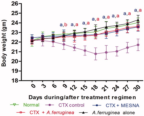
Figure 1. Effect of treatments on blood parameters in mice at various timepoints during the treatment/post-treatment period. (A) Total white blood cell [WBC] count. (B) Hemoglobin [Hb] content. Mice were given a total of 10 daily doses of CTX; every 3 days - starting prior to the first treatment (Day 0) and during/after the treatment (up to 20 days after final treatment) - blood was collected from the tail vein. From this material, total WBC counts and Hb levels were measured. Values shown are mean ± SD. ap < 0.01, bp < 0.05 for CTX only vs CTX/extract- or CTX/MESNA-co-treated. cp < 0.01, dp < 0.05 for normal versus A. ferruginea extract alone.
![Figure 1. Effect of treatments on blood parameters in mice at various timepoints during the treatment/post-treatment period. (A) Total white blood cell [WBC] count. (B) Hemoglobin [Hb] content. Mice were given a total of 10 daily doses of CTX; every 3 days - starting prior to the first treatment (Day 0) and during/after the treatment (up to 20 days after final treatment) - blood was collected from the tail vein. From this material, total WBC counts and Hb levels were measured. Values shown are mean ± SD. ap < 0.01, bp < 0.05 for CTX only vs CTX/extract- or CTX/MESNA-co-treated. cp < 0.01, dp < 0.05 for normal versus A. ferruginea extract alone.](/cms/asset/f8dd0478-c3a9-426f-a1cf-99a895f4a666/iimt_a_914988_f0001_c.jpg)
Hemoglobin levels on Day 15 of the treatment/post-treatment period (the same timepoint at which maximal depression in total WBC levels were noted) were 15.7 [± 0.3] gm% for control mice (). This value was unaffected by administration of extract alone (i.e. 15.7 [± 0.4] gm%), but significantly reduced in hosts that were treated with the CTX only (i.e. 13.6 [± 0.4] gm%). Co-treatment with A. ferruginea extract and CTX led to a mean hemoglobin content of 15.2 [± 0.5] gm%), a value clearly greater relative to that in the CTX-only hosts. Again, these protective effects remained apparent to the end of the entire treatment/post-treatment period (to Day 30).
Effects of the various treatment regimens on body weight (BW) are shown in . The results indicate significant differences in host BW values between the CTX/extract- or CTX/MESNA-co-treated vs CTX only mice over the study period (starting at Day 9). As a representative timepoint, a comparison of the BW values on Day 15 showed that the BW of control mice was 22.9 [± 0.4] g. This value was unaffected by administration of the extract alone (i.e. 23 [± 0.3] g). In contrast, treatment with the CTX alone led to significantly reduced BW values (i.e. 21.1 [± 0.4] g). The co-treatment with A. ferruginea extract or with MESNA led to a significant reversal of this outcome, i.e. mice given the CTX/MESNA- or CTX/extract regimens had BW values of 22.7 [± 0.3] and 22.6 [± 0.4] g, respectively. These protective effects remained apparent to the end of the entire treatment/post-treatment period (to Day 30).
Figure 2. Effect of treatments on body weight (BW) of mice at various timepoints during the treatment/post-treatment period. Every 3 days - starting prior to the first treatment (Day 0) and during/after the treatment (up to 20 days after the final treatment, i.e. Day 30) - the BW of each animal was measured. ap < 0.01, bp < 0.05 for CTX only versus CTX/extract- or CTX/MESNA-co-treated.
Treatment effects on relative organ weight and bone marrow cellularity/α-esterase activity
reports the relative organ weights of spleen, thymus, lungs, kidney, and liver of the treated mice on Days 7 and 11. Administration of CTX resulted in minor reductions in relative organ weights of the thymus and spleen compared to values for the control mice. Co-treatment with A. ferruginea extract led to a considerable enhancement in the weights of the thymus and spleen (compared to those in the CTX-only mice). No significant changes were observed in organ weight of lungs due to any of the treatments. Administration of CTX alone cause a slight increase in the relative weights of the kidney, liver, and urinary bladder compared to values for the control animals; these changes were reversed by extract co-treatment.
Table 1. Effect of treatments on organ indices on Days 7 and 11.
Bone marrow cellularity and levels of α-esterase+ cells were reduced by treatment with the CTX alone (on both Days 7 and 11; ) as compared to the values associated with the control mice. Each of these was significantly reversed by co-treatment with the A. ferruginea extract or with MESNA.
Table 2. Effect of treatments on bone marrow cellularity and α-esterase activity in mice on Days 7 and 11.
Effects of treatments on serum IL-2, IFNγ, GM-CSF, and TNFα
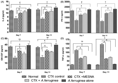
The effects of the various treatments on Day 11 serum TNFα, IL-2, IFNγ, and GM-CSF levels are shown in . Relative to values in naïve control mice sera, CTX-only-injected animals had decreases in IL-2, IFNγ, and GM-CSF, and marked increases in TNFα, levels. Co-treatment with the A. ferruginea extract significantly reversed these trends. Specifically, serum levels of TNFα, IL-2, IFNγ, and GM-CSF in CTX only-treated mice were, respectively, 147.3 [± 5.0], 7.0 [± 0.6], 1140.0 [± 84.5], and 19.6 [± 2.9] pg/ml, whereas in extract co-treated hosts these were now 71.3 [± 2.4], 11.0 [± 0.8], 1961.5 [± 59.0], and 28.3 [± 1.0] pg/ml. Similar results were obtained for MESNA co-treated mice. In mice that received only extract, except for TNFα, treatment only led to slight increases in IL-2, IFNγ, and GM-CSF (12.8 [± 0.7], 2301.4 [± 82.2], and 39.6 [± 2.3] pg/ml, respectively) vs values seen in the normal control mice.
Figure 3. Serum IL-2, IFNγ, GM-CSF, and TNFα after the respective treatments. Serum from the mice euthanized on Days 7 and 11 was assessed for each product by a specific ELISA. Values shown are mean ± SD. ap < 0.01 for CTX only versus CTX/extract- or CTX/MESNA-co-treated. cp < 0.01 for normal versus A. ferruginea extract alone.
Effect of treatments on serum markers of tissue damage and bladder LPO and GSH levels
Effects of the various treatments on host serum AST, ALT, and ALP levels are presented in . Serum AST, ALT, and ALP levels on Days 7 and 11 were significantly increased in mice that received only CTX compared to values in controls hosts (Group I). In extract-co-treated mice, these levels were significantly reduced relative to those in the CTX only mice. These results were comparable to those in the mice that received the MESNA co-treatment. Values of serum AST, ALT, and ALP levels in mice that received just the extract did not differ significantly from naïve untreated hosts.
Table 3. Effect of treatments on serum AST, ALT, and ALP in mice on Days 7 and 11.
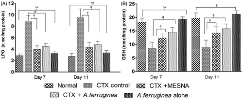
Due to CTX, elevated levels of LPO (8.9 [± 1.1] and 9.6 [± 1.5] nmol/mg protein) were found, respectively, in the bladder homogenates on Days 7 and 11; control mice values were just (2.9 [± 0.3] and 2.7 [± 0.4] nmol/mg protein). On Day 11, these elevated levels were significantly reduced to 4.5 [± 0.5] and 4.0 [± 0.6] nmol/mg protein, respectively, by co-treatment with A. ferruginea extract or MESNA (). Similar reductions were also apparent at Day 7. Use of extract alone had little impact on this parameter relative to values in the naïve controls.
Figure 4. Bladder LPO and GSH levels after the respective treatments. On Days 7 and 11, six mice from each group were euthanized and their bladders collected. An homogenate was prepared and used for measures of LPO and GSH. Values shown are mean ± SD. ap < 0.01 for CTX only versus CTX/extract- or CTX/MESNA-co-treated. cp < 0.01, dp < 0.05 for normal versus A. ferruginea extract alone.
GSH levels in bladder homogenates were reduced in the CTX-injected mice on Days 7 and 11 (8.5 [± 2.4] and 8.9 [± 2.7] nmol/mg protein, respectively) compared to naïve control values (18.3 [± 1.2] and 19.74 ± 1.74 nmol/mg protein) (). This too was reversed by co-treatment with extract (14.6 [± 1.8] and 15.8 [± 1.7] nmol/mg protein) on, respectively, Days 7 and 11. A similar effect was noted with the MESNA co-treated hosts (12.4 [± 1.5] and 14.2 [± 2.3] nmol/mg protein, respectively, on Days 7 and 11). Among mice treated with the extract alone, there was a slight (albeit significant) increase in bladder GSH levels (21.2 [± 1.3] nmol/mg protein) compared to in the naïve controls by Day 11.
Effect of treatments on small intestine and urinary bladder morphologies
Morphologic analysis of the small intestine collected 24 h after the final treatment (Day 11) revealed differences between control and treated mice (). In CTX-injected mice, the small intestine appeared severely inflamed and red, while the bladder evidences severe inflammation, dark red coloration, and hemorrhaging. In mice co-treated with A. ferruginea extract, there was only slight inflammation and hemorrhaging in the two tissues. Use of the uroprotective agent MESNA produced appreciable results as well in the bladders.
Treatment effects on small intestine and urinary bladder histopathology
The morphologies of the intestine and urinary bladder inflammation were examined. and present representative micrographs of each site’s histopathology at the end of the experiment periods. Histopathologic analysis of the small intestine (jejunal region) of CTX-injected animals showed severe damage to the villi compared, i.e. villi lengths were markedly reduced and crypt architecture largely destroyed. This effect was mitigated in hosts co-treated with A. ferruginea extract and MESNA along with CTX. The bladder in control mice had normal squamous epithelial lining, narrow lamina propria, and normal wall structure. In CTX-injected mice, there was severe damage to the urothelium, including mucosal erosion, vasodilation, inflammatory cell infiltration, widened lamina propria, fibrin deposition, necrosis, and multiple ulcerations. In contrast, in animals co-treated with the extract or MESNA along with CTX, there was reduced bladder damage characterized by urothelium preservation, only a mild thickening of the lamina propria (caused by edema), and reduced evidence of leukocyte infiltration and tissue ulceration.
Figure 5. Histology of small intestine due to the treatments. A representative micrograph from a mouse in each regimen is shown. (a) Naïve control showing normal mucosa. (b) CTX only (25 mg/kg BW) showing necrosis with damaged crypt architecture. (c) A. ferruginea extract (10 mg/kg BW) + CTX showing normal muscularis propria. (d) MESNA (25 mg/kg BW) + CTX with randomly damaged architecture. (e) Extract only showing normal architecture. All treatments were by IP injection of each treatment agent daily for 10 consecutive days. In the groups that received CTX or extract only, the volume of any missing component from the co-treatment groups was replaced by gum acacia-PBS injection so that all mice received the same volumes of injection each day. Magnification = 40×.
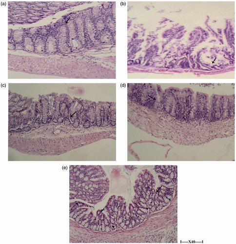
Figure 6. Histology of the urinary bladder due to treatments. A representative micrograph from a mouse in each regimen is shown. (a) Naïve control showing normal epithelium. (b) CTX only (25 mg/kg BW) showing damaged epithelium with edematous area. (c) A. ferruginea extract (10 mg/kg BW) + CTX showing normal lamina propria. (d) MESNA (25 mg/kg BW) + CTX showing normal mucosa. (e) Extract only showing normal architecture. All treatments were by IP injection of each treatment agent daily for 10 consecutive days. In all cases, the volume of any missing component for a given Group was replaced by phosphate buffer saline such that all mice received the same volume of injection each day. Magnification = 40×.
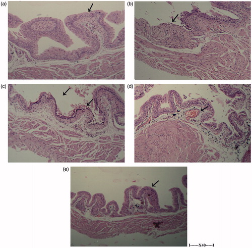
Table 4. Effect of treatments on small intestine and urinary bladder histology.
Discussion
This study examined the potential protective effect of an extract of A. ferruginea against cyclophosphamide (CTX)-induced immunosuppression. CTX is a commonly used anti-neoplastic drug for treating malignancies; it is also commonly used as an immunosuppressant. Among the deleterious effects from CTX administration are immunotoxicity (suppression of white blood cells), urotoxicity (including bladder hemorrhagic cystitis), gonadal atrophy, as well as geno-, hepato-, and nephrotoxicity. As such, there is a need to develop agents that can help protect hosts against CTX-induced toxicity while not negating the expected therapeutic effects of the CTX.
The present study sought to analyze whether A. ferruginea (as an extract) could prevent toxicity induced by CTX caused by metabolites of the drug. The initial activation of CTX is due to the microsomal oxidation system in the liver; this gives rise to the cytotoxic metabolite 4-hydroxy-CTX that diffuses from hepatocytes into the plasma, and is then distributed throughout the body. This metabolite is further converted to other cytotoxic metabolites (i.e. acrolein, phosphoramide mustard) that can cause myelosuppression (bone marrow suppression) (Grochow, Citation1996). To study the effect of A. ferruginea extract on any CTX-induced leukopenia and myelosuppression, here, host total leucocyte counts (TLC) as well as bone marrow cellularity and α-esterase activity was assessed. When compared with mice that received only CTX, total leukocyte counts were increased by co-treatment with the extract. The mice that received the extract also showed enhanced levels of bone marrow cellularity/α-esterase+ cells, suggesting to us the extract may stimulate the hematopoietic system and maturation of monocytes. Protection of bone marrow cells after cytotoxic therapy would be important in protecting hosts from CTX toxicity (Pratheeshkumar & Kuttan, Citation2010). In keeping with this ‘immunoprotective effect’, use of the A. ferruginea extract prevented the CTX-induced loss of splenic and thymic weight. The extract alone was able to increase the weights of both immune system organs, against suggesting to us that it might by itself act as an immunostimulant.
As immune system cells execute many of their functions through production of numerous cytokines (Dinarello, Citation1996), it was also important in this study to ascertain how the immunosuppressant CTX impacted on formation of select key cytokines and if the extract could mitigate this toxicity. Here, due to CTX administration, serum levels of TNFα become drastically increased, reflecting a state of inflammation in the treated hosts. This outcome was significantly reduced due to co-treatment with the A. ferruginea extract. Several studies reported that IL-2, which plays a major role in maturation/development of lymphocytes and monocytes and stimulates natural killer (NK) cell secretion of IFNγ (Theze et al., Citation1996) - which in turn stimulates macrophage activation and T-cell differentiation (Borish & Rosenwasser, 1996), is impacted by CTX treatment (Guo et al., Citation2012). Similarly, GM-CSF, a hematopoietic growth factor pivotal for the regulation of bone marrow progenitor cell proliferation (Gasson, Citation1991), was also shown to be affected by CTX treatment (Shalit et al., Citation2001). Expectedly, here, CTX drastically reduced host serum levels of IL-2, IFNγ, and GM-CSF; these were significantly increased due to co-treatment with the extract.
Hepatic dysfunction is another common toxicity in CTX-treated patients. In general, changes in serum levels of alkaline phosphatase (ALP), aspartate transaminase (AST), and alanine transaminase (ALT) are associated with damage to liver parenchymal cells and serve as specific indices of liver inflammation (Kumar et al., Citation2005). Apart from the liver, cells lining the intestine and kidney also produce a small amount of ALP and elevated levels of intestinal ALP in patients with renal impairment have been reported (Alpers et al., Citation1988; Travis et al., Citation1995). As expected, in this study, treatment of mice with the CTX caused marked elevations in serum ALP, AST, and ALT. Co-administration of the A. ferruginea extract led to significant reductions in ALP, AST, and ALT levels, suggesting the extract imparted a protective effect for the hepato-biliary system.
As the bladder is the site for urine storage, the content of these toxic metabolites is higher than in other areas of the urinary tract, making the bladder most susceptible to any potential damage. In this study, it was seen that the combined treatment of A. ferruginea extract and CTX blocked/mitigated CTX-induced toxicities in the bladder. The pro-oxidative metabolites of CTX are responsible for induction of lipid peroxidation (LPO) and further generation of free radicals that, in turn, give rise to inflammation and shifts in overall redox cycling in the bladder (Kehrer & Biswal, Citation2000). Likewise, administration of CTX causes reductions in bladder GSH levels; this outcome is primarily a result of interactions between acrolein with GSH at the site (Manesh & Kuttan, Citation2005). A depletion of GSH has been reported to increase the susceptibility of cells lining the bladder and intestine to apoptosis (Bhatia et al., Citation2006); these changes could rise to many of the histopathologic changes that were noted here. While mice here that received CTX alone evinced significant changes in oxidative stress including LPO and decreases in GSH in, respectively, the liver and intestinal mucosa, A. ferruginea extract co-treated mice had significantly reduced LPO levels and increased GSH level. Histomorphological findings in the bladder and small intestine also provided evidence of an ameliorating effect, one that was equal to or more effective than the known uroprotector MESNA.
It is still unclear which of the myriad of constituents (or combinations therein) in the methanolic extract of A. ferruginea are responsible for its ability to counter toxicities (immune, bladder, liver, etc.) induced by CTX. Our preliminary phytochemical analyses of the extract revealed a presence of numerous flavonoids, phenolics, steroids, terpenoids, alkaloids, saponins, and tannins (Sakthivel & Guruvayoorappan, Citation2013). More specifically, those GC/MS and LC/MS studies also revealed the A. ferruginea extract contained significant amounts of quinone, quinoline, imidazolidine, pyrrolidine, pyrazole, thiazole, cyclopentenone, catechin, and coumarin derivatives. Evidence in the literature has indicated that some of these agents (primarily the flavonoids) can enhance the activity of human lymphocytes (i.e. proliferation) and their secretion of IFNγ (Chiang et al., Citation2003). Other studies have shown that flavonoids/phenolics can stimulate leukocyte proliferation and increase the activity of macrophages and helper T-cells, including production of IL-2 and IFNγ by the latter, thereby mitigating immune dysfunction (Chiang et al., Citation2003; Kawakita et al., Citation2005). Flavonoids in combination with chemoprotective agents have been proven to increase the effectiveness of CTX as well as to reduce the toxicity of the drug to normal cells (Edy et al., Citation2012; Fimognari et al., Citation2006). It is clear that our future and ongoing studies will need to more clearly ascertain which, if any, of the compounds we showed were present in the A. ferruginea methanolic extract could be responsible for the protective effects produced in situ.
Conclusions
The results here suggest to us that A. ferruginea extract could potentially be used to increase total leukocyte counts as well as proliferation and differentiation of bone marrow cells, even during an ongoing toxicity from exposure to CTX. Similarly, reversals of effects of CTX on host anti-oxidant and select cytokine levels indicate an ability of the extract to help the host mitigate/reverse and immunotoxicity induced by the CTX. Further studies using isolated compounds from the extract are in progress to identify the active principle and to determine the exact mechanism of action. Lastly, we wish to note for the readers that we are cognizant that studies need to be done with tumor-bearing hosts, given both CTX and the extract to better ascertain if the co-treatment mitigates/abrogates the anti-cancer efficacy of CTX. Such studies are already planned and will begin shortly.
Declaration of interest
The authors report no conflicts of interest. The authors alone are responsible for the content and writing of the paper.
Acknowledgements
The authors wish to thank Origin Laboratories, Coimbatore for Histopathology analysis. The valuable suggestions from Dr. M. Patrick Gomez, Director, School of Biotechnology and Health Sciences and Dr Jannet Vennila, Head, Department of Biotechnology, Karunya University, are gratefully acknowledged.
References
- Ahmad, S., Mika, D., and Guruvayoorappan. C. 2012. Chemoprotective and immunomodulatory effect of Acacia nilotica during cyclophosphamide induced toxicity. J. Exp. Ther. Oncol. 10:83–90
- Alpers, D. H., DeSchryver-Kecskemeti, K., Goodwin, C. L., et al. 1988. Intestinal alkaline phosphatase in patients with chronic renal failure. Gastroenterology 94:62–67
- Amudha, G., Josephine, A., Mythili, Y., et al. 2007. Therapeutic efficacy of lipoic acid on cyclosporine A induced renal alterations. Eur. J. Pharmacol. 571:209–214
- Bachaya, H. A., Iqbal, Z., and Khan, M. N. 2009. Anthelmintic activity of Ziziphus nummularia (bark) and Acacia nilotica (fruit) against Trichostrongylid nematodes of sheep. J. Ethnopharmacol. 123:325–329
- Bancroft, J. D., and Cook, H. F., (Eds.). 1984. Manual of Histological Techniques. London: Churchill Livingston, pp. 171–174
- Bhatia, K., Kaur, M., Atif, F., et al. 2006. Aqueous extract of Trigonella foenum-graecum L. ameliorates additive urotoxicity of buthio-nine sulfoximine and cyclophosphamide in mice. Food Chem. Toxicol. 44:1744–1750
- Borish, L., and Rosenwasser, L. J. 1996. Update on cytokines. J. Allergy Clin. Immunol. 97:719–734
- Chiang, L. C., Ng, L. T., Chiang, W., et al. 2003. Immunomodulatory activities of flavonoids, monoterpenoids, triterpenoids, iridoid glycosides, and phenolic compounds of Plantago species. Planta Med. 69:600–604
- Dinarello, C. A. 1996. Biological basis for IL-1 in disease. Blood 87:2095–2147
- Edy, M., Adam, H., and Anindyajati, T. 2012. Natural products for cancer targeted therapy: Citrus flavonoids as potent chemopreventive agents. Asian Pac. J. Cancer Prev. 13:427–436
- Eldeen, I. M., Heerden, F. R., and Staden, J. 2010. In vitro biological activities of niloticane, a new bioactive cassane diterpene from bark of Acacia nilotica subsp. Kraussiana. J. Etnopharmacol. 128:555–560
- Fimognari, C., Nusse, M. N., Lenzi, M., et al. 2006. Sulforaphane increases the efficacy of doxorubicin in mouse fibroblasts characterized by p53 mutations. Mutat. Res. 601:92–101
- Gasson, J. C. 1991. Molecular physiology of granulocyte-macrophage colony-stimulating factor. Blood 77:1131–1145
- Grochow, L. B. 1996. Covalent DNA-binding drugs. In: The Chemotherapy Source Book (Perry, M. C., Ed.). Baltimore, MD: Williams & Wilkins, pp. 297–299
- Guo, Q., Zhangchun, L., Qihong, F., et al. 2012. IFNγ-producing T-cells contribute to increase of myeloid-derived suppressor cells in tumor-bearing mice after cyclophosphamide treatment. Int. J. Immuno-pharmacol. 12:425–432
- Kalaivani, T., and Mathew, L. 2011. Free radical scavenging activity from leaves of Acacia nilotica (L.) Wild. ex Delile, an Indian medicinal tree. Food Chem. Toxicol. 48:298–305
- Kawakita, S. W., Giedlin, H. S., and Nomoto, K. 2005. Immunomodulators from higher plants. J. Nat. Med. 46:34–38
- Kehrer, J. P., and Biswal, S. S. 2000. The molecular effects of acrolein. Toxicol. Sci. 57:6–15
- Kirtikar, K. R., and Basu, B. D., (Eds.). 2003. Indian Medicinal Plants, Vol. 4. Second Edition. Deharadun, India: Oriental Enterprises, pp. 1298–1300
- Kiuchi, H., Takao, T., Yamamoto, K., et al. 2009. Sesquiterpene lactone parthenolide ameliorates bladder inflammation and bladder over-activity in cyclophosphamide-induced rat cystitis model by inhibiting NF-κB phosphorylation. J. Urol. 181:2339–2348
- Kumar, G., Banu, G. S., Kannan, V., and Pandian, M. R. 2005. Anti-hepatotoxic effect of β-carotene on paracetamol induced hepatic damage in rats. Indian J. Exp. Biol. 43:351–355
- Lopes, V., Moraes, R., and Araujo, B. 2009. Physicochemical and anti-fungal properties of protease inhibitors from Acacia plumosa. Phytochemistry 70:871–879
- Manesh, C., and Kuttan, G. 2005. Effect of naturally occurring isothiocyanates in the inhibition of cyclophosphamide-induced urotoxicity. Phytomedicine 12:487–493
- Meena, P. D., Kaushik, P., and Shukla, S. 2006. Anti-cancer and anti-mutagenic properties of Acacia nilotica (Linn.) on 7,12-dimethylbenz(a) anthracene-induced skin papillomagenesis in Swiss albino mice. Asian Pac. J. Cancer Prev. 7:627–632
- Moron, M. S., Depierre, J. W., and Mannervik, B. 1979. Levels of glutathione, glutathione reductase, and glutathione-S-transferase activities in rat lung and liver. Biochim. Biophys. Acta 582:67–78
- Ohkawa, H., Ohishi, N., and Yagi, K. 1979. Assay for lipid peroxides in animal tissues by thio-barbituric acid reaction. Anal. Biochem. 95:351–358
- Ozcan, A., Korkmaz, A., Oter, S., and Coskun, O. 2005. Contribution of flavonoid anti-oxidants to preventive effect of MESNA in cyclophosphamide-induced cystitis in rats. Arch. Toxicol. 79:461–465
- Papaldo, P., Lopez, M., Marolla, P., et al. 2005. Impact of five prophylactic filgrastim schedules on hematologic toxicity in early breast cancer patients treated with Epirubicin and cyclophosphamide. J. Clin. Oncol. 23:6908–6918
- Parrotta, J. A., (Ed.). 2001. Healing Plants of Peninsular India. Oxfordshire, UK: CABI Publishing, pp. 323–324
- Pass, G. J., Carrie, D., Boylan, M., et al. 2005. Role of hepatic cytochrome P450s in the pharmacokinetics and toxicity of cyclophosphamide: Studies with the hepatic cytochrome P450 reductase-null mouse. Cancer Res. 65:4211–4217
- Pratheeshkumar, P., and Kuttan, G. 2010. Ameliorative action of Vernonia cinerea L. on cyclophosphamide-induced immunosuppression and oxidative stress in mice. Inflammopharmacology 18:197–207
- Rajbir, S., Singh, B., and Singh, S. 2010. Umbelliferone - An anti-oxidant isolated from Acacia nilotica (L.) Willd. Ex. Del. Food Chem. 120:825–830
- Sakthivel, K. M., and Guruvayoorappan, C., 2013. Acacia ferruginea inhibits tumor progression by regulating inflammatory mediators TNFα, iNOS, COX-2, IL-1β, IL-6, IFNγ, IL-2, GM-CSF, and pro-angiogenic growth factor VEGF. Asian Pac. J. Cancer Prev. 6:3909–3919
- Shalit, I., Kletter, Y., Halperin, D., et al. 2001. Immunomodulatory effects of moxifloxacin in comparison to ciprofloxacin and G-CSF in a murine model of cyclophosphamide-induced leukopenia. Eur. J. Haematol. 66:287–296
- Shathish, K., Reena, D., and Guruvayoorappan, C. 2012. Chemoprotective effect of Decalepis hamiltonii against cyclophosphamide-induced toxicity. J. Exp. Ther. Oncol. 9:291–301
- Suresh, G., and Rao, J. V. 1999. Inter-cropping sorghum with nitrogen fixing trees in semiarid India. Agroforest. Syst. 92:181–194
- Theze, J., Alzari, P. M., and Bertoglio, J. 1996. IL 2 and its receptors: Recent advances and new immunological functions. Immunol. Today 18:487–492
- Travis, L. B., Curtis, R. E., Glimelius, B., et al. 1995. Bladder and kidney cancer following cyclophosphamide therapy for non-Hodgkin's lymphoma. J. Natl. Cancer Inst. 87:524–531
- Tung, Y. T., Wu, J. H., and Hsieh, C. Y. 2008. Free radical-scavenging phytochemicals of hot water extracts of Acacia confusa leaves detected by on-line screening method. Food Chem. 115:1019–1024
- Vinod Prabhu, V., and Guruvayoorappan, C. 2012. Evaluation of immunostimulant activity and chemoprotective effect of mangrove Rhizophora apiculata against cyclophosphamide induced toxicity in Balb/c mice. Immunopharmacol. Immunotoxicol. 34:608–615