
Abstract
While T-lymphocytes are the major cell type responsible for host responses to a virus (including induction of inflammatory responses to aid in ultimate removal of virus), other cells, including macrophages, epithelial and dendritic cells also have key roles. Endothelial cells also play important roles in physiologic/pathologic processes, like inflammation, during viral infections. As endothelial cells can be activated to release various endogenous compounds, including some cytokines, ex vivo measures of cytokine formation by the cells can be used to indirectly assess any potential endothelial dysfunction in situ. The research presented here sought to investigate potential immunolomodulatory effects of five saponins on endothelial cells: Saikosaponins A (SSA) and D (SSD), Panax Notoginseng Saponin (PNS) and Notoginsenoside R1 (SR1) and Anemoside B4 (AB4). For this, cells (porcine iliac artery endothelial line) were challenged with a virus isolate PCV2-AH for 24 h and then treated with the test saponin (at 1, 5 or 10 μg/ml) for an additional 24 h at 37 °C. The culture supernatants were then collected and analyzed for interleukin (IL)-2, -4 and -10, as well as interferon (IFN)-γ by ELISA. The results revealed that PNS and SR1 inhibited the production of IL-4; PNS, SR1 and AB4 inhibited the secretion of IL-10; SSA, SSD and PNS up-regulated IL-2 expression; SSA and SSD increased the level of IFNγ. All these changes were significant. Taken together, the data suggested these saponins might potentially have a capacity to regulate immune responses in vivo via changes in production of these select cytokines by infected endothelial cells. Nevertheless, the impact of these agents on other key cell types involved in anti-viral responses, including T-lymphocytes, remains to be determined.
Introduction
Porcine circoviruses (PCV, in family Circoviridae) are non-enveloped viruses with single-stranded circular DNA genomes (Mankertz et al., Citation2000). PCV2 was demonstrated to be a causative agent of porcine circovirus-associated disease (PCVAD), which includes porcine multi-systemic wasting syndrome (PMWS), porcine dermatitis and nephropathy syndrome (PDNS), porcine respiratory disease complex (PRDC), congenital tremor (CT) and reproductive failure (Chae, Citation2005; Opriessnig et al., Citation2007). Since its emergence in the early 1990s, PCVAD has continuously been a threat to the global swine industry, causing high economic losses.
Many Chinese herbal medicines can effectively suppress and kill viral pathogens, in addition to eliminating fever and clearing toxic materials (Wang & Liu, Citation2014). They are also widely used to prevent/cure other non-viral infectious diseases and show high efficacy, lower toxicity, few side-effects and lower residual levels than many commonly-used drugs (Na-Bangchang & Karbwang, Citation2014). Accordingly, many studies have recently focused on the use of Chinese herbal medicines to treat/prevent PCV2-induced health effects (Wei et al., Citation2012).
Saikosaponin A (SSA) and Saikosaponin D (SSD) are major triterpenoid saponins derived from Bupleurum falcatum L. (Umbelliferae), commonly prescribed by Chinese and Japanese medical doctors for inflammatory and infectious diseases. These active components are reported to impart immunomodulatory, anti-inflammatory, anti-bacterial, anti-viral and anti-cancer effects (Tundis et al., Citation2009). Recently, it has been shown that SSD could exhibit an anti-proliferative effect in activated T-lymphocytes, in part via suppression of NF-κB, NF-AT and AP-1 signaling (Wong et al., Citation2009).
The major active components of notoginseng are panax notoginseng saponins (PNS), which consist of >30 different types of saponins; among these, ginsenosides Rg1 and Rb1 are found at high levels. Notoginsenoside R1 is a component unique to notoginseng (Chen et al., Citation2012). PNS and its major components exhibit anti-cancer activities and have been shown to be effective against a variety of malignancies including colorectal, lung, gastric, skin, prostate and liver cancers (Sengupta et al., Citation2004). Notoginsenoside R1 (SR1) has been shown to be a promising compound for protecting the heart from septic shock and to impart anti-inflammatory effects in mice (Sun et al., Citation2013). Pulsatilla koreana Nakai, with Anemoside B4 (AB4) as its main pharmacological effective compound, is known to have imparted numerous biological effects in vitro, including enhancing hypoglycemic, anti-tumor, neuroprotective and anti-angiogenic activity (Liu et al., Citation2014).
Endothelial cells play important roles in a number of physiologic and pathologic processes, such as inflammation, fever, diarrhea, coagulation and shock (Pate et al., Citation2010). Regarding initiation/development of immune responses, endothelial cells can be activated by pathogens, leading to their release of various endogenous compounds that modulate vascular relaxation/constriction, including some cytokines (i.e. NO, ET-1, IL-1) (Hu et al., Citation2009). As such, measures of cytokine levels can be used to indirectly assess if there is endothelial dysfunction in situ. As it has been confirmed that endothelial cells are important primary targets for systemic diseases (Lehle et al., Citation2010), iliac artery endothelial cells (including those from pigs, i.e. PIEC) represent good models for such studies of induced dysfunction.
In the present study, the effects of five saponins, e.g. Saikosaponin A (SSA), Saikosaponin D (SSD), panax notoginseng saponins (PNS), Notoginsenoside R1 (SR1) and Anemoside B4 (AB4), on endothelial cells were evaluated through measures of their secretion of four key cytokines: interleukin (IL)-4, IL-10, IL-2 and interferon (IFN)-γ. The goal of this study was to investigate pharmacologic mechanisms of these saponins for potential use in treatment of PCV2-induced diseases. At same time, it was hoped the studies would provide a theoretical basis for further research on saponins and the development of new drugs for host immune enhancement.
Materials and methods
Reagents
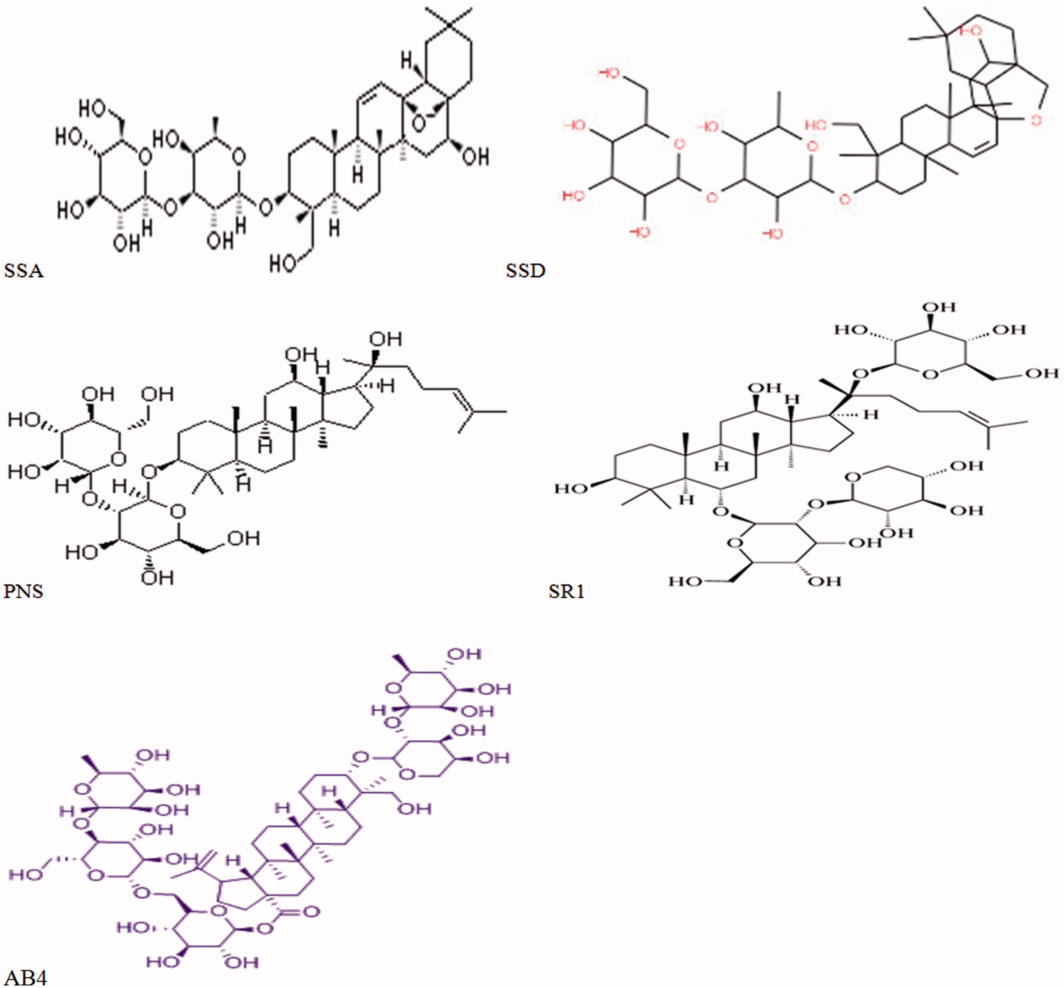
SSA, SSD, PNS, SR1 and AB4 (each at 20 mg/vial; lot numbers: 110777, 110778, 110745, 110870 and 111766, respectively; ) were each purchased from the National Institute for Control of Pharmaceutical and Biological Products (Beijing, China). All ingredients were individually diluted to three concentrations (10, 50 and 100 μg/ml) with maintenance medium containing 10% fetal bovine serum (FBS; Sigma, St. Louis, MO), 100 U penicillin/ml and 10 μg streptomycin/ml (Gibco, Grand Island, NY). The diluted solutions were then filtered through a 0.22-μm membrane and stored at 4 °C. Before use, the agents were diluted 10-fold with medium.
Figure 1. Structures of the test saponins.
The Zoonotic Prevention and Control Laboratory at Jiangsu Academy of Agricultural Sciences (Nanjing, China) provided the Virus isolate PCV2-AH. PCV2-AH has previously been shown to induce PMWS (Li et al., Citation2012). The PCV2-SH stock titers were 5 × 10⁵ TCID50 (50% tissue culture infective dose)/ml as determined by titration on PK-15 cells using an immunofluorescence assay.
Experimental protocol
Porcine iliac artery endothelial cells were obtained from the Committee on Type Culture Collection of Chinese Academy of Sciences (Shanghai, China); the providers indicate the line was developed from a single host animal. When cultured single-layer endothelial cells reached confluence in a cell incubator (BB15; Thermo Scientific, Chicago, IL), they were allocated into 26 groups. In the blank control group, complete medium was replaced with maintenance medium without the PCV2 virus or any saponins. In the PCV2 virus control group, medium was replaced with medium containing PCV2 virus at 105 TCID50. Selection of this dose of virus was based on preliminary experiments where it was found that high levels (≫105 TCID50) could affect the morphological structure and secretory function of the endothelial cells and even produce cell death. In contrast, a low level of the virus (i.e. 105 TCID50) still impacted on secretion of products from these cells (on order of 10–40%, depending on endpoint being assessed). Therefore, the 105 TCID50 dose was used here, as it was not overtly toxic yet still provided ample stimulation of product formation/release that could then be assessed for changes induced by the test saponins.
In the high-, middle- and low-dose groups of the five saponins, the cells were initially challenged with PCV2 virus at 105 TCID50, followed by addition (after 24 h) of a given saponin to final concentrations of 1, 5 or 10 μg/ml. In the drug control group, the medium was replaced with 10 μg/ml of a given saponin. Each treatment was repeated 4-times. The cells were then incubated at 37 °C for an additional 24 h, then centrifuged at 100 x g for 10 min. Thereafter, the supernatants were collected and analyzed for IL-4, IL-10, IL-2 and IFNγ. Each experiment was repeated independently 4-times with fresh cell cultures.
The choice of the doses of each saponin used here was based upon findings from in vivo studies using SSA and SSD. Specifically, SSA plasma concentrations in rats dosed with 5 mg SSA/kg were determined by LC-ESI-MS; after a single intravenous treatment, a Cmax of 1.907 µg/ml was attained (Liu et al., Citation2009). In mice given SSD in a single oral dosage of 22.55 mg SSD/kg, a plasma level (as determined by RP-HPLC) Cmax of 83.81 µg/ml was achieved (Wang et al., Citation2005). While there is no comparable data available for porcine hosts, we believe that the Cmax values from these and other studies provide justification for the doses of SSA and SSD used here. Clearly, more research is required to elucidate the pharmacokinetics of the other saponins evaluated here to determine if similar Cmax values are obtained in hosts treated in a similar manner as in the above-noted studies. However, in the absence of such information, use of doses similar to the ones for SSA and SSD is the only option to permit comparisons across the test saponins to be made with regard to each endpoint evaluated here.
Determinations of IL-4, IL-10, IL-2 and IFNγ
IL-4, IL-10, IL-2 and IFNγ levels in the supernatants were measured using commercial ELISA kits (all from RapidBio Laboratories, Calabasas, CA) according to manufacturer instructions. Aliquots (0.1 ml) of serially-diluted standards and samples were added to ELISA plates and incubated at 37 °C for 1.5 h. After washing, 0.1 ml kit-provided antibody solution was added to each well (except control wells) and the plate incubated at 37 °C for 1 h. After washing, 0.1 ml of a kit-provided solution of 3,3’,5,5’-tetramethylbenzidine [TMB] and hydrogen peroxide was added to each well and the plate incubated at 37 °C for 0.5 h. The reaction in each well was stopped by addition of 0.1 ml 1 N sulfuric acidaq. Optical density (OD) in each well was then measured at 450 nm in an ELx800 microplate reader (BioTek, Winooski, VT). Levels of each cytokine (in pg/ml) in the samples were then extrapolated from the standard curves. Kit sensitivity levels for IL-4, IL-10, IL-2 and IFNγ were, respectively, 5.0, 7.5, 10.0 and 10.0 pg/ml, as reported by the manufacturer.
Statistical analyses
All data were expressed as means ± SD. All data were tested for normal distribution using a Shapiro-Wilk test and for homogeneity of variances using a Levene’s test. If the data were normally distributed and had similar variances, then a one-way analysis of variance (ANOVA) using a Type III sum of squares was performed to compare means among all measured variables. When ANOVA results were significant, multiple comparisons of means were performed using a Tukey HSD post-hoc analysis. If the data did not have similar variances, a non-parametric Kruskal-Wallis test for comparing the median was applied as was a Mann-Whitney test (or a Two-Sample Kolmogorov-Smirnov test) for multiple comparisons among the different groups if the results of the Kruskal-Wallis test showed significant differences (at p < 0.05 level). All statistics were performed using statistical software SPSS v17.0 (SPSS Inc., Chicago, IL).
Results
IL-4
The IL-4 levels in all treatment groups are shown in . The IL-4 level in the PCV2 control group was significantly increased compared to that in the blank control group. Compared with the PCV2 control, IL-4 levels were significantly decreased for the high- and middle-dose PNS groups and with the high- and middle-dose SR1 groups. The low doses of both of those agents failed to significantly impact on IL-4 induction caused by the PCV2. The formation/secretion of IL-4 was increased 45.07% (relative to control production) in the PCV2 group; this effect was (maximally) reduced by 11.9 and 10.2% with, respectively, the high-dose PNS and SR1 treatments. There were no significant differences between the values for production among the various PNS (and similarly, SR1) treatment groups. No doses of SSA, SSD or AB4 had any impact on the PCV2-induced effect on IL-4 levels.
Table 1. IL-4 contents (pg/ml) in culture supernatants after 24 h.
IL-10
The IL-10 levels in all treatment groups are shown in . The IL-10 level in the PCV2 control group was significantly increased compared with that in the blank control. Compared with the PCV2 control, IL-10 levels were significantly decreased for the high- and middle-dose PNS groups, with the high- and middle-dose SR1 groups and the high-dose AB4 group. The low doses of all three agents failed to induce a significant reduction. Formation/secretion of IL-10 was increased 29.6% (relative to control production) in the PCV2 group; this effect was (maximally) reduced by 17.9, 15.4 and 13.0% with, respectively, the high-dose PNS, SR1 and AB4 treatments. There were no significant differences between the values for production among the various PNS, SR1 or AB4 treatment groups. No doses of SSA or SSD had any impact on the PCV2-induced effect on IL-10 levels.
Table 2. IL-10 contents (pg/ml) in culture supernatants after 24 h.
IL-2
The IL-2 levels in all treatment groups are shown in . The IL-2 level in the PCV2 control group was significantly increased compared with that in the blank control group. Compared with the PCV2 control, IL-2 levels were significantly increased further in the high- and middle-dose SSD groups and with the high-dose PNS and SSA groups. The low doses of all three agents failed to significantly enhance the IL-2 production above that caused by the PCV2 alone. IL-2 formation/secretion was increased 11.5% (relative to control production) in the PCV2 group; this effect was (maximally) enhanced by 10.3, 10.7 and 12.8% with, respectively, the high-dose PNS, SSA and SSD treatments. Again, although the middle- (in the case of SSA and PNS) or the low-dose regimen (in all three cases) outcomes did not significantly differ from the PCV2 only sample values, there were no significant differences between production values within each of the various PNS, SSA or SSD treatment groups. No doses of SR1 or AB4 had any impact on the PCV2-induced effect on IL-2 levels.
Table 3. IL-2 contents (pg/ml) in culture supernatants after 24 hr.
IFNγ levels
The IFNγ levels in all treatment groups are shown in . The IFNγ level in the PCV2 control group was significantly increased compared with that in the blank control. Compared with the PCV2 control, IFNγ levels were significantly increased further in the high- and middle-dose SSD and SSA groups. The low doses of both agents failed to significantly enhance production above that caused by the PCV2 alone. IFNγ formation/secretion was increased 10.9% (relative to control production) in the PCV2 group; this effect was (maximally) enhanced by 12.4 and 16.3% with, respectively, the high-dose SSA and SSD treatments. In this case, only the low-dose regimen (in both cases) outcomes did not differ significantly from the PCV2-only samples. Again, there were no significant differences between production values within each of the various SSA or SSD groups. No doses of PNS, SR1 or AB4 had any impact on the PCV2-induced effect on IFNγ levels.
Table 4. IFNγ contents (pg/ml) in culture supernatants after 24 h.
Discussion
During viral infections, several immune cell types are activated. While T-lymphocytes are the major cell type responsible for host responses to the virus (including induction of inflammatory responses to aid in ultimate removal of the virus), other cells, including macrophages, epithelial and dendritic cells also have key roles. Endothelial cells also play important roles in physiologic/pathologic processes, like inflammation, associated with viral infections (Czuprynski, Citation2009; Muruve, Citation2004; Sahni, Citation2007). Endothelial cells can be activated by pathogens, leading to their release of various endogenous compounds, including some cytokines; thus, measures of cytokine levels can be used to indirectly assess if there is endothelial dysfunction in situ.
Porcine circovirus Type 2 genome or antigen can be detected in several tissues and cell types such as macrophage/monocyte lineage cells, dendritic cells and sporadically respiratory, renal and intestinal epithelial cells, hepatocytes, enterocytes, vascular endothelium and lymphocytes. Chinese herbal medicine can regulate the balance of cellular functions and improve the physiological conditions of organism in many ways, especially for microvascular endothelial cells. Existing data show that PCV2 has immunosuppressive characteristics that can cause an imbalance in a variety of cytokine expressions (Darwich et al., Citation2003; Segales et al., Citation2004). Cytokines are important immunoregulatory factors as signal molecules between cells in the immune network. IL-2 and IFNγ are important T-helper (TH)-1-type cytokines with important roles in cellular immune response; IL-4 and IL-10 are important TH2 cytokines key to humoral responses. During tolerance induction to an allograft, there is a decrease in TH1 cytokines like IL-2 and IFNγ and increases in TH2 cytokines including IL-4 and IL-10. Because studies have already shown there is significant expression of IL-4 (Hocke et al., Citation2006), IL-10 (Niu et al., Citation2011; Verma et al., Citation2006) and IFN (Hu et al., Citation2010, Citation2012; Niu et al., Citation2011) in several different non-leukocyte cell types, including endothelial cells (Sprague & Khalil, Citation2009), use of the porcine iliac artery cells here provided an appropriate model to investigate potential immunoregulatory effects of the test saponins.
IL-4 aids in antigen-stimulated polarization of naive TH cells into TH2 effector cells and also helps to propagate TH2 responses by binding its receptor (IL-4Rα) and activating signal transducer and activator of transcription (STAT)-6 signaling pathways (Nelms et al., Citation1999; Shimoda et al., Citation1996). STAT6, through induction of zinc-finger transcription factor GATA3 (GATA-binding protein-3), can directly suppress TH1 cell development by silencing IFNγ expression (Takeda et al., Citation1996). Studies also indicate that IL-4 enhances TH2 immunity by inhibiting TH1 responses through the repression of IL-12 signaling (Ouyang et al., Citation1998). IL-4 also induces formation of inducible T-regulatory (Treg) cells from naive CD4+ T-lymphocytes. The present study showed that endothelial cell secretion of IL-4 was significantly suppressed by some of the higher doses (i.e. 10 µg/ml) of PNS and SR1 tested. The increases in IL-4 expression by the endothelial cells – and subsequent increase in IL-10 release – could lead to excessive vasodilation, hyperemia and microvascular injury. The ability to induce IL-4 formation after PCV2 exposure was much higher than what was noted in the control group; this outcome was in accordance with our previous animal experiments.
It is tempting to suggest that thymic depletion and atrophy in PMWS pigs could be caused by over-expression of IL-10 mRNA in the thymus. Many studies have revealed that the induction of IL-10 up-regulation is a basic characteristic of PCV2 infection (Darwich et al., Citation2008; Stevenson et al., Citation2006) and that excessive up-regulation of IL-10 may cause immunosuppression. IL-10 is a multi-functional cytokine produced by lymphoid and non-lymphoid cells and displays many kinds of biological effects in the cytokine network, including an ability to inhibit production of tumor necrosis factor (TNF)-α, IL-8, tissue factor and matrix metalloproteinase (MMP)-9. IL-10 can also mediate a negative feedback of TH1 and TH2 immune responses (Muraille & Leo, Citation1998), regulate NK and B-cell growth (Levy & Brouet, Citation1994), downregulate MHC-II expression on antigen-presenting cells (APC) (Moore et al., Citation2001) and affect MHC-I expression by modulating expression of transporter proteins associated with antigen processing (Salazar-Onfray et al., Citation1997). Although anti-inflammatory processes are characteristic of defense reactions, it can also – if unchecked – cause endothelial cell dysfunction, increased permeability and tissue damage. In the present study, we found that endothelial cell secretion of IL-10 was significantly suppressed by PNS, SR1 and AB4, with PNS having the greatest effect. Such findings are in line with those of other studies. For example, it was shown that PNS produces robust protective effects in spinal cord ischemia-reperfusion injury and that this might be mediated by changes in the levels of IL-10 in the hosts (Ning et al., Citation2012). Similarly, PNS was shown to impart a therapeutic effect against hepatic fibrosis, probably by causing shifts in the balance between pro- and anti-inflammatory/-fibrotic cytokines in situ (Peng et al., Citation2009).
IL-2 – produced after antigen activation – is a pleiotropic cytokine that plays pivotal roles in the immune response. First described as a T-cell growth factor, IL-2 also promotes CD8+ T-cell and NK cell cytolytic activity and modulates T-cell differentiation programs in response to antigen, promoting naive CD4+ T-lymphocyte differentiation into TH1 and TH2 cells while inhibiting TH17 and T-follicular helper (TFH) cell differentiation. Moreover, IL-2 is essential for development/maintenance of Treg cells and for activation-induced cell death, thereby mediating tolerance and limiting inappropriate immune reactions. It has been reported that Tripterygium wilfordii Hook F significantly inhibited activation-induced T-lymphocyte IL-2 production, IL-2R expression and proliferation responses
In the present study, expression of IL-2 by endothelial cells was dramatically up- regulated in response to PCV2. In addition, secretion of IL-2 was further significantly increased by treatments with SSA, SSD or PNS. This impact on IL-2 formation/release could represent a mechanism by which saponins could be of use in treatment of select (auto)immune pathologies. As SSD was shown here (and elsewhere) to promote IL-2 production and IL-2R expression, this showed that SSD could modulate T-cell function and that at least one target of the action of SSD was located at or before c-fos gene transcription and after T-cell receptor/CD3-mediated protein tyrosine kinase activation (Kato et al., Citation1994). It is also possible SSD up-regulated IL-2 production through activation of a “receptor-bypassed” pathway in the cells; Kato et al. (Citation1995) noted a similar effect in thymocytes and suggested that SSD could (among various saponins) have a unique cell type-dependent immunomodulatory action. Similar information on activities of SSA and PNS are lacking in the literature; thus, their potential modes of action remain to be discerned with respect to effects on IL-2 formation.
It is known that IFNγ drives TH1 responses, activating cytotoxic effector cells to eliminate infected cells and promoting virus-specific antibody production. In some cases, PCV2 infection elicits an antibody response together with virus-specific IFNγ-secreting (CD4+ and CD8+) cells (Fort et al., Citation2009; Steiner et al., Citation2009). During a viral infection, down-regulated IFNγ production may result in decreases in CD8+ cytolytic T-cell function (Kienzle et al., Citation2002). In the current study with PIEC, there was up-regulation of IFNγ expression by SSA or SSD (moreso the latter) after direct stimulation with PCV2. Previous findings have shown that a part of the immunomodulatory activity of SSA and SSD against activated T-cells is related to changes in the ability of NF-κB, NF-AT and AP-1 (c-Fos) signaling pathways to be induced (Sun et al., Citation2009; Wong et al., Citation2009). Ongoing studies in our laboratories will need to ascertain if the effects of SSA and SSD (and other saponins tested) modulate these pathways in the PIEC and if this is directly linked to the changes seen in inducible IFNγ expression.
The findings from these in vitro studies are in keeping with more global observations of the effects of these saponins in vivo in PCV2-infected hosts (Lin et al., Citation2013; Yi et al., Citation2009). Those previous animal experiments suggested SSA, SSD, PNS, SR1 and AB4 could lead to reductions in host mortality and lessen the incidence/severity of specific pathologies, e.g. body temperature lowering, weight loss, reductions in levels of leukocytes and platelets, internal organ swelling and lesions, in mice challenged with PCV2. The findings here that some of the test saponins caused reductions in virus-stimulated endothelial cell formation/release of down-regulators of immune responses/promotors of antibody responses (i.e. TH2 cytokine IL-10) and increases in the expression of up-regulating factors paramount to optimal virus clearance (i.e. TH1 cytokines IFNγ and IL-2, key to generation/activation of CD8+ CTL responses) (Karupiah, Citation1998) suggested to us that, in infected hosts, viral titers were likely affected as a result of the augmented activity of immune cells activated by these saponins. Still, how each of the test saponins acted to impact on formation/release of these specific cytokines remains to be determined.
Conclusions
The five saponins may exert some therapeutic actions on the immune system and counter immunomodulatory effects/related syndromes induced by virus/pathogen/toxicant exposure, at least by modulating the expression of IL-4, IL-10, IL-2 and IFN-γ to prevent PCV2-induced damage to endothelial cells. Taken together, the data suggested these saponins might potentially have a capacity to regulate immune responses in vivo via changes in production of these select cytokines by infected endothelial cells. Nevertheless, the impact of these agents on other key cell types involved in anti-viral responses, including T-lymphocytes, remains to be determined.
Declaration of interest
The authors report no conflicts of interest. The authors alone are responsible for the content and writing of the paper.
Acknowledgments
This work was supported by the Innovation of Agricultural Sciences in Jiangsu province (cx5030), Special Fund for Public Welfare Industry of Chinese Ministry of Agriculture (201503150 and 201403051), National Natural Science Foundation of China (31072155 and 31302071). The authors greatly appreciate the help we received from all of our colleagues and collaborators in these experiments.
References
- Chae, C. 2005. A review of porcine circovirus 2-associated syndromes and diseases. Vet. J. 169:326–336
- Chen, H., Yin, J., Deng, Y., et al. 2012. Protective effects of ginsenoside Rg1 against hypertension target-organ damage in spontaneously hypertensive rats. BMC Comp. Alt. Med. 12:53
- Czuprynski, C. J. 2009. Host response to bovine respiratory pathogens. Animal Health Res. Rev. 10:141–143
- Darwich, L., Balasch, M., Plana-Duran, J., et al. 2003. Cytokine profiles of peripheral blood mononuclear cells from pigs with postweaning multi-systemic wasting syndrome in response to mitogen, superantigen, or recall viral antigens. J. Gen. Virol. 84:3453–3457
- Darwich, L., Segales, J., Resendes, A., et al. 2008. Transient correlation between viremia levels and IL-10 expression in pigs subclinically infected with porcine circovirus Type 2 (PCV2). Res. Vet. Sci. 84:194–198
- Fort, M., Fernandes, L. T., Nofrarias, M., et al. 2009. Development of cell-mediated immunity to porcine circovirus Type 2 (PCV2) in caesarean-derived, colostrum-deprived piglets. Vet. Immunol. Immunopharmacol. 129:101–107
- Hocke, A. C., Ermert, M., Althoff, A., et al. 2006. Regulation of IL-4, IL-13, IL-10, and their downstream components in LPS-exposed rat lungs. Comparison of constitutive expression between rats and humans. Cytokine 33:199–211
- Hu, G., Xue, J., Duan, H., et al. 2010. IFNγ induces IFNα and IFNIβ expression in cultured rat intestinal mucosa microvascular endothelial cells. Immunopharmacol. Immunotoxicol. 32:656–662
- Hu, G., Xue, J. Z., Liu, J., et al. 2012. Baicalin Induces IFNα/β and IFNγ expression in cultured mouse pulmonary microvascular endothelial cells. J. Integrative Agric. 11:646–654
- Hu, Y., Chen, X., Duan, H., et al. 2009. Pulsatilla decoction and its active ingredients inhibit secretion of NO, ET-1, TNFα, and IL-1α in LPS-induced rat intestinal microvascular endothelial cells. Cell. Biochem. Funct. 27:284–288
- Karupiah, G. 1998. Type 1 and Type 2 cytokines in anti-viral defense. Vet. Immunol. Immunopathol. 63:105–109
- Kato, M., Pu, M. Y., Isobe, K., et al. 1994. Characterization of the immunoregulatory action of Saikosaponin-D (SSD). Cell. Immunol. 159:15–25
- Kato, M., Pu, M. Y., Isobe, K., et al. 1995. Cell type-oriented differential modulatory actions of saikosaponin-d on growth responses and DNA fragmentation of lymphocytes triggered by receptor-mediated and receptor-bypassed pathways. Immunopharmacol. Immunotoxicol. 29:207–213
- Kienzle, N., Buttigieg, K., Groves, P., et al. 2002. A clonal culture system demonstrates that IL-4 induces a sub-population of non-cytolytic T-cells with low CD8, perforin, and granzyme expression. J. Immunol. 168:1672–1681
- Lehle, K., Straub, R. H., Morawietz, H., and Kunz-Schughart, L. A. 2010. Relevance of disease- and organ-specific endothelial cells for in vitro research. Cell Biol. Intl. 34:1231–1238
- Levy, Y., and Brouet, J. C. 1994. IL-10 prevents spontaneous death of germinal center B-cells by induction of the bcl-2 protein. J. Clin. Invest. 93:424–428
- Li, B., Ma, J., Liu. Y., et al. 2012. Complete genome sequence of a highly prevalent porcine circovirus 2 isolated from piglet stool samples in China. J. Virol. 86:4716–4723
- Lin, X. L., Lin, B. Q., Pan, S. L., et al. 2013. Effect of TCM on growth and immune - response to hog cholera vaccine of weaned piglets carrying naturally acquired porcine circovirus Type 2. Fujian J. Agric. Sci. 28:957–961
- Liu, M., Zhao, X., Xiao, L., et al. 2014. Cytotoxicity of the compounds isolated from Pulsatilla chinensis saponins and apoptosis induced by 23-hydroxybetulinic acid. Pharm. Biol. 15:1–9
- Liu, S. J., Ju, W. Z., Liu, Z. X., et al. 2009. Pharmacokinetic study of saikosaponin-A in rat plasma by LC ESI-MS. CPB 25:1380–1383
- Mankertz, A., Domingo, M., Folch, J. M., et al. 2000. Characterisation of PCV-2 isolates from Spain, Germany, and France. Virus Res. 66:65–77
- Moore, K. W., de Waal Malefyt, R., Coffman, R. L., and O'Garra, A. 2001. IL-10 and the IL-10 receptor. Annu. Rev. Immunol. 19:683–765
- Muraille, E., and Leo, O. 1998. Revisiting the TH1/TH2 paradigm. Scand. J. Immunol. 47:1–9
- Muruve, D. A. 2004. The innate immune response to adenovirus vectors. Human Gene Ther. 15:1157–1166
- Na-Bangchang, K., and Karbwang, J. 2014. Traditional herbal medicine for control of tropical diseases. Trop. Med. Health 42:3–13
- Nelms, K., Keegan, A. D., Zamorano, J., et al. 1999. The IL-4 receptor: Signaling mechanisms and biologic functions. Annu. Rev. Immunol. 17:701–738
- Ning, N., Dang, X., Bai, C., et al. 2012. Panax notoginsenoside produces neuroprotective effects in rat model of acute spinal cord ischemia-reperfusion injury. J. Ethnopharmacol. 139:504–512
- Niu, Q. X., Zheng, J., Chen, Z. Y., and Lin, J. L. 2011. Influence of trypsin on IL-10, IL-12, and IFNγ release from endothelial cells. Hainan Med. J. 22:1–3
- Opriessnig, T., Meng, X., and Halbur, P. G. 2007. Porcine circovirus Type 2-associated disease: Update on current terminology, clinical manifestations, pathogenesis, diagnosis, and intervention strategies. J. Vet. Diag. Invest. 19:591–615
- Ouyang, W., Ranganath, S. H., Weindel, K., et al. 1998. Inhibition of TH1 development mediated by GATA-3 through an IL-4-independent mechanism. Immunity 9:745–755
- Pate, M., Damarla, V., Chi, D. S., et al. 2010. Endothelial cell biology: Role in inflammatory response. Adv. Clin. Chem. 52:109–130
- Peng, X. D., Dai, L. L., Huang, C. Q., et al. 2009. Relationship between anti-fibrotic effect of Panax notoginseng saponins and serum cytokines in rat hepatic fibrosis. Biochem. Biophys. Res. Commun. 388:31–34
- Sahni, S. K. 2007. Endothelial cell infection and hemostasis. Thrombosis Res. 119:531–549
- Salazar-Onfray, F., Charo, J., Petersson, M., et al. 1997. Down-regulation of expression and function of transporter associated with antigen processing in murine tumor cell lines expressing IL-10. J. Immunol. 159:3195–3202
- Segales, J., Domingo, M., Chianini, F., et al. 2004. Immunosuppression in post-weaning multi-systemic wasting syndrome-affected pigs. Vet. Microbiol. 98:151–158
- Sengupta, S., Toh, S. A., Sellers, L. A., et al. 2004. Modulating angiogenesis: The yin and the yang in ginseng. Circulation 110:1219–1225
- Shimoda, K., van Deursen, J., Sangster, M. Y., et al. 1996. Lack of IL-4-induced TH2 response and IgE class switching in mice with disrupted STAT6 gene. Nature 380:630–633
- Sprague, A. H., and Khalil, R. A. 2009. Inflammatory cytokines in vascular dysfunction and vascular disease. Biochem. Pharmacol. 78:539–552
- Steiner, E., Balmelli, C., Gerber, H., et al. 2009. Cellular adaptive immune response against porcine circovirus Type 2 in subclinically infected pigs. BMC Vet. Res. 5:45
- Stevenson, L. S., McCullough, K., Vincent, I., et al. 2006. Cytokine and C-reactive protein profiles induced by porcine circovirus Type 2 experimental infection in 3-wk-old piglets. Viral Immunol. 19:189–195
- Sun, B., Xiao, J., Sun, X. B., and Wu, Y. 2013. Notoginsenoside R1 attenuates cardiac dysfunction in endotoxemic mice: An insight into estrogen receptor activation and PI3K/Akt signaling. Br. J. Pharmacol. 168:1758–1770
- Sun, Y., Cai, T. T., Zhou, X. B., and Xu, Q. 2009. Saikosaponin-A inhibits proliferation and activation of T-cells through cell cycle arrest and induction of apoptosis. Intl. Immunopharmacol. 9:978–983
- Takeda, K., Tanaka, T., Shi, W., et al. 1996. Essential role of STAT6 in IL-4 signaling. Nature 380:627–630
- Tundis, R., Bonesi, M., Deguin, B., et al. 2009. Cytotoxic activity and inhibitory effect on nitric oxide production of triterpene saponins from the roots of Physospermum verticillatum (Waldst & Kit) (Apiaceae). Bioorgan. Med. Chem. 17:4542–4547
- Verma, S., Nakaoke, R., Dohgu, S., and Banks, W. A. 2006. Release of cytokines by brain endothelial cells: A polarized response to LPS. Brain. Behav. Immun. 20:449–455
- Wang, S. C., Zhao, H. P., and Huang, M. J. 2005. Pharmacokinetic of saikosaponin D in Wuling capsules in mouse. Chinese Trad. Patent Med. 27:809–811
- Wang, X., and Liu, Z. 2014. Prevention and treatment of viral respiratory infections by traditional Chinese herbs. Chinese Med. J. Peking 127:1344–1350
- Wei, Y. Y., Hu, T. J., Su, Z. J., et al. 2012. Immunomodulatory and anti-oxidant effects of carboxymethylpachymaran on the mice infected with PCV2. Int. J. Biol. Macromol. 50:713–719
- Wong, V. K., Hu, T. J., Zhou, H., et al. 2009. Mechanistic study of Saikosaponin-D (SSD) on suppression of murine T-lymphocyte activation. J. Cell. Biochem. 107:303–315
- Yi, D. M., Zhang, M. L., Yu, P. S., et al. 2009. Curative effects of the Chinese herbal compound on the mixture infection of PCV-2 and bacteria. Prog. Vet. Med. 30:122–124
- Yin, L., Hu, G., Suo, Z. W., et al. 2007. ConA induces intestinal mucous microvascular endothelial cells to secrete IFNγ. Acta Anat. Sinica 38:486–488