Abstract
Numerous studies have sought to assess stent patency by cardiac computer tomographic angiography (CCTA) in comparison with invasive coronary angiography in patients who had undergone percutaneous coronary stenting. Even with newer generation scanners, CCTA has been of limited value in the assessment of the revascularized patient. The main reason being blooming artifact from metallic stents often obscures stent luminal dimension, making the stented segment unassessable. We report on a novel finding of good visibility of TITAN-2 coronary stents demonstrable on CCTA for 2 patients and discuss the possible mechanism and potential implications of this observation.
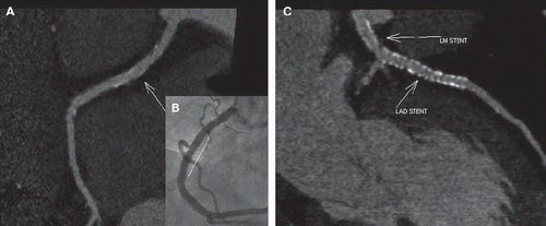
A 59 year old previously healthy male presented with chest pain. Electrocardiogram showed ST elevation over the inferior leads. Emergency coronary angiography showed thrombotic occlusion in mid right coronary artery (RCA), 70% stenosis in proximal left anterior descending (LAD) artery, moderate 60% stenoses in mid left circumflex artery and ostial left main (LM). He successfully underwent primary percutaneous coronary intervention (PCI) of mid RCA () with implantation of a 4.0 × 22 mm TITAN-2 coronary stent. A staged PCI of proximal LAD was successfully performed a month later with implantation of a 3.5 × 33 mm Cypher stent. The ostial LM was found to be obstructive by intravascular ultrasound (IVUS) evaluation and was subsequently stented with a 4.0 × 11 mm Biomatrix stent. He remained well after PCI and underwent a follow-up cardiac computer tomographic angiography (CCTA) assessment at six months. All the implanted coronary stents were widely patent. Nevertheless, the TITAN-2 coronary stent in mid RCA was strikingly more ‘visible’ than the other two stents () in the left coronary system. There was minimal blooming artifact and RCA lumen within the stented segment was clearly seen.
Figure 1. (A) CCTA of RCA showing widely patent mid RCA stent with minimal blooming artifact (site of stent denoted by arrow); image generated with a sharp kernel (B46f, Heartview, Siemens). (B) ICA of mid RCA post stenting (white line denoting site of stent). (C) CCTA of left coronary system showing stents in LM and LAD with some blooming artifact (site of stents denoted by arrow); image generated with a sharp kernel (B46f, Heartview, Siemens).

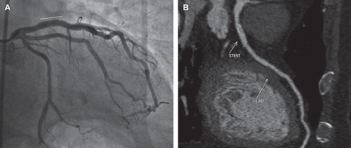
A 78 year old male patient presented with unstable angina. He was a chronic smoker with history of diabetes mellitus and hyperlipidemia. Coronary angiography showed single vessel disease with 65% stenosis in proximal LAD. IVUS evaluation revealed heavy plaque burden with significant narrowing of coronary lumen. PCI of proximal LAD was successfully performed with implantation of a 3.5 × 16 mm TITAN-2 coronary stent (). He presented five months later with atypical chest pain and underwent a CCTA assessment (). The LAD stent was widely patent. Again like the former case, minimal blooming artifact of LAD stent was noted and LAD lumen within the stented segment was easily visualized.
Figure 2. (A) ICA of LAD post stenting (white line denoting site of stent). (B) CCTA of LAD showing widely patent proximal LAD stent with minimal blooming artifact (site of stent denoted by arrow); image generated with a sharp kernel (B46f, Heartview, Siemens).

From the two cases, we demonstrated for the first time the striking feature of TITAN-2 coronary stent of having good visibility on CCTA. Both patients were scanned on a 256-slice dual source scanner (Siemens SOMATOM Definition Flash) using conventional scanning protocol. The TITAN-2 coronary stent (1) (Hexacath, Rueil-Malmaison, France) is a tubular thin-strut (0.07–0.09 mm) balloon expandable stent made of stainless steel and specially coated with a titanium nitride oxide (TNO) compound. Besides completely preventing discharge of nickel, chromium and molybdenum ions, the TNO coating also minimizes inflammatory arterial response to stent implant. These features are thought to enhance endothelialization and thus reduce rate of stent-related restenosis and thrombosis. We attribute the good visibility of TITAN-2 coronary stents demonstrable on CCTA to its unique TNO coating as this particular feature is absent in other coronary stent systems.
PCI remains the preferred method of revascularization for patients with obstructive coronary artery disease and in-stent restenosis (ISR) remains its major Achilles heel. Patients who present with recurrence of chest pain post PCI often have to undergo invasive coronary angiography (ICA) to exclude ISR. A non-invasive detection of ISR will be of clinical importance in the follow-up of patients with prior stent implantation. CCTA is advantageous as it is less invasive than ICA, much quicker, reduces the mental stress and potential complications for patients who require repeat angiography. With the advent of drug-eluting stents (DES), the rate of ISR has been significantly reduced and its clinical use has been extended to more complex lesion subsets in the real world setting. A routine non-invasive follow-up of these patients with a reported ISR rate > 10% would be preferred.
Numerous studies have sought to assess stent patency by CCTA in comparison with ICA in post PCI patients (2–4). Even with newer generation scanners, CCTA has been of limited value in the assessment and follow-up of the revascularized patient. The main reason being blooming artifact from metallic stents often obscures some 30% of stent luminal dimension, making the stented segment unassessable.
From our two cases, we demonstrated that TITAN-2 coronary stents produced minimal imaging artifact on CCTA and the stented segments were easily visible. With clearly visible stents seen on CCTA, this will be potentially beneficial to revascularized patients who could now be evaluated more confidently on follow-up. Clinical trials evaluating new stent platforms can also opt for CCTA evaluation to assess binary restenosis instead of performing routine ICA. In terms of patient's safety, radiation dose from CCTA is becoming lower (<2 millisievert) with newer generation scanners (5), thus making CCTA assessment an attractive option. For our 2 patients, we employed prospective electrocardiograph-triggered scanning and the radiation doses for both patients were 0.85 and 0.4 millisieverts respectively. We believe the unique TNO coating on TITAN-2 coronary stent enhances its visibility on CCTA and this technology could be incorporated in other existing coronary stent systems. Both patients had large stents implanted and whether our observation is replicated in smaller coronary vessels (≤3.0mm) will require further investigation.
Declaration of interest: The authors report no conflicts of interest. The authors alone are responsible for the content and writing of the paper.
References
- Ran Kornowski. The titanium nitride oxide coated stent. Catheter Cardiovasc Interv. 2010;76:288–90.
- Ehara M, Kawai M, Surmely JF, Matsubara T, Terashima M, Tsuchikane E, . Diagnostic accuracy of coronary in-stent restenosis using 64-slice computed tomography. J Am Coll Cardiol. 2007;49:951–9.
- Sheth T, Dodd JD, Hoffmann U, Abbara S, Finn A, Gold HK, . Coronary stent assessability by 64 slice multi-detector computed tomography. Catheter Cardiovasc Interv. 2007;69:933–8.
- Seifarth H, Heindel W, Maintz D. Imaging of coronary stents using multislice computed tomography. Radiologe. 2010;50: 507–13.
- Alkadhi H, Stolzmann P, Desbiolles L, Baumueller S, Goetti R, Plass A, . Low-dose, 128-slice, dual-source CT coronary angiography: Accuracy and radiation dose of the high-pitch and the step-and-shoot mode. Heart 2010;96:933–8.
