Abstract
The present study was designed to determine the effects of ghrelin on in vivo and in vitro secretion of testosterone (T) and the expression of androgen receptor (AR) mRNA in the adult rat testis. The distribution of growth hormone secretagogue receptors (GHS-R1a) in the testis was also investigated. GHS-R1a immunoreactivity presented mainly in Sertoli and Leydig cells, primary spermatocytes, and secondary spermatocytes. Adult rats that were intracerebroventricularly (i.c.v.) administrated different dosages (1 nmol and 3 nmol) of ghrelin could significantly inhibit the secretion of T. The experession of AR mRNA in the testis was also notably reduced with 3 nmol ghrelin. Additionaly, in vitro exposure of the Leydig cells to increasing concentrations of ghrelin resulted in no obvious changes of T secretion in the culture media and AR mRNA expression of Leydig cells. Overall, our data demonstrate that the i.c.v. injection of ghrelin plays a physiological role in T secretion and AR mRNA expression in the testis, further confirming the reproductive role of ghrelin.
Introduction
Ghrelin, a 28-amino-acid peptide with an essential serine 3 n-octanoylation, is the natural ligand of the growth hormone secretagogue receptor (GHS-R), which belongs to a large family of G protein-coupled seven-transmembrane receptors [Kojima et al. Citation1999]. Ghrelin has been demonstrated to be a pleiotropic regulator involved in a large array of endocrine and non-endocrine functions, including food intake and energy balance [Wren et al. Citation2000; Horvath et al. Citation2001]. Moreover, accumulated evidence suggests that ghrelin may participate in the regulation of different aspects of reproductive function. For example, intracerebroventricular (i.c.v.) administration of 3 nmol of ghrelin evokes a significant inhibition of luteinizing hormone (LH) secretion in cyclic female rats throughout their estrous cycle [Fernandez-Fernandez et al. Citation2005]. Ghrelin has been shown to inhibit human chorionic gonadotropin (hCG) and cyclic adenosine monophosphate (cAMP)-stimulated testicular testosterone secretion in rats in vitro [Tena-Sempere et al. Citation2002]. Testosterone (T) is a critical steroid hormone that is essential for spermatogenesis, fertility, and maintenance of the male phenotype, including the outward development of secondary sex characteristics [Wang et al. Citation2009]. Such actions are mediated by the androgen receptor (AR), a member of the nuclear receptor superfamily. The impact of deficient AR in the testis includes spermatogenesis arrest and abnormal fertility [Wang et al. Citation2009; Tsai et al. Citation2006; Xu et al. Citation2007; Chang et al. Citation2004]. Increasing evidence suggests that many factors are involved in the control of AR mRNA expression, including follicle-stimulating hormone (FSH) and estrogen (E2) [Sanborn et al. Citation1991; Pelletier et al. Citation2004]. It is still not known whether ghrelin can regulate testosterone secretion and AR mRNA expression. This is addressed in the following communication.
Results
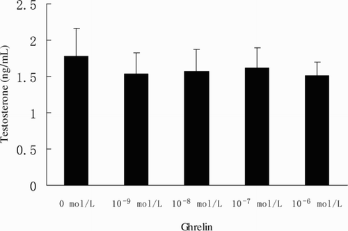
The level of serum LH was lower than that of the control after i.c.v. injection of ghrelin at dosages of 1 and 3 nmol (P < 0.05). Administration of 0.3 nmol ghrelin significantly reduced the serum T concentration (P < 0.05). FSH secretion was also significantly inhibited by 3 nmol ghrelin (P < 0.05) (). Leydig cells were cultured for 24h after the addition of ghrelin. The concentration of T in the culture medium did not change compared to the control (P > 0.05) ().
Figure 1. The effect of ghrelin on LH, FSH, and T. Serum concentrations of LH, FSH, and T in rats through intracerebroventricular administration with ghrelin. *indicates significant difference compared with the dosage of 0 nmol (P < 0.05).
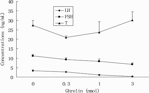
Figure 2. Effect of ghrelin on T secretion by cocultured Leydig cells.
The GHS-R1a protein showed a wide tissue distribution in rat testis, detected mainly in the Sertoli and Leydig cells. Primary spermatocytes and secondary spermatocytes also showed clear-signals, whereas spermatogonia and spermatids were not stained (). As summarized in , the AR mRNA level was significantly inhibited at the dose of 3 nmol (P < 0.05). No significant changes in AR mRNA expression were observed at doses of 0.3 and 1 nmol (P < 0.05) (). The effect of ghrelin on AR mRNA levels was also detected in vitro using primary Leydig cell cultures. Exposure of the Leydig cells to increasing concentrations of ghrelin resulted in no significant change of AR mRNA expression in the media (P > 0.05) ().
Figure 3. Histological observation of rat testis tissue by immunolocalization of GHS-R1a protein. (A) Testis: Leydig cells (arrow a), Sertoli cells (arrow b), primary spermatocytes (arrow c), secondary spermatocytes (arrow d), and spermatogonia (arrow e). (B) Testis: spermatids (arrow f). (C) negative control reaction carried out after substitution of PBS. A, B, C, Scale bar = 25 μm.
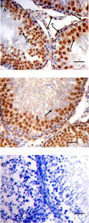
Figure 4. AR mRNA levels as a function of ghrelin. Expression of AR mRNA in the testis after injection of different dosages of ghrelin in rats. *significant differences between the control and injected group (P < 0.05).

Figure 5. Effect of ghrelin on AR mRNA expression by cocultured Leydig cells.

Discussion
Ghrelin has been shown to be highly selective of mature Leydig cells of rat testis [Tena-Sempere et al. Citation2002]. In addition, the expression of the functional ghrelin receptor, GHS-R1a, has been shown in the Sertoli and Leydig cells of the same organ [Gaytan et al. Citation2004]. Immunohistochemistry results showed clear-GHS-R1a immunostaining in primary and secondary spermatocytes. In contrast, negligible staining was observed in spermatogonia and spermatids, suggesting that GHS-R1a is expressed in a stage-dependent manner during spermatogenesis. The above suggests that ghrelin may be involved in the control of some aspects of testicular function. Testosterone secretion and the expression of AR mRNA have been demonstrated to play important roles in spermatogenesis and fertility. Whether or not ghrelin is involved in the modulation of testosterone secretion and the expression of AR mRNA remains to be established.
Ghrelin has been proven to inhibit testicular testosterone secretion by dose-dependent actions in vitro [Tena-Sempere et al. Citation2002]. The present study demonstrates that the i.c.v. injection of ghrelin in rats affects their serum testosterone and AR mRNA levels, while the assessment of the biological actions of ghrelin on T secretion and AR mRNA level by an in vitro model showed that ghrelin could not act directly on Leydig cells to change testosterone secretion and AR mRNA expression.
Interestingly, 0.3 nmol ghrelin significantly inhibited the secretion of testosterone, while 1 nmol–3 nmol ghrelin appears to have no effect. This suggests that ghrelin has an inhibitory action on testosterone release in vivo which is limited to a certain dosage range. The mechanism of the inhibitory effects of ghrelin on testosterone secretion could reflect that the inhibitory effect may be related to the proliferation of Leydig cells. Injection of ghrelin significantly decreases the proliferation activity of differentiating immature Leydig cells. Ghrelin also induces a significant decrease in stem cell factor (SCF) mRNA, a key regulator signal of Leydig cell development [Barreiro et al. Citation2004; Yan et al. Citation2000]. Second, LH may act to decrease testosterone. Testicular LH receptors are selectively expressed in Leydig cells. LH stimulates serum testosterone concentration through its functional receptor. It was observed that 1–3 nmol of ghrelin significantly inhibits LH secretion. Ghrelin may inhibit testosterone secretion by decreasing LH release. Finally, Tena-Sempere et al. [2002] found that ghrelin was significantly decreased hCG-stimulated expression of some mRNAs that encode several key steroidogenic factors, such as steroid acute regulatory protein (StAR), P450 cholesterol side-chain cleavage (P450scc), 3ß-hydroxy steroid dehydrogenase (3ß-HSD), and testis-specific 17ß-hydroxy steroid dehydrogenase (17ß-HSD III) type III. In vitro, the concentration of testosterone also showed no significant difference in all dose groups compared to a control group at 24 h culture (P > 0.05). Our results demonstrate an indirect regulatory action of ghrelin on T secretion.
Although ghrelin has been reported to regulate testosterone secretion, information regarding the control of gene expression at the mRNA level remains incomplete. Our present results demonstrate that 3 nmol ghrelin could significantly inhibit AR mRNA expression whereas 0.3–1 nmol does not. In vitro, ghrelin does not change the amount of AR mRNA when co-cultured with Leydig cells. These observations suggest that ghrelin has an inhibitory action on AR mRNA expression that is indirectly mediated by Leydig cells. Germ cell development within the mammalian testis requires testosterone stimulation of Leydig cells via interaction with intracellular AR [Xu et al. Citation2007]. AR mRNA expression levels undergo marked changes after injection of 3 nmol of ghrelin, suggesting that the modulation of AR mRNA expression is an important mechanism for germ cell development. Adult rats treated with ethane dimethane sulphonate to eliminate the presence of Leydig cells showed very low levels of testosterone in their blood and testis; the AR mRNA levels of these rats were unchanged [Blok et al. Citation1992], indicating that the inhibitory action of ghrelin on AR mRNA expression must take place in a step beyond testosterone secretion, although ghrelin could also significantly inhibit the secretion of testosterone. In past studies, FSH has been reported to increase AR mRNA in Sertoli cells [Sanborn et al. Citation1991]. An i.c.v. injection of 3 nmol ghrelin significantly inhibits the secretion of FSH. Therefore, the inhibition may be related to FSH. To date, the molecular mechanisms governing the inhibitory action of ghrelin on AR mRNA expression is not fully understood. In vitro, the expression of AR mRNA remains unaffected after exposure to increasing concentrations of ghrelin. This provided novel evidence that ghrelin regulated AR mRNA levels through other pathway and Leydig cells were not the primary target of ghrelin.
In summary, gonads are complex endocrine organs. Although tests of its effects via acute administration have suggested a possible role for ghrelin in the control of testosterone secretion and expression in AR mRNA, the mechanisms of such actions remain unclear and further research is necessary to identify the precise functional role of ghrelin.
Materials and methods
Animals and Sampling
Sprague Dawley rats (180–240 g) were purchased from Experimental Animal Center of Anhui Medical University. The rats were kept (6 per cage) under controlled conditions of light (12 h light, 12 h darkness, light at 07:00 h) and temperature (22oC), relative humidity of 50–60% and with free access to pelleted food (Experimental Animal Center of Anhui Medical University, Hefei, China) and tap water. Each experimental group consisted of eight animals. The study was approved by the Animal Care and Use Committee of Anhui Agricultural University. Ghrelin was obtained from Sigma (USA) and dissolved in saline solution immediately before use. The rats were intracerebroventricularly injected with 2 μL ghrelin at different doses (0.3 nmol, 1 nmol, and 3 nmol ), that control animals got saline only and the injection volume was 2 μL. All animals were sacrificed by decapitation 15 min after injection with ghrelin. The trunk blood of the animals was collected into centrifuge tubes without heparin and centrifuged at 1,600g at 4oC for 20 min, while their testis was rapidly dissected out and then stored at −80oC. Serum was collected and stored at −20oC. Other testes samples were taken for the immunohistochemical analysis of GHS-R1a peptide expression.
Isolation and purification of Leydig cells
Rats were humanely sacrificed by decapitation, and testes were collected. The procedure for the isolation of Leydig cells is described below. In brief, each testis was removed and placed in dissociation buffer (DMEM/F-12 with 2.5 g/L HEPES, 0% BSA, 100 U/mL penicillin, 100 μg/mL stmycin) at 4oC. Testes were decapsulated, and dissociation was continued at 34oC for 20 min in a lower concentration (0.25 mg/mL) of collagenase, with low speed shaking (80 cycles/min). Seminiferous tubules were removed by filtration through a 100-µm pore size nylon mesh. The dissociated cells in the filtrate were subjected to centrifugal elutriation for 10 min at 2,000 x g. The fraction enriched with Leydig cells was collected and centrifuged at 2,000 x g at 18oC for 30 min through a 50% percoll gradient formed in situ. Leydig cells with a density of 1.078 g/L were harvested. Cells were subsequently plated in 24-well tissue culture plates at a concentration of 1 × 10⁵ cells/well, respectively, and were incubated without or with increasing amounts of ghrelin (10−9 to 10−6 mol/L) at 37oC in a humidified atmosphere of 5%CO2/95%O2 for 24 h, then culture media was gathered for T determination and Leydig cells was collected for subsequent RNA extraction.
GHS-R1a Immunohistochemistry in Rat Testis
Detection of GHS-R1a protein was carried out in 4% paraformaldehyde fixed sections. Sections of 5 µm were cut and mounted on polylysine coated glass slides and dried overnight at 42oC. After dewaxing and rehydrating the samples and antigen retrieval in a microwave oven by treating the sample 3 times for 5 min at 700 W, endogenous peroxidase was inhibited by incubation in a 3% hydrogen peroxide solution for 10 min. After washing in phosphate buffer saline (PBS), sections were blocked with normal goat serum (Golden Bridge International, Beijing, China) and incubated overnight with anti-GHS-R1a antibody (Acris, Herford, Germany) at a 1:40 dilution. The sections were then processed according to PV-9000 two–step method detection kit instructions (Golden Bridge International), treated with polymer helper for 20 min at 37oC, washed in PBS 3 times for 5 min each time, and incubated in polyperoxidase-anti-Rabbit IgG (Golden Bridge International) for 30 min at 37oC. Antibody binding was visualized by a DAB (Golden Bridge International) detection system, with sections counterstained with hematoxylin. In all reactions, negative controls were run in parallel by omitting the primary anti-GHS-R1a antibody (substituted by PBS) to demonstrate the absence of specific immunoreactivity.
LH, FSH, and T determination
LH and FSH concentrations in the serum were measured using a double-antibody method (ELISA kits were purchased from Shanghai Westang Biological Engineering Company, China). The mean intra- and inter-assay coefficients of variation were 9.7 % for LH and 10 % for FSH, the assay sensitivity was 0.8 ng/mL for LH and 1 ng/mL for FSH, respectively. SerumT concentrations were measured using a commercially available radioimmunoassay (Furui Biological Engineering Company, Beijing, China). The mean intra- and inter-assay coefficients of variation were 9.5 % and 10.3 % for T, respectively. The assay sensitivity was 0.002 ng/mL for T.
Total RNA Isolation and Reverse Transcription
Total RNA was extracted from testis using Trizol reagent (TransGen, Beijing, China) according to the manufacturer's protocol. The quality of total RNA was assessed by formaldehyde gel electrophoresis. Samples that showed good RNA quality were selected for further reverse transcription. Reverse transcription was performed using the TransScript First-Strand cDNA Synthesis SuperMix (TransGen) according to the manufacturer's protocol. Approximately 2 μL of total RNA was used for reverse transcription.
Real-time PCR
Forward and reverse primers to detect the AR target genes and β-actin as the housekeeping internal control gene yielding 247 and 208 bp products were AGGCAGGAGCCACAACACG (AR forward) plus ATTTGGAAACCCTAATACCC (AR reverse) and CAGTGGTTGACCCTGCTA (actin forward) plus TGTTGTCCCTGTATGCCT (actin reverse). Complete target gene sequences are available at http://www.ncbi.nlm.nih.gov (GenBank Accession No. M20133.1 and AF 122902.1, respectively). All primers were designed using Primer Express 5.0 and allowed the amplification of regions that span introns. Real-time PCR was performed using SYBR Premix Ex TaqTM Kits (TaKaRa, Dalian, China) and a Rotor-Gene 6000 instrument (Corbett Life Science, Australia). Each reaction well was loaded with 2.0 μL (10 pmol/μL) of the forward and reverse primers, 12.5 μL of SYBR Premix Ex TaqTM, 2 μL of cDNA and 8.5 μL of double-distilled water. The final reaction volume was 25 μL. All samples were run in triplicate. The following PCR conditions were used: preliminary denaturation at 95oC for 30 s, followed by 40 cycles with a temperature profile of 5 s at 95oC, 20 s at 55oC, and 15 s at 72oC. Melting curves were also determined. Quantitative analysis was performed using the relative standard curve method as described in ABI User Bulletin #2. Serial dilutions of a known amount of cDNA sample prepared from testis total RNA were used to construct the standard curves for AR and for β-actin amplifications. The efficiency was 1.05 for AR, 1.04 for β-actin, and the absolute slope values of two standard curves were 3.214 and 3.237, indicating similar amplification efficiency for both AR and β-actin. For each unknown sample, the relative amount is calculated using linear regression analysis from their respective standard curves. A relative AR expression value was then obtained by dividing the value for the gene of interest (AR) by the value for the β-actin.
Statistical Analysis
Results are expressed as mean ±SD. Differences in the serum T, LH, FSH, and AR mRNA relative quantities were carried out using one-way repeated-measures analysis of variance. The level of P < 0.05 was considered statistically significant.
Abbreviations
| T: | = | testosterone |
| AR: | = | androgen receptor |
| GHS-R1a: | = | growth hormone secretagogue receptors |
| i.c.v.: | = | intracerebroventricularly |
| GHS-R: | = | growth hormone secretagogue receptor |
| LH: | = | luteinizing hormone |
| hCG: | = | human chorionic gonadotropin |
| cAMP: | = | cyclic adenosine monophosphate |
| FSH: | = | follicle-stimulating hormone |
| E2: | = | estrogen |
| SCF: | = | stem cell factor |
| StAR: | = | steroid acute regulatory protein |
| P450scc: | = | P450 cholesterol side-chain cleavage |
| 3ß-HSD: | = | 3ß-hydroxy steroid dehydrogenase |
| 17ß-HSD(III): | = | 17ß-hydroxy steroid dehydrogenase type III. |
Acknowledgments
We acknowledge support from the Anhui Provincial Natural Science Foundation (Grant No. 070411015) from the Anhui Science and Technology Department (Anhui, P. R. China).
Declaration of interest: The authors report no conflicts of interest. The authors alone are responsible for the content and writing of the paper.
References
- Barreiro, M.L., Gaytan, F., Castellano, J.M., Suominen, J.S., Roa, J., Gaytan, M., (2004) Ghrelin inhibits the proliferative activity of immature Leydig cells in vivo and regulates stem cell factor messenger ribonucleic acid expression in rat testis. Endocrinology 145:4825–4834.
- Blok, L.J., Bartlett, J.M., Bolt-De Vries. J., Themmen, A.P., Brinkmann, A.O., Weinbauer, G.F., (1992) Effect of testosterone deprivation on expression of the androgen receptor in rat prostate, epididymis and testis. Int J Androl 15:182–198.
- Chang, C., Chen, Y.T., Yeh, S.D., Xu, Q., Wang, R.S., Guillou, F., (2004) Infertility with defective spermatogenesis and hypotestosteronemia in male mice lacking the androgen receptor in Sertoli cells. Proc Natl Acad Sci USA 101:6876–6881.
- Fernandez-Fernandez, R., Tena-Semepere, M., Victor, M., Navarro, M. L., Barreiro, J. M., Castellano, A.E., Pinilla, L. (2005) Effects of ghrelin upon gonadotropin-releasing hormone and gonadotropin secretion in adult female rats; in vivo and in vitro studies. Neuroendocrinology 82:245–255.
- Gaytan, F., Barreiro, M.L., Caminos, J.E., Chopin, L.K., Herington, A.C., Morales, C., (2004) Expression of ghrelin and its functional receptor, the type 1a growth hormone secretagogue receptor, in normal human testis and testicular tumors. Clin Endocrinol Metab 89:400–409.
- Pelletier, G., Luu-The, V., Li, S., Labrie, F. (2004) Localization and estrogenic regulation of androgen receptor mRNA expression in the mouse uterus and vagina. J Endocrinol 180:77–85.
- Horvath, T.L., Diano, S., Sotonyi, P., Heiman, M., Tschop, M. (2001) Ghrelin and the regulation of energy balance-A hypothalamic perspective. Endocrinology 142:4163–4169.
- Kojima, M., Hosoda, H., Date, Y., Nalazato, M., Matsuo, H., Kangawa, K. (1999) Ghrelin is a growth-hormone-releasing acylated peptide from Stomach. Nature 402:656–660.
- Sanborn, B.M., Caston, L.A., Chang, C., Liao, S., Speller, R., Porter, L.D., Ku, C.Y. (1991) Regulation of androgen receptor mRNA in rat Sertoli and peritubular cells. Biol Reprod 45:634–641.
- Tena-Sempere, M., Barreiro, M.L., Gonzalez, L.C., Gaytan, F., Zhang, F.P., Caminos, J.E., (2002) Novel expression and functional role of ghrelin in rat testis. Endocrinology 143:717–725.
- Tsai, M.Y., Yeh, S.D., Wang, R.S., Yeh, S., Zhang, C., Lin, H.Y., (2006) Differential effects of spermatogenesis and fertility in mice lacking androgen receptor in individual testis cells. Proc Natl Acad Sci USA 103:18975–18980.
- Wang, R.S., Yeh, S., Tzeng, C.R., Chang, C. (2009) Androgen receptor roles in spermatogenesis and fertility: lessons from testicular cell-specific androgen receptor knockout mice. Endocr Rev 30:119–132.
- Wren, A.M., Small, C.J., Ward, H.L., Murphy, K.G., Dakin, C.L., Taheri, S., (2000) The novel hypothalamic peptide ghrelin stimulates food intake and growth hormone secretion. Endocrinology 141:4325–4328.
- Xu, Q., Lin, H.Y., Yeh, S.D., Yu, I.C., Wang, R.S., Chen, Y.T., (2007) Infertility with defective spermatogenesis and steroidogenesis in male mice lacking androgen receptor in Leydig cells. Endocrine 32:96–106.
- Yan, W., Kero, J., Huhtaniemi, I., Toppari, J. (2000) Stem cell factor functions as a survival factor for mature Leydig cells and a growth factor for precursor Leydig cells after ethylene dimethane sulfonate treatment: implication of a role of the stem cell factor/c-Kit system in Leydig cell development. Development Biology 227:169–182.