Abstract
In recent years concern has arisen whether carrying a cellular phone near the reproductive organs such as the testes may cause dysfunction and particularly decrease in sperm development and production, and thus fertility in men. The present study was performed to investigate the effects of a 1.95 GHz electromagnetic field on testicular function in male Sprague-Dawley rats. Five week old animals were divided into 3 groups of 24 each and a 1.95-GHz wide-band code division multiple access (W-CDMA) signal, which is used for the freedom of mobile multimedia access (FOMA), was employed for whole body exposure for 5 hours per day, 7 days a week for 5 weeks (the period from the age of 5 to 10 weeks, corresponding to reproductive maturation in the rat). Whole-body average specific absorption rates (SAR) for individuals were designed to be 0.4 and 0.08 W/kg respectively. The control group received sham exposure. There were no differences in body weight gain or weights of the testis, epididymis, seminal vesicles, and prostate among the groups. The number of sperm in the testis and epididymis were not decreased in the electromagnetic field (EMF) exposed groups, and, in fact, the testicular sperm count was significantly increased with the 0.4 SAR. Abnormalities of sperm motility or morphology and the histological appearance of seminiferous tubules, including the stage of the spermatogenic cycle, were not observed. Thus, under the present exposure conditions, no testicular toxicity was evident.
Keywords:
Introduction
Although recent rapid advances in EMF technology and communication are contributing to social and economic benefits, potential effects of EMFs on our health have become a great concern. Possible associations between EMF exposure and brain dysfunction, including tumor development in adulthood and childhood, have been a major focus of attention. However, with a wide spread of usage of cellular phones and the diversification of life style, increased variation in exposure type to EMF from cellular phones means that other sites need to be taken into account. For example, in addition to the exposure toward the side of the head during actual talking on a phone, exposure to the chest, abdomen, and testis while carrying cellular phones might have some influence. Many men keep their cell phones in trouser pockets or hung from their trouser belt. Under such circumstances, testicles may be exposed to prolonged weak EMF emitted from a phone and several studies have suggested that damage to the testis may occur, resulting in reduced fertility potential of men [Davoudi et al. Citation2002; Fejes et al. Citation2005; Agarwal et al. ; Citation2009 Citation2008]. Experimental data, however, are inconsistent. Yan et al. [2007] reported that 3-h periods of daily cellular phone emission for 18 weeks caused a significant increase in sperm cell death and abnormal clumping of sperm cells. In contrast, there were no such averse effects of microwaves emitted from cellular phone in the animal experiments carried out by Dasdag et al. [2003; 2008], Lee et al. [2006], and Wang et al. [2008b]. Thus, although concern over the effects on male fertility has been growing, no conclusive data are available.
The present study was therefore performed to assess effects of whole body exposure to 1.95GHz for 5 weeks on rat testicles. Since setting devices for small animals which would correspond to men carrying a cellular phone on their trouser belt is not feasible, we selected whole body exposure designed to provide a high SAR to the testicles.
Results
No abnormalities of condition, including animal death, were noted during either the exposure or non-exposure periods. There were no differences in body weight gain and food consumption among the groups (data not shown) and final body weights of the 3 groups were very similar (). Macroscopical lesions were noted in the kidney (renal cysts) in one rat each of the control and 0.08 W/kg groups. There were no statistical significant differences in the absolute and relative weights of the testes, epididymis, seminal vesicles, and prostate (). summarizes data for the sperm count, motility, and morphological abnormalities. No significant differences were observed among these values except for the sperm count in the testes, which was significantly greater in the high level exposure group than with the sham exposure.
Figure 1. Staging of seminiferous tubules. The entire stages categorized from I to XIV, were classified into 4 groups; I ∼ VI, VII ∼ VIII, IX ∼ XI, and XII ∼ XIV. The data represent the mean values with SD. Statistical significance were not shown using Dunnett test in all stages. P values of each stage were: Stage I-VI 0 vs. 0.08: 0.500, 0 vs. 0.4: 0.5305; Stage VII-VIII 0 vs. 0.08: 0.3159, 0 vs. 0.4: 0.3693; Stage IX-XI 0 vs. 0.08: 0.2790, 0 vs. 0.4: 0.1980; and Stage XII-XIV 0 vs. 0.08: 0.4720, 0 vs. 0.4: 0.5052.
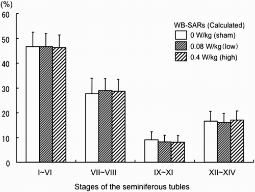
Figure 2. Exposure setup. A) Gross appearance of the inside of an exposure chamber with four animal cages; B) Schematic illustration of the exposure box.
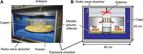
Table 1. Final body and reproductive organ weights.
Table 2. The count, mobility, and morphological abnormality of the sperm.
The seminiferous tubules of rodents show spermatogenic cycles along the length of the epithelium. In the rat, the cycle can be divided into 14 stages (I-XIV) with a unique and fixed composition of spermatogenic cells characterizing each [Leblond and Clermont Citation1952]. However, such stages are not static and with the passage of time, any given stage progresses into the next as the spermatogenic cells mature. As an easy and convenient method to evaluate any damage in the seminiferous tubules as a part of testicular toxicity, Takahashi and Matsui [1993] proposed a 4 group classification instead of the 14 stages using at least H & E staining: these respectively include Stage I-VI, VII-VIII, IX-XI, and XII-XIV as shown in . Histological details of the 4 groups were described in their report [Takahashi and Matsui Citation1993]. Histopathological evaluation of the seminiferous epithelial cycle revealed that the percentage of each group among the exposures was similar, indicating no clear shift induced by EMF (). Data for histological lesions in the testis, epididymis, prostate, and seminal vesicles are summarized in . There were no significant lesions found except multiple nucleated cells in the seminiferous tubules of one low level group animal, and slight lymphocyte infiltration in the prostate in several rats in each group.
Figure 3. FDTD models of the exposure chamber with rats.
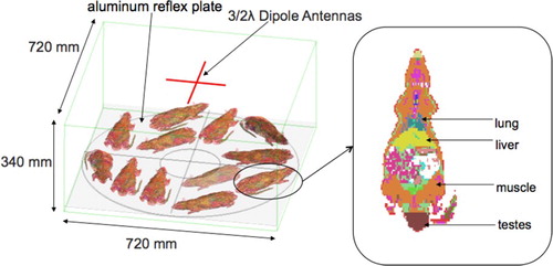
Table 3. Histopathological findings of reproductive organs.
Discussion
The present study clearly demonstrated no obvious testicular toxicities including alteration in the sperm count and motility or abnormal appearance of sperm even with EMF exposure at low and high SARs during the development period. Although a statistically significant increase in the sperm count in the testis was observed in the high level exposure group, this is considered to be not significant, because the difference was slight and no difference was evident either in the sperm count in the epididymis or in the sperm motility. From the computational dosimetry estimation of the SARs, the testis SAR (1g-average) for low-level EMF exposure condition was about 0.2 W/kg (0.19 W/kg ± 0.09) during long-term experimentation. The ratio of the testis SAR to the WB-SAR was as high as three. As a result, with the high-level EMF exposure, the testicular SAR (1g-average) was about 1.0 W/kg (0.96 W/kg ± 0.45). This value was only approximately 37 percent lower than the spatial peak SAR value over any 1g tissue for maximum permissible exposure in uncontrolled environment recommended in the ANSI/IEEE C95.1 -1991. Under such extreme EMF exposure conditions, no obvious testicular toxicity or sperm deficiency were evident.
Testicular dysfunction, particularly defective spermatogenesis is thought to be associated with many factors in our environment and dependent on our lifestyle [Agarwal et al. Citation2008; Sharpe Citation2000]. It is well-known that the lower temperature of testicles than the core body value is a fundamental condition to maintain normal testicular function. Conditions that increase the temperature of the testicles can thus affect spermatogenesis, these include a hot occupational environment, tight clothing, hot bathing, and a long sitting position at work and traveling in a train or car, as with occupational automobile driving. Steroid hormones and artificial and natural hormone-like chemical substances are also reported to be influencing factors, as well as smoking and alcohol consumption. Ionizing radiation is a good example of an agent causing deterioration in spermatogenesis. Thus many factors in our regular life possess the potential to disturb testicular function.
Agarwal et al. [2008] reviewed effects of cellular phone usage on male fertility, postulating reduced sperm motility in men using a cell phone more than 4 h/d compared to men not using at all, citing a significant rate of infertility among military men exposed to radiofrequency-electromagnetic waves (RF-EMW). However, types of exposure must be considered. There must be differences in the daily lifestyle between heavy users and non-users of cellular phones and between military workers near and distant from RF-EMW fields. In epidemiological studies to clarify RF adverse effects on male reproductive organs, many factors that can bias the final results must be carefully excluded. Quite recently, Agarwal et al. [2009] again reported a prospective pilot study in which RF-EMW radiation of semen samples collected from normal healthy men and infertile patients induced a significant decrease in sperm mortality and viability, increase in reactive oxygen species (ROS) levels, and decrease in ROS-total antioxidant capacity. They speculated that keeping the cell phone in a trouser pocket in the talk mode may exert adverse effects on testicular function. However, we must be careful to evaluate in vitro results because exposure is direct to sperm that are specifically developed cells isolated from the testes without the characteristically well-organized testicle-microenvironment. Sharpe [2000] emphasized that contributions of lifestyle and environmental factors to male infertility cannot be ignored but there are so many uncertainties. Infertility is not equal to a decrease in the amount and/or motility of sperm. The semen necessary for normal male fertility is generally considered to be a volume of 2-5 ml, with more than 20 million sperm per ml, more than 50% of which are progressively motile, more than 30% have a normal appearance, and with less than 1 million white blood cells/ml. Thus, ranges of sperm quality for normal fertility are very wide.
Yan et al. [2007] reported that two - three-h periods of daily cellular phone emissions for 18 weeks resulted in a significantly higher sperm cell death incidence and presence of abnormal clumping of sperm cells. However, several aspects require clarification. For example, exposure conditions of four types of cellular phones were used set beneath the rat holder with different modes and wide power ranges, and the data were combined; this may not necessarily provide the optimal analysis path.
In contrast, Dasdag et al. [2008] obtained evidence that radiation for 2 h/d, 7 days a week, for 10 months did not cause any apoptosis in spermatogenic cells in the testes of exposed rats. Similar negative results were obtained in the 24 month long-term whole body exposure in vivo study of Takahashi et al. [2010], the 18 month long-term exposure study by Lee et al. [2006], the 2 month study of Akdag et al. [2006], and a series of one-month studies [Dasdag et al. Citation2003; Ozguner et al. Citation2005]
Thus, the present data are in line with previously reported animal experimental results demonstrating no adverse effects of EMF on the testes [Dasdag et al. Citation2008; Citation2003; Lee,et al. 2006; Wang et al. Citation2008b].
Materials and methods
Animals
A total of 84 Crl:CD(SD) 4-w-old rats were purchased at 2 time points from Charles River Japan (Shiga, Japan). All animals were allowed a 6-d quarantine and acclimation period after each time of purchase. After confirmation of normal health status, they were subjected to experimentation at 5 weeks of age. Excess animals were eliminated. The experiment was performed in duplicate exactly in the same manner. For each, the rats were divided into 3 groups of 12 rats each. Group 1 received sham exposure, and Groups 2 and 3 were exposed to EMF at low and high levels, respectively. All rats were individually set in four translucent acrylic cages in a total of 9 exposure boxes, three boxes for each group. Individual recognition of animals was facilitated by the ear punch method.
Animals were housed in plastic cages, three per cage with W260 x L412 x H195 mm, with wood-chip bedding in an air-conditioned animal room maintained on a 12 h light/dark cycle at 22 ± 2°C and 55 ± 10% humidity and were allowed free access to irradiated (6.0kGy) pelleted diet (MF, Oriental Yeast Co., Ltd., Tokyo Japan) and drinking water. For the exposure, all animals were individually transferred to cages in the exposure chambers including wood-chip bedding. The cages in the exposure chambers were also under the environment with a 12 h light/dark cycle at 22 ± 2°C and 55 ± 10% humidity and rats were again allowed free access to the pelleted diet (MF, Oriental Yeast Co., Ltd., Tokyo, Japan) and drinking water.
Exposure apparatus for EMF
The exposure chambers used for earlier experiments with 1.95-GHz EMF [Ogawa et al. Citation2009; Shirai et al. Citation2007] were modified for the present whole-body exposure study. They were originally developed for a toxicity study of RF exposure to 2 GHz-band W-CDMA cellular base station using pregnant rats [Takahashi et al. Citation2010]. For the present experiment, a metallic plate reflector was set at the bottom of each exposure chamber in order to increase EMF exposure to the front (ventral) side of the body where the testes of rats are located. The insides of the exposure boxes (dimensions: 90 x 90 x 40 cm outside, 70 x 70 x 34 cm inside), except the ceilings, were covered with a 6-cm-thick planar RF absorber. The ceiling of the exposure chamber was made of metal mesh to prevent leakage of radiowaves to the outside and covered with translucent acryl plates (4.2 mm thickness) allowing light from fluorescent light tubes on the ceiling to shine inside. A turntable in the exposure chamber was covered with a 2 mm thick reflex plate made of aluminum, with which EMF exposure to the front side including the testes was increased. Four translucent acrylic cages in a 90 degree fan-shape were set on the floor of each box. Each cage had a translucent acryl removal cover and punctured square or slit-like holes were set on the cover and side for air ventilation. Each cage had a 200 mm depth x 190 mm height with 690.8 cm2 of floor space. A ventilation hole was provided at the center of the exposure chamber through which electric fan-forced ventilation air was introduced. A plastic water bottle was set at the center side of each cage. Additional detail of the exposure apparatus including the acrylic cages is available in our previous report [Takahashi et al. Citation2010]. For whole-body exposure to EMF, two crossing 3/2-wavelength dipole antennas (180 mm long) covered with a ABS resin cap for 1.95 GHz were set horizontally with a spacing of 40 mm from the ceiling of the exposure box in the center. The two antennas intersected at a right angle had a phase difference of 90 degrees, which realized a circular polarization in the exposure chamber. The employment of a circularly polarized antenna was to provide a constant exposure with respect to unrestrained rats. The antenna input power was maintained at a constant level during the long-term exposure period.
EMF dosimetry of the testes
The specific absorption rates (SARs) for rat testes (1g-averaged) were calculated by simulation using the finite difference time domain (FDTD) method. In order to confirm that the EMF exposure level for each rat did not exceed the designed condition, whole-body average SARs (WB-SARs) of the rats were also calculated since rats had various different locations and orientations in the cages. The rat phantom model obtained from the Brooks Air Force Database [Mason et al. Citation1995] was used for the numerical simulations. The SAR estimations involving uncertainties due to variety in the position and age-related changes of the rats in the exposure equipment were achieved with statistical analysis. In our experiment, sham, low (WB-SAR: 0.08 W/kg), and high level of EMF (WB-SAR: 0.4 W/kg) exposure conditions were employed. With this dosimetry estimation, the low EMF exposure condition was adopted. Then, an antenna input power of 1 W was selected. The high level exposure was set to be five times greater.
Computation model and parameters
The exposure equipment used in the in vivo experiment is illustrated in [Wake et al. Citation2007; Wang et al. Citation2008a]. We confirmed that, by adopting aluminum plates on the turntables in the chambers, the exposure level to the ventral body including testes was increased between 1.5 -2 times. The model used for the calculation is shown in and the computation parameters were frequency: 1.95 GHz (continuous wave), cell size: 1.05 mm–2.0 mm (non-uniform mesh), number of FDTD-grid cells: 45 Mcells, absorbing boundary condition: uniaxial perfectly matched layer (10-layers), repetition period: 100 periods, and antenna model: 3/2 dipole antenna. In the simulation, 12 rat models that had different locations and orientations in the exposure chamber were examined in order to consider the uncertainty of the estimated SAR due to the rats' positions inside the cage. The numerical rat model consisted of 31 different tissues and had a spatial resolution of 1.05 x 1.05 x 2 mm3. The modeling of the exposure box with rats was performed with a non-uniform mesh that allows flexible spatial resolution to improve the accuracy of modeling. The resolution chosen proved to be sufficient and adequate for the calculation of 2 GHz SAR distributions inside the rat body. FDTD based commercial software, SEMCAD X (SPEAG, Switzerland) was used for the dosimetry analysis. The dielectric properties of tissues of the inhomogeneous numerical rat phantom were based on measured data [Gabriel Citation2006]. For SAR estimation in the long-term in-vivo experiment, body size and weight of the model are essential parameters. Based on the body size of each age of rat that was obtained by means of measurement of actual rats, 6 rat models simulating ages of 5, 6, 7, 8, 9, and 10 weeks were made with linear shrinkage and enlargement of the original anatomical rat phantom model. Additionally, in order to consider the uncertainty of the estimated SAR due to combinations of rats' positions inside the cages, 22 pattern analyses were conducted for rats of each age. Then, 1,584 rat models for different ages, locations, and orientations were used for statistical analysis of SARs.
Animal handling and termination of the experiment
Rats were examined carefully twice a day throughout the experiment for symptoms and mortality. Body weights and food consumption were measured every day until the termination. At the end of the experiment, animals were sacrificed and blood was collected from the aorta. All organs were examined macroscopically and organ weights were also recorded. The testes were fixed with glutaldehyde formalin acetic acid and others with 10% formalin and routinely processed for embedding in paraffin and sectioning for hematoxylin and eosin staining.
All organs were histopathologically examined. PAS staining was applied to paraffin sections of the left testes for analysis of seminiferous staging, histologically classified into four groups (I ∼ VI, VII ∼ VIII, IX ∼ XI, and XII ∼ XIV). One hundred seminiferous tubules were classified in each slide [Takahashi and Matsui Citation1993]. For sperm examination, the right caudal epididymis was cut with the tip of a pair of surgical scissors and a small amount of the sperm fluid leaking out was collected and introduced into medium (M199, Invitrogen Corp., USA) to diffuse the sperm at 37 °C for 5 min. Then more than 100 sperm were assessed for motility and abnormalities of morphology. Furthermore, the right testis and epididymis were homogenized (Politoron-TP20, POLITRON) for 1 min in 10 ml water and ultrasonicated (Sine Sonic 100, Bational Electronic Industry). Counts of sperm heads were made in 1 ml chambers (Kova Glasstic Slide 10 with grids, Hycor Biomedical Inc.), following the method of Meistrich [1989] .
Data analysis methods
Statistical comparisons between group 1 (sham exposure) and exposed groups (groups 2 and 3) for average data (body weight, organ weight, and number of sperm) were performed using Bartlett's test. If data were homogeneous, one-way analysis of variance was used. When significant differences were observed, Dunnett's multiple parametric comparison test was applied. When uniformity was not confirmed, non-parametric Steel method was applied instead. The significance of inter-group differences in incidences on gross pathological examination and/or histopathological observation was also assessed using the Fisher's exact test (one sided), and of the grade using the Wilcoxon Rank sum test (two sided). In all cases, significance was concluded at p < 0.05.
Animal care and protocols
The experimental design was approved by the Institutional Animal Care and Use Committee of Nagoya City University School of Medical Sciences and also by an intra-institutional committee. The animal facilities at the DIMS Institute Medical Science, Inc. where the tests were conducted are fully accredited as compliant with the GLP Standards by the Ministry of Health and Welfare of Japan, the Organization for Pharmaceutical Safety and Research, and the Ministry of Agriculture, Forestry and Fisheries of Japan.
Abbreviations
| W-CDMA: | = | wide-band code division multiple access |
| EMF: | = | electromagnetic field |
| SARs: | = | specific absorption rates |
| FOMA: | = | the freedom of mobile multimedia access |
| FDTD: | = | the finite difference time domain |
| WB-SARs: | = | whole-body average SARs |
| RF-EMW: | = | radiofrequency-electromagnetic waves |
| ROS: | = | reactive oxygen species. |
Declaration of Interest: This study was performed with support from the Association of Radio Industries and Businesses (ARIB), Japan. The authors report no conflicts of interest. The authors alone are responsible for the content and writing of the paper.
References
- Agarwal, A., Desai, N.R., Makker, K., Varghese, A., Mouradi, R., Sabanegh E., (2009) Effects of radiofrequency electromagnetic waves (RF-EMW) from cellular phones on human ejaculated semen: an in vitro pilot study. Fertil Steril 92:1318–1325.
- Agarwal, A., Desai, N.R., Ruffoli, R., and Carpi, A. (2008) Lifestyle and testicular dysfunction: a brief update. Biomed Pharmacother 62:550–553.
- Akdag, M.Z., Dasdag, S., Aksen, F., Isik, B., and Yilmaz, F. (2006) Effect of ELF magnetic fields on lipid peroxidation, sperm count, p53, and trace elements. Med Sci Monit 12:BR366–371.
- Dasdag, S., Akdag, M.Z., Ulukaya, E., Uzunlar, A.K., and Yegin, D. (2008) Mobile phone exposure does not induce apoptosis on spermatogenesis in rats. Arch Med Res 39:40–44.
- Dasdag, S., Zulkuf Akdag, M., Aksen, F., Yilmaz, F., Bashan, M., Mutlu Dasdag, M., (2003) Whole body exposure of rats to microwaves emitted from a cell phone does not affect the testes. Bioelectromagnetics 24:182–188.
- Davoudi, M., Brossner, C., and Kuber, W. (2002) The influence of electromagnetic waves on sperm motility. Urol Urogynacol 19:18–22.
- Fejes, I., Zavaczki, Z., Szollosi, J., Koloszar, S., Daru, J., Kovacs, L., (2005) Is there a relationship between cell phone use and semen quality? Archives of Andrology 51(5):385–393.
- Gabriel, C. and Peyman, A. (2006) Dielectric measurement: error analysis and assessment of uncertainty. Phys Med Biol 51:6033–6046.
- Leblond, C.P., and Clermont, Y. (1952) Definition of the stages of the cycle of the seminiferous eiptheliumin the rat. Ann. NY. Acad Sci 55:548–573.
- Lee, H.J., Kim, S.H., Choi, S.Y., Gimm, Y.M., Pack, J.K., Choi, H.D., (2006) Long-term exposure of Sprague Dawley rats to 20 kHz triangular magnetic fields. Int J Radiat Biol 82:285–291.
- Mason, P.A., Walters, T.J., Fanton, J.W., Erwin, D.N., Gao, J.-H., Roby, J.W., (1995) Database created from magnetic resonance images of a Sprague–Dawley rat, rhesus monkey, and pigmy goat. Fed. Amer. Soc. Exper. Biol. J 9:434–440.
- Meistrich, M.L. (1989) Evaluation of reproductive toxicity by testicular sperm head count. Int J Toxicol 8:551–567.
- Ogawa, K., Nabae, K., Wang, J., Wake, K., Watanabe, S., Kawabe, M., (2009) Effects of gestational exposure to 1.95-GHz W-CDMA signals for IMT-2000 cellular phones: Lack of embryotoxicity and teratogenicity in rats. Bioelectromagnetics 30:205–212.
- Ozguner, M., Koyu, A., Cesur, G., Ural, M., Ozguner, F., Gokcimen, A., (2005) Biological and morphological effects on the reproductive organ of rats after exposure to electromagnetic field. Saudi Med J 26:405–410.
- Sharpe, R.M. (2000) Lifestyle and environmental contribution to male infertility. Br Med Bull 56:630–642.
- Shirai, T., Ichihara, T., Wake, K., Watanabe, S., Yamanaka, Y., Kawabe, M., (2007) Lack of promoting effects of chronic exposure to 1.95-GHz W-CDMA signals for IMT-2000 cellular system on development of N-ethylnitrosourea-induced central nervous system tumors in F344 rats. Bioelectromagnetics 28:562–572.
- Takahashi, M. and Matsui, H. (1993) Mechanisms of testicular toxicity. J Toxicol Pathol 6:161–174.
- Takahashi, S., Imai, N., Nabae, K., Wake, K., Kawai, H., Wang, J., (2010) Lack of adverse effects of whole-body exposure to a mobile telecommunication electromagnetic field on the rat fetus. Radiat Res 173:362–372.
- Wake, K., Mukoyama, A., Watanabe, S., Yamanaka, Y., Uno, T. and Taki, M. (2007) An exposure system for long-term and large-scale animal bioassay of 1.5-GHz digital cellular phones. IEEE Trans. on Microwave Theory and Techniques 55:343–350.
- Wang, J., O., F., Kawai, H., K., W. and Watanabe, S. (2008a) Development and Dosimetry Analysis of a 2-GHz Whole-Body Exposure Setup for Unrestrained Pregnant and Newborn Rats. IEEE Trans. on Microwave Theory and Techniques 56:2008–2013.
- Wang, X.W., Ding, G.R., Shi, C.H., Zhao, T., Zhang, J., Zeng, L.H., (2008b) Effect of electromagnetic pulse exposure on permeability of blood-testicle barrier in mice. Biomed Environ Sci 21:218–221.
- Yan, J.G., Agresti, M., Bruce, T., Yan, Y.H., Granlund, A., and Matloub, H.S. (2007) Effects of cellular phone emissions on sperm motility in rats. Fertil Steril 88:957–964.