Abstract
Efforts have been made for the isolation and characterization of human stem spermatogonia (SG) which would be of major interest for fertility preservation in oncologic patients. We evaluated the expression of mammalian SG stem cell markers, KIT, OCT4, integrin alpha 6 (ITGA6), and integrin beta 1 (ITGB1) as possible indicators for the isolation of those cells in humans. Two different types of SG were individually isolated by micromanipulation from testicular biopsies of men with conserved spermatogenesis. Expression of mRNA showed the absence of KIT and ITGB1 markers in SG. By immunocytochemistry (IC), protein expression for KIT and integrins revealed two types of SG populations, negative (type-1) and positive (type-2). By immunohistochemistry (IH), protein expression for KIT and ITGB1 also revealed two kinds of SG populations, negative (SG A-dark) and positive (SG A-pale). Results suggest that in humans it may be possible to obtain pure populations of stem SG by using negative KIT(-)/ITGB1(-) sorting.
Introduction
In humans, sperm cell precursors coexist at a specific section of the seminiferous tubules (St) since they are present not only from the base to the lumen, but also circumferentially, due to the specific three dimensional arrangement of the different spermatogenic stages (in patches). Rodents also contain sperm cell precursors that are present from the base to the lumen. However, due to the specific three dimensional arrangement of the different spermatogenic stages (in segments), circumferential diversity is not present. Despite this, in rodents, spermatogenesis presents a predominance of a particular cell type at different postnatal ages [Nebel et al. Citation1961]. This facilitated the development of methodologies for the separation of specific germ cell stages and the identification of germ cell-specific genes. On the contrary, in human St, different cell types coexist during spermatogenesis at all ages [Clermont Citation1963; 1966] hampering the isolation and study of human germ cells in each cell stage. Thus, the mass isolation of human stem spermatogonia (SG) in a safe way is not yet a feasible task [Goossens and Tournaye Citation2013]. Notwithstanding, several experiments enabled the purification of human SG using integrin alpha 6 (ITGA6) by using fluorescence-activated cell sorting (FACS) or magnetic-activated cell separation (MACS) [Geens et al. Citation2007], MACS [Conrad et al. Citation2008], or selection by selective matrix adhesion [Geens et al. Citation2011]. The G protein-coupled receptor 125 (GPR125) [He et al. Citation2010], the stage-specific embryonic antigen-4 (SSEA-4) [Izadyar et al. Citation2011], and v-Kit Hardy-Zuckerman 4 Feline Sarcoma Viral Oncogene Homolog (KIT) [von Schonfeldt et al. 1999] have also been successfully used for the isolation of human testicular stem SG by MACS. Studies have also demonstrated their contamination with cancer cells [Geens et al. Citation2007; Geens et al. Citation2011; Jahnukainen et al. Citation2011]. This has hampered their use since a human threshold of contaminating malignant cells that can cause a relapse after transplantation to the testis is still unknown [Goossens and Tournaye Citation2013].
Spermatogonia and Sertoli cells (SC) are adherent to the basal lamina. Sertoli cells are the support and nutritive cells, with intimate interdigitations and junctions with germ cells. The basal region of the St mainly contains SG, whereas meiosis and spermiogenesis occur in the adluminal compartment. Integrins mediate the binding of cells to the extracellular matrix or surrounding cells in physiological processes. According to data from immunocytochemistry (IC) and immunohistochemistry (IH) studies, ITGA6 [Conrad et al. Citation2008; He et al. Citation2010; Izadyar et al. Citation2011] as well as integrin beta 1 (ITGB1) [Virtanen et al. Citation1997] are expressed in SG of the human seminiferous epithelium. On the contrary, ITGA6 and ITGB1 were not immunologically detected in SG of testis cryosections [Schaller et al. Citation1993]. In the mouse, ITGB1 and ITGA6 were identified as surface markers of SG [Shinohara et al. Citation1999; Kanatsu-Shinohara et al. Citation2008] which allowed the selection of cell fractions rich in SG stem cells by MACS and FACS [Shinohara et al. Citation1999; Shinohara et al. Citation2000; Kubota et al. Citation2003; Phillips et al. Citation2010].
Octamer-binding transcription factor 4 (OCT4) is encoded in humans by the POU5F1 gene [Schöler et al. Citation1990; Takeda et al. Citation1992]. It presents two splicing variants, isoforms A and B, and is known to be critically involved in the self-renewal of undifferentiated embryonic stem cells [Takeda et al. Citation1992]. In mammals, OCT4 is a key element in maintaining the undifferentiated stage of cells during early development and it is an important regulator of germ cell differentiation [Pesce et al. Citation1999]. In humans, the expression of OCT4 can be found in various types of stem cells: embryonic stem cells isolated from the inner cell mass of blastocysts [Verlinsky et al. Citation1998; Hansis et al. Citation2000; Reubinoff et al. Citation2000], embryonic germ cells isolated from fetal tissue, being down regulated during differentiation [Goto et al. Citation1999; Rajpert-De Meyts et al. Citation2004], in SG stem cell lines from human adult testis [Kossak et al. 2009], in SG of the adult testis by reverse transcriptase polymerase chain reaction (RT-PCR), IC, and IH [Bhartiya et al. Citation2010], and in SG stem cell colonies [Koruji et al. Citation2012]. Octamer-binding transcription factor 4 is thus a possible marker of SG progenitor cells of the human male germ line [Conrad et al. Citation2008; Izadyar et al. Citation2011; Liu et al. Citation2011].
The KIT receptor is a proto-oncogene which encodes for a transmembrane receptor protein with tyrosine kinase activity [Yarden et al. Citation1987]. Upon binding its ligand, stem cell factor, KIT forms an active dimer that autophosphorylates itself and activates a signaling cascade that induces cell growth [Blume-Jense et al. 1991]. In the human adult testes, KIT expression is resumed in puberty and was found at low levels in various stages of spermatogenesis [Sandlow et al. Citation1996; Sandlow et al. Citation1997; von Schonfeldt et al. 1999; Unni et al. Citation2009], only in SG [Stoop et al. Citation2008; von Kopylow et al. Citation2010; von Kopylow et al. Citation2012; Izadyar et al. Citation2011] or totally absent from SG [He et al. Citation2010].
These markers thus might serve to isolate mass germ cell-specific populations, by MACS or FACS, which would allow the use of adult stem cells for autologous transplantation programs in oncology and gene therapy in cases of male infertility of genetic cause [Wyns et al. Citation2010]. Cell purification with further differentiation in vitro could also contribute to the study of the molecular mechanisms that control SG proliferation, spermatocyte (ST) meiosis, and spermatid differentiation, as well as to develop better in vitro culture systems of cells involved in human spermatogenesis for male infertility treatments. The possible contamination of male germ cells with leukemic cells during auto transplantation programs constitutes an obstacle to the application of this technology to young cancer patients [Geens et al. Citation2007; Geens et al. Citation2011; Jahnukainen et al. Citation2011]. To overcome this problem, the use of surface markers expressed exclusively in certain cell types might enable the positive selection of SG or the negative selection of leukemic cells, or to improve the performance of germ cell fraction purity.
Due to some contradictory findings, the present work aimed to further investigate OCT4, KIT, ITGA6, and ITGB1 in sperm cell precursors in order to identify the marker combination present or absent in the male germ stem cell in human adult testis. By analysis of gene and protein expression, the present work aimed to provide the framework for subsequent studies using large scale isolation such as FACS or MACS that could be of therapeutic value. Additionally, it aimed to study the contamination of isolated cell fractions with lymphocytes (Ly). This makes the search for the right methodology within this context more relevant to human fertility preservation.
Results
Characterization of gene expression
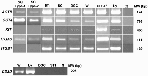
We determined the gene expression levels for human molecular markers SG, OCT4, KIT, ITGA6, and ITGB1 by RT-PCR in samples of type-1 and type-2 SG, primary ST (ST1), SC, diploid germ cell (DGC) fractions, total testicular tissue, umbilical cord blood CD34+, and peripheral blood Ly (). Octamer-binding transcription factor 4 and ITGA6 mRNA were detected in all cells studied (). Transcripts of KIT and ITGB1 were detected in all cell types except in SG (). The CD3 delta (CD3D) marker was used to assess the level of contamination of the DGC fractions with Ly, and transcripts were only detected in samples of total testicular tissue and peripheral blood Ly (). Expression of the house-keeping gene, human beta actin (ACTB) was detected in all samples, demonstrating the presence of mRNA in detectable amounts in all cases (internal control of RT-PCR) (). The controls without cDNA were all negative, ensuring the absence of contamination in RT-PCR ().
Figure 1. Example of live cells images taken in an inverted microscope after individual cell isolation by micromanipulation. SC: Sertoli cells; SGA: spermatogonia: type-1 spermatogonia elongated and fusiform (arrows) and type-2 spermatogonia elongated (white arrowhead) and round (black arrowhead); ST1: primary spermatocytes; CD34+: umbilical cord blood CD34+ cells. Bars: = 20 μm (SC, SGA, ST1) and = 10 µm (CD34+)
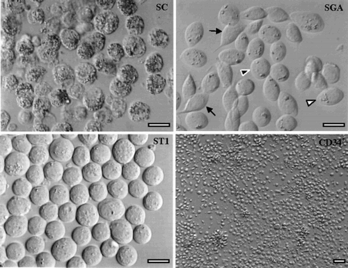
Figure 2. Reverse transcriptase-polymerase chain reaction characterization of OCT4, KIT, ITGA6, ITGB1, and CD3D in human testicular and hematopoietic cells. The subsequent code was employed to assign testicular and hematopoietic cells: SG: spermatogonia; ST1: primary spermatocytes; SC: Sertoli cells; DGC: diploid germ cell suspensions; W: whole-testicular cell suspensions; CD34+: umbilical cord blood CD34+ cells; Ly: lymphocytes. MW indicates the molecular weight marker of 100bp (Invitrogen). No bands were detected in the negative control (N). Human beta actin (ACTB) was used as house-keeping gene. OCT4: octamer-binding transcription factor 4; KIT: v-Kit Hardy-Zuckerman 4 Feline Sarcoma Viral Oncogene Homolog; ITGA6: integrin alpha 6; ITGB1: integrin beta 1; CD3D: CD3 delta
Characterization of protein expression
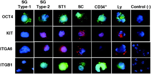
By IC, using individually isolated cells by micromanipulation, the OCT4 protein was detected in type-1 and type-2 SG, ST1, SC, CD34+, and Ly (). Protein expression of KIT was also detected in all the cells studied, with the exception of type-1 SG being negative (KIT(–)) and type-2 SG being positive (KIT(+)) (). Protein expression of ITGA6 was similar to KIT results, with the presence of two types of staining for SG, with type-1 SG being negative (ITGA6(–)) and type-2 SG being positive (ITGA6(+)) (). Integrin beta 1 protein levels were similar to KIT and ITGA6, with staining in SG, with type-1 SG being negative (ITGB1(-)) and type-2 SG being positive (ITGB1(+)) (). Umbilical cord blood CD34+ cells, peripheral blood Ly, and SC stained positive for both integrins (). Negative controls (peripheral blood Ly without primary antibody) were always negative, showing the absence of nonspecific labeling ().
Figure 3. Immunocytochemical localization of OCT4, KIT, ITGA6, and ITGB1 in adult human testicular cells and hematopoietic cells. The subsequent code was employed to assign testicular and hematopoietic cells: SG: spermatogonia; ST1: primary spermatocytes; SC: Sertoli cells; CD34+: umbilical cord blood CD34+ cells; Ly: lymphocytes. Replacement of primary antibody with phosphate buffered saline (PBS) in Ly provided the negative controls. OCT4: octamer-binding transcription factor 4; KIT: v-Kit Hardy-Zuckerman 4 Feline Sarcoma Viral Oncogene Homolog; ITGA6: integrin alpha 6; ITGB1: integrin beta 1
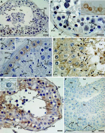
Human testicular tissue was taken for immunohistochemical examination (paraffin sections) to better understand the localization of SG marker in human adult testicular cells. Octamer-binding transcription factor 4 displayed positive staining in SG, ST1, and SC (A, B). v-Kit Hardy-Zuckerman 4 Feline Sarcoma Viral Oncogene Homolog was detected in the membrane and cytoplasm of SG A-pale (progenitor SG) but no labeling was found in SG A-dark (stem SG), ST1, or SC (C). The expression of ITGA6 was traced in the membrane of the two types of SG, ST1, and SC. Labeling was also observed in the cytoplasm of SC and ST1, but not in SG (D). ITGB1 was observed in the cytoplasmic membrane of SG A-pale, ST1, and SC, with no labeling found in SG A-dark. Cytoplasmic staining was observed only in SC ( E). In negative controls, with antibody omission no staining was observed (F).
Figure 4. Immunohistochemical localization of cell markers in paraffin sections of adult human testis. A, B inset-section without hematoxylin counterstaining) OCT4; C, inset) KIT; D) ITGA6; E, inset) ITGB1; and F) example of a negative control with primary antibody omission. BL: basal lamina; PTC: peritubular cells; SC: Sertoli cells; SG: spermatogonia; Ad: spermatogonia A-dark; Ap: spermatogonia A-pale; ST1: primary spermatocytes; Sa: round spermatids; Sd: elongated spermatids; Sz: spermatozoa; De: scaled cells in the lumen of the seminiferous tubule; Lu: lumen of the seminiferous tubule. Scale bars = 5 µm (B, C, E), 10 µm (D, F), and 20 µm (A).
Discussion
In human St different cell types coexist during spermatogenesis [Clermont Citation1963; 1966], which makes it very difficult for the isolation of pure populations of SG stem cells. Although several experiments enabled the purification of human SG using surface markers such as ITGA6 [Geens et al. Citation2007; Geens et al. Citation2011; Conrad et al. Citation2008], GPR125 [He et al. Citation2010], stage-specific embryonic antigen-4 (SSEA-4) [Izadyar et al. Citation2011], and KIT [von Schonfeldt et al. 1999]. Other studies have also shown that when these strategies are used they often yield a population of cells that are contaminated with cancer cells [Geens et al. Citation2007; Geens et al. Citation2011; Jahnukainen et al. Citation2011].
Expression of alpha 6 and beta 1 integrins in the human seminiferous epithelium
Previous studies in human St have detected ITGA6 protein expression in ST, spermatids, and testicular spermatozoa, but not in SG and SC [Schaller et al. Citation1993], in SG and SC [He et al. Citation2010], in SG (with presence of positive and negative populations) [Izadyar et al. Citation2011], in SG both for ITGA6 and ITGB1 [Virtanen et al. Citation1997], in SG stem cell lines [Conrad et al. Citation2008] and in SG stem cell cultures [Koruji et al. Citation2012]. In the present work the expression of integrins was detected in all cell stages including those that are not in contact with basal lamina, which suggests that these molecules also mediate binding to SC until spermiation. Two populations of SG for ITGA6 and ITGB1 were identified by IC, one negative (type-1) and another positive (type-2), as previously observed for ITGA6 [Izadyar et al. Citation2011]. In relation to ITGA6, this dual labeling was not found at the mRNA level or at IH. On the contrary, for ITGB1 these two SG populations were consistently found in both protein expression studies. These results thus suggest that by itself ITGA6 might not be a suitable marker for the isolation of stem SG, in contrast with previous reports in humans [Conrad et al. Citation2008; Geens et al. Citation2007; Geens et al. Citation2011] and rodents [Shinohara et al. Citation1999; Shinohara et al. Citation2000; Kubota et al. Citation2003; Kanatsu-Shinohara et al. Citation2008].
Expression of OCT4 in the human seminiferous epithelium
In mice, Oct4 has been used as a marker of stem germ cells of the seminiferous epithelium, revealing a specific protein and gene expression profile of undifferentiated cells in type-A SG of adult St [Pesce et al. Citation1998; Ohbo et al. Citation2003]. In humans, OCT4 immunolocalization was positive in SG of adult St [Bhartiya et al. Citation2010; Izadyar et al. Citation2011], in isolated SG stem cells, and in embryonic stem cells [Conrad et al. Citation2008]. In the testis, two populations of SG, negative and positive, were also observed [Bhartiya et al. Citation2010; Izadyar et al. Citation2011], with no labeling in the other germ cells and in SC [Bhartiya et al. Citation2010]. On the contrary, in other studies, although positive in fetal life, no positive labeling in germ cells was found after birth [Rajpert-De Meyts et al. Citation2004; He et al. Citation2010]. The results presented in this study show both gene and protein expression for OCT4, and showed staining in SG as well as in other germ cells and SC.
We did not exclude germ cell contamination in the SC samples by amplification of germ cell-specific transcript, for example with VASA [Castrillon et al. Citation2000; Noce et al. Citation2001] or RBMY [Elliot Citation2004]. However, we have previously demonstrated the specific expression of SC with KITGL and the absence of expression with MAGEA1, MLH1, HSPA2, and SPANXA1 [Sá et al. Citation2008]. Additionally, the purity was confirmed as cells were individually isolated by micromanipulation, and the distinct cell features were evaluated by published morphological criteria [Holstein and Roosen-Runge 1991]. The finding of OCT4 in SC is thus novel.
A special note for OCT4 is required. There are two isoforms of OCT4, A (expressed in pluripotent cells) and B (expressed in multipotent cells). We assayed both isoforms and found that both were expressed in testicular tissue, DGC fractions, ST1, and Ly (data not shown). However, there is a possibility of OCT4 primers simultaneously amplify genomic cDNA due to the similarity of gene sequences with pseudogene on chromosome 8 and a similar gene on chromosome 12. The data obtained from protein expression, whereby IC cells were plated with a high level of purity and by IH using paraffin sections suggests that this is not the case.
Expression of KIT in the human seminiferous epithelium
In rodents, KIT expression is found in SG (but not in other germ cells or SC) [Manova et al. Citation1990; Shinohara et al. Citation1999], in isolated SG [Morena et al. Citation1996], in differentiated SG, and in SC [Schrans-Stassen et al. Citation1999]. In humans, KIT protein expression has only been observed in SG [Natali et al. Citation1992], in SG and spermatids [Sandlow et al. Citation1996; Sandlow et al. Citation1997], only in SG [Stoop et al. Citation2008], in SG, ST, round spermatids, and SC [Unni et al. Citation2009]. In contrast, labeling was found in ST and round spermatids but not in SG and SC [He et al. Citation2010]. In other studies, two types of SG staining, KIT(-) and KIT(+), were also observed [Izadyar et al. Citation2011; von Kopylow et al. Citation2010; von Kopylow et al. Citation2012].
In the present study, results obtained by RT-PCR revealed expression of KIT in all germ cells but not in SG. By IC (isolated cells), KIT was present in SC and in all germ cells with the exception of the presence of two types of SG staining, type-1 negative and type-2 positive as found for integrins. By IH (paraffin sections), KIT was negative in stem SG A-dark and positive in progenitor SG A-pale, as for ITGB1. However, contrary to ITGB1, staining was limited to SG. It is thus possible that our isolated type-1 SG corresponds to stem SG A-dark, rendering KIT as a suitable marker for the isolation of stem SG as previously suggested [von Schonfeldt et al. 1999]. In SC, KIT mRNA was detected by RT-PCR, presented a positive staining in isolated cells by IC and a negative labeling in paraffin sections. The presence of labeled SC was previously reported [Unni et al. Citation2009; Izadyar et al. Citation2011].
Of note regarding positive and negative SG, for ITGA6, positive and negative SG was found only by IC (protein expression in isolated cells). For ITGB1 and KIT, the dual expression was found in both methods of protein analysis but was negative by RT-PCR. The absence of mRNA could not be due to the small amount of cells used, once it was positive in the same conditions for ITGA6 and OCT4. Perhaps the time at which the cells were isolated, ITGB1 and KIT were transcriptionally repressed in type-2 SG. Notwithstanding, the presence of very small amounts of mRNA cannot be discarded. Regarding ITGA6, the difference found only in IC could be due to the fact that isolated cells are easier permeabilized or due to different antibody specificity. Differences could also be due to technical differences, such as during paraffin embedding and paraffin removal procedures the structure of epitopes in human testes might have been changed.
Spermatogonia stem and progenitor cells: two distinct populations
In rodents, SG KIT(-) were identified as the truly stem cells (type A-single, which is similar to A-dark SG in men), whereas KIT(+) SG corresponded to the progenitor cells (type A-aligned, which correspond to human A-pale SG) [Shinohara et al. Citation1999; Schrans-Stassen et al. Citation1999; Izadyar et al. Citation2003; Singh et al. Citation2011]. The KIT receptor has, indeed, been considered as a marker of differentiation as the Erk 1/2 pathway can be activated by stem cell factor in KIT expressing SG to stimulate their proliferation [Singh et al. Citation2011].
The same was observed in humans [Izadyar et al. Citation2011; von Kopylow et al. Citation2012]. We detected on isolated cells by IC two populations of human SG, type-1 SG that were KIT(-), ITGA6(-), and ITGB1(-), and type-2 SG that were KIT(+), ITGA6(+), and ITGB1(+). In paraffin sections both KIT(-) and ITGB1(-) corresponded to SGA-dark. On one hand KIT staining was confined to SGA-pale, ITGB1 staining was also present in the other cells of the St. On the the other hand, for ITGA6 both SGA-dark and SGA-pale appeared stained. Contrarily to previous observations, two types of SG populations for OCT4, OCT4(-), and OCT4(+) [Bhartiya et al. Citation2010; Izadyar et al. Citation2011], were not found in all assays ().
Table 1. Marker positivity (+) in germ cells and Sertoli cells.
Expression of all four markers in human peripheral blood lymphocytes and umbilical cord blood CD34+ cells
In the present study we detected the expression (by RT-PCR and IC) of all four markers both in peripheral blood Ly and in CD34+. The expression of ITGA6 and ITGB1 had already been described in peripheral blood Ly [Shimizu et al. Citation1990; Pilling et al. Citation1998] and for ITGB1 in CD34+ [Ramírez et al. Citation2001] but had not yet been described for ITGA6. The presence of the OCT4 transcript and protein in peripheral blood Ly, confirms previous results [Zangrossi et al. Citation2007]. The fact that OCT4 was detected in differentiated cells confirms what had already been established by several authors with respect to this as a useful marker in different adult human differentiated cells [Takeda et al. Citation1992; Zangrossi et al. Citation2007].
The expression of the markers studied both in isolated Ly in CD34+ cells must be taken into consideration when using these markers for germline stem cell isolation by MACS or FACS. It does run a risk of contamination of cellular fractions with Ly, unwanted in the case of autologous transplantation in cancer patients. In the present study, CD3D was only detected in samples of total esticular tissue and Ly suggesting that the isolation method used in this study might be efficient. Furthermore, it was observed that in the cell cognate of the human testicular microenvironment (IH) or in isolated cells and cell suspensions (RT-PCR and IC assays) the profile was maintained. This leads us to believe that our isolation method is quite efficient.
In conclusion, from the data presented in this work, it is suggested that KIT(–) / ITGB1(-) might be a suitable marker for the isolation of human stem SG. Future studies will have to be conducted with these markers using isolated populations of germ cells from human testis to evaluate the efficacy of stem cell isolation by MACS and sorting, followed by in vitro maturation or transplantation into mice. It is however necessary to note some caution regarding the extrapolation of the present results to large scale isolation mass. Scale-up may reveal additional difficulties to removing minor contaminants or uncover additional subpopulations not fitting the initial model. Other genes could be screened to identify additional specific surface markers of stem/progenitor cells in human St.
Materials and Methods
Ethical considerations and biological material
According to the National Law on Medically Assisted Procreation (PMA, Law 32/2006) and the guidelines of the National Council on Medically Assisted Procreation (CNPMA, 2008), data bases and testicular material were used after patient informed and written consent from five cases with secondary obstructive azoospermia and anejaculation, who needed an open testicular biopsy to recover testicular spermatozoa for intracytoplasmic sperm injection treatment cycles. In all cases, a small fragment of testicular tissue was taken for hemalumen-eosin (HE) staining to confirm the diagnosis. All patients, aged 31-45 years, had normal karyotypes and absence of cystic fibrosis transmembrane condunctance regulator mutations, Yq11.2 microdeletions, and DAZ 1, 2 (deleted in azoospermia) gene copies deletions.
Patient characteristics were as follows: three had obstructive azoospermia (OAZ) due to epididymis injury after inguinal surgery; one had retrograde ejaculation (RE) and the other neurological anejaculation due to diabetes mellitus insulin dependent (DMID). These patients were selected from cases with conserved spermatogenesis after treatment testicular biopsy and HE staining. This was an expected finding due to the types of pathologies involved: OAZ was not due to an infectious disease; RE is a special case where the production of sperm and ejaculation is still ongoing; and in DMID the patient cannot form an erection or ejaculate due to peripheral neuropathy.
Patients had normal epididymis (except patients with obstructive azoospermia whose epididymis were difficult to evaluate), normal testicular volume, and normal serum FSH levels. An open testicular biopsy for testicular sperm extraction was performed under spermatic cord block [Sousa et al. Citation2002a]. In two cases the right testicle was used, whereas in the other three cases it was the left testicle used (4-6 samples each). Each testicle sample consisted of 2-3 mm3 St fragments, and all showed abundant germ cells and 2-4 testicular sperm with in situ or slow progressive movements when inspected at x40 in an inverted microscope (Nikon DIAPHOT 200, Nikon, Tokyo, Japan), equipped with thermal stage (33°C) and Hoffman optics (Nikon).
Biological replicates for RT-PCR, IC, and IH, of at least three independent experiments from each patient were performed. RT-PCR, used approximately1,000 cells of each cell-type isolated by micromanipulation for each independent RT-PCR. In IC, about 100 cells of each cell-type in each experiment were scored in duplicate. For IH all St per section were scored per patient in duplicate from two nonconsecutive sections. In every case, all cell types were analyzed at the same time.
Human umbilical cord blood was harvested by umbilical vein puncture after normal full-term delivery under previous informed consent from the pregnant woman before delivery, in accordance to the guidelines of the local Ethics Committee. Sample was collected in 150cc sterile bags, containing 21mL of citrate-phosphate dextrose (CDP; MacoPharma, Tourcoing, France), and kept at room temperature for less than 12h. We only used one umbilical cord blood unit but three independent experiments were performed.
Following the guidelines of the local Ethics Committee, Ly were obtained by informed and written consent from peripheral blood of a healthy man, showing a normal karyotype.
Isolation of testicular cells
For tissue digestion, testicular St fragments, collected in Sperm Preparation Medium (SPM-HEPES; Medicult Origio, Jyllinge, Denmark), were fragmented and squeezed. The resultant fluid fraction was washed in SPM (2x5 min, 300 x g) and incubated in 2ml erythrocyte-lysing buffer. The suspension was then washed (2x5 min, 500 x g) and finally submitted to enzymatic treatment [Crabbé et al. Citation1998]. The suspension was separated (5 min, 50 x g) into supernatant (haploid germ cells) and pellet (DGC) fractions [Sousa et al. Citation2002b; Sá et al. Citation2008]. The fluid fractions were washed with SPM (2x5 min, 1,000 x g), and the final pellets were resuspended in 100 µl in in-vitro fertilization medium (Universal IVF Medium, Medicult) and incubated at 32°C. Sertoli cells were recovered from the initial pellet fraction.
Individual germ cells were isolated by micromanipulation pursuant to the procedures described previously [Sousa et al. Citation2002a; Sousa et al. Citation2002b; Sá et al. Citation2008]. Cell stages were confirmed by morphological criteria [Holstein and Roosen-Runge Citation1991; Sousa et al. Citation2002b]. Sertoli cells were large cells, had a cytoplasm filled with lipid droplets and a nucleus that exhibited an elevated border with a large nucleolus. We isolated two types of SG. Type-1 was less frequent and had an ellipsoidal shape with terminal fine extensions. Type-2 were more frequent and had an elliptical shape without terminal extensions with several evolving to a round shape after isolation. Primary spermatocytes were the largest cells with about a19-24 µm diameter (). In addition to distinct morphological cell features, the purity of these cell types, which were individually isolated by micromanipulation, were previously characterised by FISH analysis, mRNA expression, and electron microscopy [Sá et al. Citation2008].
Isolation of hematopoietic cells
Lymphocytes were isolated by gradient centrifugation with Ficoll-Histopaque (Sigma, St. Louis, MO, USA), with some samples being cultured under phytohemagglutinin stimulation to induce lymphoblastoid cells. Human CD34+ were isolated as previously described [Pinho et al. Citation2011]. Briefly, mononuclear cells were isolated from umbilical cord blood by gradient centrifugation and then subjected to a MACS system (Miltenyi Biotec, Auburn, CA, USA), using a CD34 epitope QBEND/10 to immunomagnetically select the CD34+ cells. The purity of the CD34+ fraction was determined by flow cytometry, being consistently above 90%.
RNA expression studies
For RNA expression studies, total mRNA was isolated, converted to cDNA and amplified by RT-PCR. Total RNA was extracted from the testicular isolated cells, peripheral blood Ly, and CD34+ using the RNeasy Mini Kit (QIAGEN GmbH, Hilden, Germany). To avoid contamination with genomic DNA, samples were treated with DNase (RNase-Free Dnase Set, QIAGEN). The cDNA synthesis was performed using the Superscript Reverse Transcription Kit (Invitrogen, Carlsbad, USA) with random primers according to the manufacturer's instructions. RNA samples were converted to cDNA and the cDNA product was precipitated with two volumes of ethanol (Panreac, Barcelona, Spain) and 2.5 M sodium acetate pH 6.0 (Merck, Darmstadt, Germany) overnight at -20°C. The precipitated cDNA was resuspended in 20 µl of diethylpyrocarbonate (DEPC)-treated RNase free water (Promega, Wisconsin, USA). Aliquots of ≤ 500ng/reaction of cDNA were used as a template for PCR amplification using QuantiTect SYBR Green PCR kit (QIAGEN) (the use of this kit did not aim to analyze the samples in real time, but only improve the efficiency of PCRs) with 25 pmol/μl specific primer sets (100 pmol/μl Primers, Thermo Electron, Ulm, Germany,) for ACTB, CD3D, OCT4, KIT, ITGA6, and ITGB1. The oligonucleotide primer pairs used are represented in . The primers used for human genes KIT and OCT4 [Ohbo et al. Citation2003], CD3D [Ziyyat et al. Citation1999], and ACTB [Honke et al. Citation2002] were based on previous reports and analyzed in National Center for Biotechnology Information (NCBI; http://www.ncbi.nlm.nih.gov/tools/primer-blast/). The primers for the human genes of ITGA6 and ITGB1 were designed using the OLIGO 4.0 software program according to location of at least one of the primers in two different exons (to avoid co-amplification of genomic DNA [Shinohara et al. Citation1999]. PCR cycling conditions were as follows: initial activation step (5 min at 95°C) then an initial denaturation at 95°C for 1 min followed by 45 cycles (denaturation at 95°C for 1 min, annealing for 1 min at 56°C for ACTB, 62°C for OCT4, 55°C for CD3D, 60°C for KIT, 61°C for ITGA6, and 58°C for ITGB1, extension for 1 min at 72°C) and a final extension step of 5 min at 72°C (Gene Amp, PCR System 9700, Applied Biosystem, Foster City, CA, USA). The number of amplification cycles was increased up to 45 to ensure that less abundant transcript would amplify, and 45 cycles were also used for all the transcripts in order to homogenize the results obtained. The PCR products were loaded onto a 1.5% agarose gel, stained with ethidium bromide, and observed in the transiluminator (Image Master VDS, Pharmacia Biotech, Fall River, MA, USA). Negative controls included a sample lacking DNA, which was replaced by water.
Table 2. Forward and reverse primers used for RT-PCR.
Protein expression studies
For analysis of protein levels, IC was applied to isolated cells and IH was applied to paraffin sections of testicular biopsies fixed with paraformaldehyde from the same patients as well as from archive testicular biopsies. Two protein measures were undertaken. The former was performed on samples of intact cells that had all of their surrounding extracellular matrix removed as they were isolated by micromanipulation from a cell suspension. Immunohistochemistry was performed in samples that result from paraffin sections of biological tissue, where each cell is surrounded by tissue architecture and neighbouring cells (within a specialized microenvironment) normally found in the intact tissue. The antibodies employed in isolated cells are different from those employed in paraffin sections because they were IC or IH specific.
Immunocytochemistry
Isolated cells were fixed with paraformaldehyde diluted in PBS (Sigma) (germ cells: 0.2%; Ly and CD34+: 4%) on glass slides coated with poly-L-lysine (Menzel-Glaser-Polysine, Saarbruckener, Germany) and processed immunocytochemically according to a protocol adapted from the Center Medical Genetics and Molecular, Institute of Oncologic Research of Barcelona, Spain. Following a 5 min wash with PBS, the cells were subsequently permeabilized and blocked for 1h in PBS containing 0.2% Triton X-100 (Sigma) and 20% fetal bovine serum (Mod.Fetalclone1-HyClone, Logan, UT, USA) in a dark moist chamber at room temperature. Then the cells were incubated with primary antibodies for KIT (Ab 81): mouse monoclonal IgG; ITGA6 (CD49f; BQ16): mouse monoclonal IgG; ITGB1 (CD29; N-20): goat polyclonal IgG; and OCT4 (C-20): goat polyclonal IgG), at a 1:25 dilution in a dark moist chamber, overnight at 4°C. After three washes in PBS, cells were incubated with secondary antibodies, marked with red and green fluorochromes (fluorescein isothiocyanate (FITC)-conjugated mouse anti-goat IgG or rhodamine-conjugated goat anti-mouse IgG1-R cat), at a 1:100 dilution in a dark moist chamber for 60 min at room temperature. The secondary antibodies used were labeled with fluorochromes, green in the case of ITGB1 and OCT4, and red in the case of ITGA6 and KIT (FITC-conjugated and rhodamine-conjugated, respectively). The nuclei was stained with 4',6'-Diamidino-2-phenylindole (DAPI-Vectashield, Vector Laboratories, Burlingame, CA, USA) and the cells were observed for epifluorescence using a Nikon Eclipse E400 Epifluorescence Microscope (Nikon). All the primary and secondary antibodies were purchased from Santa Cruz Biotechnology, Inc. (Santa Cruz, CA, USA). Replacement of primary antibody with PBS was used as a negative control [He et al. Citation2010].
Immunohistochemistry
For IH, 3 µm sections on glass slides coated with Vectabond Reagent (Vector) were dewaxed in xylene and rehydrated through a series of graded alcohols. Antigen retrieval was performed using the antigen unmasking solution (Vector), and the endogenous peroxidase activity was quenched with 3% hydrogen peroxide (Merck). After a 5 min wash in PBS and blocking with blocking-serum (Vector), the sections were incubated with avidin and biotin solutions in order to produce the most intense staining and the least background staining. Afterwards, the sections were incubated with primary antibodies (AB1), including rabbit polyclonal AB1 to OCT4 (Cell Signalling Technology, MA, USA) at a 1:100 dilution, mouse monoclonal AB1 to KIT (Dakocytomation, CA, USA) at a 1:300 dilution, polyclonal AB1 to ITGA6 (Santa Cruz) at a 1:50 dilution, and AB1 clone 7F10 to ITGB1 (Novocastra, Newcastle, UK) at a 1:40 dilution, in a dark moist chamber for 1h. To determine the expression of markers, the sections were incubated with the universal biotinylated second antibody (Vector) for 10 min. Next, streptavidin-peroxidase enzyme conjugate (Vector) was added to the cells and reaction product was generated by the addition of 3-3' diaminobenzidine (Vector) as the chromogen. After immunostaining, testis sections were counterstained with hematoxylin (Vector) and examined under a light microscope with CCD camera and IM50 software (Leica Microsystems Ltd, Heerbrugg, Germany). Replacement of primary antibody with PBS was used as a negative control.
According to previous morphological studies in histological sections, SG A-dark and A-pale adhere to the basal lamina and are elliptical. Spermatogonia A-dark are considered the stem cell pool and are recognized by a pale region inside the nucleus. Spermatogonia A-pale are the progenitor type and do not display that nuclear feature. Spermatogonia B are derived from A-pale, have a round appearance, and are above the SG cell line although linked to the basal lamina by a peduncle [Holstein and Roosen-Runge Citation1991].
Abbreviations
| ACTB: | = | human beta actin |
| CD3D: | = | CD3 delta |
| CD34+: | = | umbilical cord blood CD34+ cells |
| DGC: | = | diploid germ cell |
| DMID: | = | diabetes mellitus insulin dependent |
| FACS: | = | fluorescence-activated cell sorting |
| FITC: | = | fluorescein isothiocyanate |
| GPR125: | = | G protein-coupled receptor 125 |
| HE: | = | hemalumen-eosin |
| IC: | = | immunocytochemistry |
| IH: | = | immunohistochemistry |
| ITGA6: | = | integrin alpha 6 |
| ITGB1: | = | integrin beta 1 |
| KIT: | = | v-Kit Hardy-Zuckerman 4 Feline Sarcoma Viral Oncogene Homolog |
| Ly: | = | lymphocyte |
| MACS: | = | magnetic-activated cell sorting |
| OAZ: | = | obstructive azoospermia |
| OCT4: | = | octamer-binding transcription factor 4 |
| RE: | = | retrograde ejaculation |
| RT-PCR: | = | reverse transcriptase polymerase chain reaction |
| SC: | = | Sertoli cell |
| SG: | = | spermatogonia |
| SPM: | = | sperm preparation medium |
| St: | = | seminiferous tubule |
| ST: | = | spermatocyte |
| ST1: | = | primary ST |
| SSEA-4G: | = | stage-specific embryonic antigen-4. |
The authors appreciate the contribution of Maria João Pinho, PhD (Department of Genetics, Faculty of Medicine of University of Porto) for CD34+ preparation; Susana Fernandes, PhD (Department of Genetics, Faculty of Medicine of University of Porto) for RT-PCR teaching; Luis Ferraz, MD, Urologist (Director of Department of Urology, Hospital of Vila Nova de Gaia) for treatment testicular biopsies; António Couceiro, MD; Pathologist (Department of Pathology, Hospital of Vila Nova de Gaia) for archive testicular biopsies; Sónia Oliveira, MD, Gynecologist (Department of Gynecology and Obstetrics) for human umbilical cord blood; Joaquina Silva, MD, Senior Clinical Embryologist-ESHRE (CGR A. Barros) for IVF supervision; Paulo Viana, BSc and Mariana Cunha, BSc, Clinical Embryologists-ESHRE (CGR A. Barros) for biopsy preparation, as well as the support provided by a PhD grant to Rosália Sá from the Foundation for Science and Technology, Ministry of Science, Technology and Superior Education, Portugal.
Declaration of interest: PhD grant to RS (SFRH/BD/23616/2005) from the Foundation for Science and Technology, Ministry of Science, Technology and Superior Education, Portugal. The authors report no conflicts of interest. The authors alone account for the content and writing of the manuscript.
Author contributions: Full data acquisition, analysis, interpretation, critical discussion, and manuscript writing: RS; Initial data acquisition, analysis, interpretation, discussion, and text writing: CM; Co-supervision of the PhD of RS and text review: FC; Patient recruitment and ART supervision: AB; Study conception and design, data acquisition, analysis and interpretation, critical discussion, and final text review: MS.
References
- Bhartiya, D., Kasiviswanathan, S., Unni, S.K., Pethe, P., Dhabalia, J.V., Patwardhan, S., (2010) Newer insights into premeiotic development of germ cells in adult human testis using Oct-4 as a stem cell marker. J Histochem Cytochem 58:1093–1106.
- Blume-Jensen, P., Claesson-Welsh, L., Siegbahn, A., Zsebo, K.M., Westermark, B. and Heldin, C.H. (1991) Activation of the human c-kit product by ligand-induced dimerization mediates circular actin reorganization and chemotaxis. EMBO J 10:4121–4128.
- Castrillon, D.H., Quade, B.J., Wang, T.Y., Quigley, C. and Crum, C.P. (2000) The human VASA gene is specifically expressed in the germ cell lineage. Proc Natl Acad Sci USA 97:9585–9590.
- Clermont, Y. (1963) The cycle of the seminiferous epithelium in man. Am J Anat 112:35-51. Clermont, Y. (1966) Renewal of spermatogonia in man. Am J Anat 118:509–524.
- Conrad, S., Renninger, M., Hennenlotter, J., Wiesner, T., Just, L., Bonin, M., (2008) Generation of plutipotent stem cells from adult human testis. Nature 456:344–349.
- Crabbé, E., Verheyen, G., Silver, S., Tournaye, H., Van de Velde, H., Goossens, A., (1998) Enzymatic digestion of testicular tissue may rescue the intracytoplasmic sperm injection cycle in some patients with non-obstructive azoospermia. Hum Reprod 13:2791–2796.
- Elliot, D.J. (2004) The role of potential splicing factors including RBMY, RBMX, hnRNPG-T and STAR proteins in spermatogenesis. Int J Androl 27:328–334.
- Geens, M., Goossens, E. and Tournaye, H. (2011) Cell selection by selective matrix adhesion is not sufficiently efficient for complete malignant cell depletion from contaminated human testicular cell suspensions. Fertil Steril 95:787–791.
- Geens, M., Van de Velde, H., De Block, G., Goossens, E., Van Steirteghem, A.C. and Tournaye, H. (2007) The efficiency of magnetic-activated cell sorting and fluorescence-activated cell sorting in the decontamination of testicular cell suspensions in cancer patients. Hum Reprod 22:733–742.
- Goossens, E. and Tournaye, H. (2013) Adult stem cells in the human testis. Semin Reprod Med 31:39–48.
- Goto, T., Adjaye, J., Rodeck, C.H. and Monk, M. (1999) Identification of genes expressed in human primordial germ cells at the time of entry of the female germ line into meiosis. Mol Hum Reprod 5:851–860.
- Hansis, C., Grifo, J.A. and Krey, L.C. (2000) Oct-4 expression in inner cell mass and trophectoderm of human blastocysts. Mol Hum Reprod 6:999–1004.
- He, Z., Kokkinaki, M., Jiang, J., Dobrinski, I. and Dym, M. (2010) Isolation, characterization, and culture of human spermatogonia. Biol Reprod 82:363–372.
- Holstein, A.F. and Roosen-Runge, E.C. (1991) Atlas of Human Spermatogenesis. Grosse Verlag, Berlin, Germany.
- Honke, K., Hirahara, Y., Dupree, J., Suzuki, K., Popko, B., Fukushima, K., (2002) Paranodal junction formation and spermatogenesis require sulfoglycolipids. Proc Natl Acad Sci USA 99:4227–4232.
- Izadyar, F., Den Ouden, K., Creemers, L.B., Posthuma, G., Parvinen, M. and de Rooij, D.G. (2003) Proliferation and differentiation of bovine type A spermatogonia during long-term culture. Biol Reprod 68:272–281.
- Izadyar, F., Wong, J., Maki, C., Pacchiarotti, J., Ramos, T., Howerton, K., (2011) Identification and characterization of repopulating spermatogonial stem cells from the adult human testis. Hum Reprod 26:1296–1306.
- Jahnukainen, K., Ehmcke, J., Hou, M. and Schlatt, S. (2011) Testicular function and fertility preservation in male cancer patients. Best Pract Res Clin Endocrinol Metab 25:287–302.
- Kanatsu-Shinohara, M., Takehashi, M., Takashima, S., Lee, J., Morimoto, H., Chuma, S., (2008) Horning of mouse spermatogonial stem cells to germline niche depends on beta 1-integrin. Cell Stem Cell 3:533–542.
- Koruji, M., Shahverdi, A., Janan, A., Piryaei, A., Lakpour, M.R. and Sedighi, M.A.G. (2012) Proliferation of small number of human spermatogonial stem cells obtained from azoospermic patients. J Assist Reprod Genet 29:957–967.
- Kossack, N., Meneses, J., Shefi, S., Nguyen, H.N., Chavez. S., Nicholas, C., (2009) Isolation and characterization of pluripotent human spermatogonial stem cell-derived cells. Stem Cells 27:138–149.
- Kubota, H., Avarbock, M.R. and Brinster, R.L. (2003) Spermatogonial stem cells share some, but not all, phenotypic and functional characteristics with other stem cells. Proc Natl Acad Sci USA 100:6487–6492.
- Liu, S., Tang, Z., Xiong, T. and Tang, W. (2011) Isolation and characterization of human spermatogonial stem cells. Reprod Biol Endocrinol 9:141–149.
- Manova, K., Nocka, K., Besmer, P. and Bachvarova, R.F. (1990) Gonadal expression of c-kit encoded at the W locus of the mouse. Development 110:1057–1069.
- Morena, A.R., Boitani, C., Pesce, M., De Felici, M. and Stefanini, M. (1996) Isolation of highly purified type A spermatogonia from prepubertal rat testis. J Androl 17:708–717.
- Natali, P.G., Nicotra, M.R., Sures, I., Santoro, E., Bigotti, A. and Ullrich, A. (1992) Expression of c-kit receptor in normal and transformed human nonlymphoid tissues. Cancer Res 52:6139–6143.
- Nebel, B., Amarose, A. and Hackett, E. (1961) Calendar of gametogenic development in the prepubertal male mouse. Science 134:832–833.
- Noce, T., Okamoto-Ito, S. and Tsunekawa, N. (2001) Vasa homolog genes in mammalian germ cell development. Cell Struct Funct 26:131–136.
- Ohbo, K., Yoshida, S., Ohmura, M., Ohneda, O., Ogawa, T., Tsuchiva, H., (2003) Identification and characterization of stem cells in prepubertal spermatogenesis in mice. Dev Biol 258:209–225.
- Pesce, M., Wang, X., Wolgemuth, D.J. and Schöler, H.R. (1998) Differential expression of the Oct-4 transcription factor during mouse germ cell differentiation. Mech Dev 71:89–98.
- Pesce, M., Anastassiadis, K. and Schöler, H.R. (1999) Oct-4: lessons of totipotency from embryonic stem cells. Cells Tissues Organs 165:144–152.
- Phillips, B.T., Gassei, K. and Orwig, K.E. (2010) Spermatogonial stem cell regulation and spermatogenesis. Philos Trans R Soc Lond B Biol Sci 365:1663–1678.
- Pilling, J.E., Galvin, A., Robins, A.M., Sewell, H.F. and Mahida, Y.R. (1998) Expression of α5 (CD49e) and α6 (CD49f) integrin subunits on T cells in the circulation and the lamina propria of normal and inflammatory bowel disease colonic mucosa. Scand J Immunol 48:425–428.
- Pinho, M.J., Punzel, M., Sousa, M. and Barros, A. (2011) Ex vivo differentiation of natural killer cells from human umbilical cord blood CD34+ progenitor cells. Cell Commun Adhes 18:45–55.
- Rajpert-De Meyts, E., Hanstein, R., Jorgensen, N., Graem, N., Vogt, P.H. and Skakkebaek, N.E. (2004) Developmental expression of POU5F1 (OCT-3/4) in normal and dysgenetic human gonads. Hum Reprod 19:1338–1344.
- Ramírez, M., Segovia, J.C., Benet, I., Arbona, C., Güenechea, G., Blaya, C., (2001) Ex vivo expansion of umbilical cord blood (UCB) CD34+ cells alters the expression and function of α4β1 and α5β1 integrins. Br J Haematol 115:213–221.
- Reubinoff, B.E., Pera, M.F., Fong, C.Y., Trouson, A. and Bongso, A. (2000) Embryonic stem cell lines from human blastocysts: somatic differentiation in vitro. Nat Biotechnol 18:399–404.
- Sá, R., Neves, R., Fernandes, S., Alves, C., Carvalho, F., Silva, J., (2008) Cytological and expression studies and quantitative analysis of the temporal and stage-specific effects of follicle-stimulating hormone and testosterone during cocultures of the normal human seminiferous epithelium. Biol Reprod 79:962–975.
- Sandlow, J.I., Feng, H.L., Cohen, M.B. and Sandra, A. (1996) Expression of c-KIT and its ligand, stem cell factor, in normal and subfertile human testicular tissue. J Androl 17:403–408.
- Sandlow, J.I., Feng, H.L. and Sandra, A. (1997) Localization and expression of the c-KIT receptor protein in human and rodent testis and sperm. Urology 49:494–500.
- Schaller, J., Glander, H.J. and Dethloff, J. (1993) Evidence of β1 integrins and fibronectin on spermatogenic cells in human testis. Hum Reprod 8:1873–1878.
- Schöler, H.R., Dressler, G.R., Balling, R., Rohdewohld, H. and Gruss, P. (1990) Oct4: a germline-specific transcription factor mapping to the mouse t-complex. EMBO J 9:2185–2195.
- Schrans-Stassen, B.H., van de Kant, H.J., de Rooij, D.G. and van Pelt, A.M. (1999) Differential expression of c-kit in mouse undifferentiated and differentiating type A spermatogonia. Endocrinology 140:5894–5900.
- Shimizu, Y., Van Seventer, G.A., Horgan, K.J. and Shaw, S. (1990) Regulated expression and binding of three VLA (β1) integrin receptors on T cells. Nature 345:250–253.
- Singh, S.R., Burnicka-Turek, O., Chauhan, C. and Hou, S.X. (2011) Spermatogonial stem cells, infertility, and testicular cancer. J Cell Mol Med 15:468–483.
- Shinohara, T., Avarbock, M.R. and Brinster, R.L. (1999) β1- and α6- integrin are surface markers on mouse spermatogonial stem cells. Proc Natl Acad Sci USA 96:5504–5509.
- Shinohara, T., Orwig, K.E., Avarbock, M.R. and Brinster, R.L. (2000) Spermatogonial stem cell enrichment by multiparameter selection of mouse testis cells. Proc Natl Acad Sci USA 97:8346–8351.
- Sousa, M., Cremades, N., Alves, C., Silva, J. and Barros, A. (2002a) Developmental potential of human spermatogenic cells co-cultured with Sertoli cells. Hum Reprod 17:161–172.
- Sousa, M., Cremades, N., Silva, J., Oliveira, C., Ferraz, L., Teixeira da Silva, J., (2002b) Predictive value of testicular histology in secretory azoospermic subgroups and clinical outcome after microinjection of fresh and frozen-thawed sperm and spermatids. Hum Reprod 17:1800–1810.
- Stoop, H., Honecker, F., van de Geijn, G.J.M., Gillis, A.J.M., Cools, M.C., de Boer, M., (2008) Stem cell factor as a novel diagnostic marker for early malignant germ cells. J Pathol 216:43–54.
- Takeda, J., Seino, S. and Bell, G.I. (1992) Human Oct3 gene family: cDNA sequences, alternative splicing, gene organization, chromosomal location, and expression at low levels in adult tissues. Nucleic Acids Res 20:4613–4620.
- Unni, S.K., Modi, D.N., Pathak, S.G., Dhabalia, J.V. and Bhartiya, D. (2009) Stage-specific localization and expression of c-kit in the adult human testis. J Histochem Cytochem 57:861–869.
- Verlinsky, Y., Morozov, G., Verlinsky, O., Koukharenko, V., Rechitsky, S., Goltsman, E., (1998) Isolation of cDNA libraries from individual human preimplantation embryos. Mol Hum Reprod 4:571–575.
- Virtanen, I., Lohi, J., Tani, T., Korhonen, M., Burgeson, R.E., Lehto, V.P., (1997) Distinct changes in the laminin composition of basement membranes in human seminiferous tubules during development and degeneration. Am J Pathol 150:1421–1431.
- von Kopylow, K., Kirchhoff, C., Jezek, D., Schulze, W., Feig, C., Primig, M., (2010) Screening for biomarkers of spermatogonia within the human testis: a whole genome approach. Hum Reprod 25:1104–1112.
- von Kopylow, K., Staege, H., Spiess, A.N., Schulze, W., Will, H., Primig, M., (2012) Differential marker protein expression specifies rarefaction zone-containing human Adark spermatogonia. Reproduction 143:45–57.
- von Schönfeldt, V., Krishnamurthy, H., Foppiani, L. and Schlatt, S. (1999) Magnetic cell sorting is a fast and effective method of enriching viable spermatogonia from Djungarian hamster, mouse, and marmoset monkey testes. Biol Reprod 61:582–589.
- Wyns, C., Curaba, M., Vanabelle, B., Van Langendonckt, A. and Donnez, J. (2010) Options for fertility preservation in prepubertal boys. Hum Reprod Update 16:312–328.
- Yarden, Y., Kuang, W.J., Yang-Feng, T., Coussens, L., Munemitsu, S., Dull, T.J., (1987) Human proto-oncogene c-kit: a new cell surface receptor tyrosine kinase for unidentified ligand. EMBO J 6:3341–3351.
- Zangrossi, S., Marabese, M., Broggini, M., Giordano, R., D'Erasmo, M., Montelatici, E., (2007) Oct-4 expression in adult human differentiated cells challenges its role as a pure stem cell marker. Stem Cells 25:1675–1680.
- Ziyyat, A., Lassalle, B., Testart, J., Briot, P., Amar, E., Finaz, C., (1999) Flow cytometry isolation and reverse transcriptase-polymerase chain reaction characterization of human round spermatids in infertile patients. Hum Reprod 14:379–387.