Abstract
The aim of this study was to analyze pathophysiological changes after testicular sperm aspiration (TESA) and microsurgical epididymal sperm aspiration (MESA) procedures. Twenty four mature male Wistar albino rats with a proven breeding history, weighing approximately 200–250 gm were used for the study. Animals were randomly divided into four groups (n = 6), i.e., control, sham-control, unilateral TESA, and MESA. Using a 22G needle, the aspiration procedures were done in testis or caudal epididymis. At the end of 60 days of survival, blood samples were collected and processed for antisperm antibody detection by enzyme-linked immunosorbent assay (ELISA). After euthanasia, testes and epididymides were collected and processed for paraffin embedding. Sections were stained with hematoxylin and eosin, and TUNEL technique. Serum antisperm antibody titer significantly increased in TESA (P < 0.001) when compared to MESA. Histomorphometric analysis indicated testicular alterations in TESA and MESA, with significant damage in TESA in both testes (P < 0.001). Following the MESA procedure, ipsilateral caudal and carpus epididymis showed significant alterations (P < 0.001) and no such alterations were seen in the ipsilateral caput and intact contralateral epididymis. TUNEL staining revealed an up-regulation of apoptosis in both contra- and ipsilateral testes of TESA. Needle prick had produced drastic and irreversible alterations in testis of TESA. Ensuing processes of immunological and inflammatory reaction had the potential to disrupt spermatogenesis and increase germ cell apoptosis. However, extrapolating conclusions from the experimental model to the clinic needs to be done cautiously.
Introduction
The use of intracytoplasmic sperm injection (ICSI) in clinical practice revolutionized the treatment of patients with severe male factor of infertility [Palermo et al. Citation1993; Devroey et al. Citation1995; Glina et al. Citation2003]. While initially this type of treatment was limited to patients with obstructive azoospermia [Craft et al. Citation1997; Schoysman et al. Citation1993], later studies reported the successful recovery of mature sperm in patients with non-obstructive azoospermia [Devroey et al. Citation1995] leading to the first successful pregnancies and deliveries in this later group of infertile patients [Tournaye et al. 1995; Lewin et al. Citation1996].
Several surgical sperm retrieval methods were introduced, this includes open surgery aimed at the testis by testicular biopsy called testicular sperm extraction (TESE) [Silber et al. Citation1995a; Silber et al. Citation1995b; Abuzeid et al. Citation1995] or testicular sperm aspiration (TESA) [Craft et al. Citation1997; Westlander et al. Citation2001]. Similarly, procedures aimed at epididymis by micro-surgical epididymal sperm aspiration (MESA) were also developed [Tournaye et al. 1994; Zenke et al. 2004]. These procedures can cause physiological consequences leading to several side effects, such as inflammation and haematomas [Schlegel and Su Citation1997; Ramasamy et al. Citation2005], progressive and irreversible damage to the architecture of the tubules, fibrotic changes [Shufaro et al. Citation2002], testicular atrophy and impaired Leydig cell function [Tash and Schlegel Citation2001; Bouloux et al. Citation2002; Schill et al. Citation2003]. The pathological changes increased when the procedure was repeated on the same subject [Glina et al. Citation2003]. Surprisingly, published reports indicate that there is lack of consensus to recommend any particular sperm retrieval technique [Van Peperstraten et al. Citation2006; Donoso et al. Citation2007; Rupin Citation2011; Esteves et al. Citation2011]. However, based on these studies it appears that the simplest and least invasive technique should be tried first and the choice falls on TESA or MESA. Thus analyzing the effects of these relatively less invasive procedures would be worthwhile to study and can help avoid more aggressive techniques. In the present study, we have compared the histological and serum antibody levels of TESA and MESA procedures on testis using the albino rat experimental animal model.
Results
Enzyme-linked immunosorbent assay (ELISA)
The mean absorbance on ELISA for serum antisperm antibodies of TESA and MESA was compared with control. Analysis of variance (One way ANOVA) showed no significant difference in the serum antisperm antibody levels between the control and sham-control groups. The anitisperm antibody titer in the TESA group was significantly elevated in comparison with MESA or control animals (P < 0.001). In the MESA group, the mean values for antisperm antibody were higher than those calculated for the control and sham-control animals. However the differences were not statistically significant (). No significant change was observed in the testosterone and estradiol levels in any of the experimental groups (data not shown).
Figure 1. Comparison of serum anti-sperm antibody levels in control, sham-control, TESA, and MESA groups. Each bar indicates mean absorbance on ELISA (optical density at 414 nm) and the error bars represent the mean ± SEM of six animals. Significant at ***(P < 0.001). Data indicates no significant difference in the serum antisperm antibodies levels between the control and sham-control groups. The titer in TESA group was significantly elevated in comparison with MESA or control animals (P < 0.001). However, in MESA group titer was higher than control and sham-control animals; but the differences were not statistically significant. TESA: testicular sperm aspiration; MESA: microsurgical epididymal sperm aspiration
Histopathological changes in the testis
The gross measurements of the testes are given in . Significant changes were observed in weight and volume of TESA ipsilateral testes. No significant changes in gross measurements were observed in the rest of the groups. A normal histo-testicular architecture was seen in control and sham-control rats, whereas in the case of testis that underwent TESA or MESA procedures, severe testicular damage was noted. However, the extent of damage varied within the ipsilateral and contralateral side testis. In the TESA group the testes showed characteristic degenerative changes in ipsilateral side, such as interstitial fibrosis, karyopyknosis, karyolysis of immature germ cells, and depletion of the epithelial component. Some of the tubules were predominantly lined by flattened Sertoli cells. There were many empty seminiferous tubules with an undulated tubular wall.
Table 1. Tabulated values of weight and volume of the testis taken from control and other experimental groups.
Both the TESA contralateral and MESA ipsilateral testes showed a similar pattern of degeneration, which includes coagulative necrosis, germ cell aplasia, and tubular damage. However, tubular hyalinization or tubular sclerosis was observed in MESA testis. In the TESA and MESA groups there were tubules with premature spermatocytes and round spermatids in their lumen, intermingled with some tubules with normal spermatogenesis (). The histomorphometric values of the various testicular parameters studied are presented in . No significant change was noted in the number of Leydig cells (counted per unit area (mm3) of the testis) in any these of groups.
Figure 2. Photomicrograph of testis stained with hematoxylin and eosin taken from various experimental groups. Photomicrograph from control and sham-control rat testes demonstrating normal pattern of spermatogenesis. Histological picture from both ipsi and contralateral testes indicates alteration in TESA and MESA group. However, TESA group testis showing more damage especially ipsilateral testis was severely damaged and devoid of sperm cells in the majority of the tubules after TESA procedure. TESA: testicular sperm aspiration; MESA: microsurgical epididymal sperm aspiration
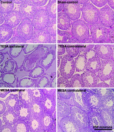
Figure 3. Histomorphometric analysis (relative values) of testicular components in control and experimental groups (A, B, C, D). Data showed significant increase in the number of tubular cross sections (numerical density) per unit area in the TESA testes (A) this could be due to the reduction in seminiferous tubular diameter as indicated by data shown in B, along with loss of epithelial height (C). D) indicates the reduction in the volume of various testicular components (tubules, epithelium, and connective tissues), these findings correspond to the significant decrease in testicular weight and volume of the gross measurements in corresponding groups. These data indicate a significant loss of germinal epithelium in both TESA and MESA with a significant damage in TESA ispsilateral testis. Each bar represents mean ± SEM of six animals. Significant at **(P < 0.01) and ***(P < 0.001). TESA: testicular sperm aspiration; MESA: microsurgical epididymal sperm aspiration
Histopathological changes in the epididymis
Normal histological features were seen in the epididymides of control and sham-control, TESA and MESA contralateral sides. In the MESA group, inflammatory reactions in the interstitium in the ipsilateral epididymis and characteristic thickening of the basement membrane, inflammatory cells in the interstitium. This was marked by massive mononuclear cell aggregations or perivascular cuffing in and around capillaries. The histomorphometric values of various epididymal parameters studied are presented in .
Figure 4. Histomorphometric values (relative values) of epididymis in control and experimental groups are shown in A, B, C, D, E, F (height of the epithelium, diameter of tubules, number of tubules, and volume of various epididymal components in caput, corpus, and cauda region, respectively). Histometric data indicates that apart from the change in epithelium (E) and connective tissue (F) in MESA-ipsilateral epididymis, there was no obvious change in the rest of the groups. Each bar represents mean ± SEM of six animals. Significant at *** (P < 0.001); TESA: testicular sperm aspiration; MESA: microsurgical epididymal sperm aspiration
In-situ cell death detection (TUNEL)
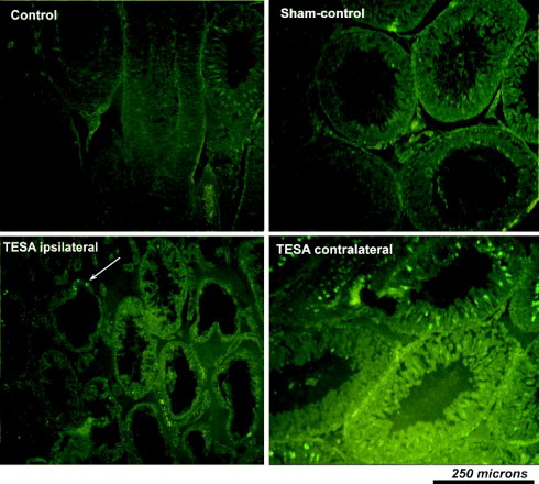
TUNEL staining to demonstrate in-situ cell death revealed apoptotic up-regulation in the contralateral side testis of the TESA group when compared to the control and sham-control. Whereas in the case of ipsilateral testis there were fewer apoptotic cells seen with TUNEL staining. This was due to severe epithelial degeneration leaving very few germ cells or apoptotic positive cells in tubular lumen (). However, TUNEL staining showed no noticeable change in apoptotic cells in testis taken from the MESA group when compared to control testis.
Figure 5. Photomicrograph of testis taken from various experimental groups after staining with TUNEL technique to demonstrate apoptotic positive cells. No obvious apoptotic cells seen in control and sham-control. However, increased apoptosis of germ cells was seen in TESA testis (predominantly in the regions of germ/sperm cells). Though it appears that the number of apoptotic positive cells were relatively reduced in TESA ipsilateral testis when compared to other groups and contralateral side, actually it was because of the loss of seminiferous epithelia and sperm cells in majority of the tubules after TESA. TESA: testicular sperm aspiration; MESA: microsurgical epididymal sperm aspiration
Discussion
The present study demonstrates severe damage and inflammatory complication after needle prick injury, the procedure used to aspirate sperm from testis. Following the unilateral TESA procedure, there was severe destruction of the tubules and spermatogenesis was severally altered in both ipsilateral and contralateral testes after 60 days. The secondary damage might have been caused by inflammatory and immunological reactions rather than trauma caused by this procedure. It is often very difficult to find a suitable method for sperm aspiration and none of the currently available techniques fulfils desirable criteria, i.e., minimal testicular trauma and the retrieval of a sufficient amount of motile spermatozoa [Donoso et al. Citation2007]. Testicular alteration appears to be progressive in nature within the postoperative period [Shufaro et al. Citation2002]. Histomorphometric analysis of testis showed a significant increase in the number of tubular cross sections (numerical density) per unit area in the TESA testis when compared to controls. This could result from a reduction in tubular diameter after epithelial cell depletion, thereby permittng tubules to come closer together. On the whole the quantitative data obtained from TESA testes showed a reduction in the volume of the testicular components, i.e., tubules, epithelium, and connective tissues, which correlate to the significant decrease in gross measurements of testicular weight and volume.
In the case of the unilateral MESA procedure there was a microscopic inflammatory reaction in the interstitium of epididymis, with slight alterations in their corresponding testis. However, the severity of the complications in the testis following the MESA procedure seems to be less compared to the TESA procedure. Our results confirm the involvement of both immunological and paracrine mechanisms in triggering immune reactions and acute inflammatory changes in the testis that are followed by testicular alteration. The histomorphometric observations indicate a significant loss in both sides of germinal epithelium in TESA and MESA. Significant damage was recorded in the testicular components of TESA ipsilateral side testes. Surprisingly, spermatic granuloma formation was not seen in testis or epididymis in any of these groups. The Leydig cell population and the testosterone and estradiol levels were not altered in any of the groups up to 60 days when compared with controls. It has been suggested in previous reports that these procedures have less impact on sexual and hormonal functions [Lewin et al. Citation1999; Westlander et al. Citation2001; Komori et al. Citation2004]. Conversely, a decrease in the concentrations of testosterone after testicular sperm aspiration procedures has been reported (Manning et al. 1998; Schill et al. Citation2003]. These reports indicate an unclear picture with respect to the hormonal level. Hence, future studies focusing on complete hormone profiling or analyzes of hypothalmo-hypophysial gondal axis after these procedures could be interesting.
The serum anti-sperm antibody titer levels increased in TESA and MESA animals, indicating the potential role of immuno-regulatory factors in degenerative changes. However, in the TESA group the titer was significantly higher than the MESA group. Though there was an increase in titer of MESA animals this seems to be insufficient to induce testicular damage around 60 days of survival. With regard to the appearance of anti-sperm antibodies, previous studies have shown fluctuation (appearance and reduction) in the development of anti-sperm antibody during various time periods (1–12 weeks) in vasectomized rats [Flickinger et al. Citation2000]. The clinical consequences of the anti-sperm antibody in infertile couples is still not clear and remains controversial. There is no direct causal relationship between the amount of testicular damage and levels of anti-sperm antibodies [Mazumdar and Levine Citation1998; Chehval et al. Citation2002]. In our study, the damage inflicted to the testis in MESA group was less than the TESA group. If the MESA procedure was repeated on the same subject the chance of inducing testicular damage would increase [Vernaeve et al. Citation2006].
The lack of an inflammatory reaction in testis after traumatic injuries or injection of testicular substances has been reported [Itoh et al. Citation1999]. This has been attributed to the lack of infiltration in testis. It could reflect the large population of resident-type macrophages in testis, exhibiting immunosuppressive activity [Hedger and Meinhardt Citation2003], faster lymphatic drainage in testis [Itoh et al. Citation1999], immunosuppressive effect of Leydig cells [Born and Wekerle Citation1982], and blood–testis barrier [Hedger and Meinhardt Citation2003]. Despite these possibilities, the infiltration and inflammatory reactions do occur in the testis after TESA, indicating that the injury/trauma inflicted by this procedure was more than the withstanding or repairing ability of the testis.
Spermatogenesis is a dynamic process of germ cell proliferation and differentiation from spermatogonia to spermatozoa. Testicular germ cell apoptosis occurs normally and continuously throughout adult life [Bartke Citation1995; Billig et al. Citation1995]. Massive testicular germ cell loss is known to result from toxicant exposure [Blanchard et al. Citation1996; Richburg and Boekelheide Citation1996], depletion of growth factors [Yoshinaga et al. Citation1991], alterations of hormonal support (testosterone or pituitary hormones including FSH and LH) [Russell et al. Citation1987; Sinha Hikim et al. Citation1995], heat exposure [Miraglia and Hayashi Citation1993], and radiation or treatment with chemotherapeutic compounds [Meistrich Citation1993]. In the majority of these conditions, germ cells are known to undergo apoptosis [Bartke Citation1995; Billig et al. Citation1995], indicating that a specific pathway is activated when the testicular environment is unable to support spermatogenesis. In this study, up-regulation of germ cell apoptosis was seen in testicular needle prick, i.e., TESA in both contralateral and ipsilateral testes suggesting paracrine and immunological factor involved in germ cell apoptosis. The TUNEL staining of MESA testes showed inconsistent results, indicating variation in testicular response to the epididymal trauma. This could reflect the variation in potential adverse effects on the epididymis that can be overshadowed. Some of the possible mechanisms (apart from immunological and paracrine) that result in seminiferous tubular alteration [Hess Citation1998] include fluid reabsorption, sperm stasis, followed by leukocyte chemotaxis and fibrosis. However, germ cell apoptotic up-regulation and duct system pathology could possibly increase the number of defective sperm and sperm with poor DNA integration. This could affect fertility in subsequent trials. Our previous study had shown that the pathobiology of the duct system plays a major role in the etiology of defective sperm [Suresh et al. Citation2010]. In general, when compared to the testicular sperm retrieval procedure, the pathophysiological consequences of epididymal sperm retrieval procedure in animal model remains to be explored.
In conclusion, the present observations showed that even in a single trial fewer punctures in any one of the testis can cause traumatic damage in rat testis. This could decrease the success rate of sperm retrieval and indicates the importance of these procedures in assisted reproduction in men with very limited baseline fertility who are under high risk of becoming totally infertile. Both these procedures challenge the testicular immuno-privilege nature by breaching the blood–testis barrier. Thus, the process of the inflammatory reaction in the testicular interstitium has the potential to disrupt spermatogenesis. However, extrapolating conclusions from an experimental model to the clinic needs to be done cautiously.
Materials and Methods
Animals
Twenty-four mature male Wistar albino rats with a proven breeding history, weighing around 200–250 gm were used for the study. Animals were randomly divided into four groups (n = 6), i.e., control, sham-control, unilateral TESA, and MESA. Another eight animals were used for immunization procedure (n = 6), two animals were utilized for isolating sperm required for immunization (n = 1), and ELISA plate coating (n = 1). The quarantine procedures and animal maintenance have been carried out according to the International (Canadian Council) and Local body (Laboratory animals, India). The Institutional Animal Ethical Committee, University of Madras, has approved the protocol of the work (IAEC No.01/015/03). Details of the animal maintenance are given elsewhere [Suresh et al. Citation2009; Citation2010].
TESA/MESA procedures
All the (TESA and MESA) procedures were performed under xylazine (10 mg/kg i.m.) (Indian Immunological Limited, India) and ketamine (80 mg/kg i.p.) (Neon Laboratories Limited, India) anesthesia. TESA was performed as described by Shufaro et al. [2002]. Scalp vein set with 22 gauge needle attached to a 20 mL syringe was used as the aspiration device. While holding the testicle between the index finger and the thumb, five multidirectional punctures were made in different regions of the right testicle applying strong negative pressure with the syringe. Hemostasis was achieved by the application of gentle pressure on each site after aspiration. Similarly, MESA was performed by needle aspiration as described by Schroeder-Printzen et al. [2000] in caudal epididymis, three multi-directional punctures were made. These procedures were done unilaterally on right side leaving left-side testis or epididymis undisturbed. In sham-control, animal testis, and epididymis were manipulated similar to the above procedures, but without making needle aspiration. Postoperative period was uneventful.
Serum sample
At the end of the postoperative period blood samples were collected by retro-orbital venous plexus from all the experimental groups at the 60th day of the postoperative period. The blood samples were collected and allowed to coagulate for 1 h at 4°C. The samples were centrifuged at 2,000 x g for 15 min and the supernatant was removed and the procedure was repeated. The resulting serum was stored in aliquots at -70°C or processed further. Stored serum was used to estimate testosterone and estradiol using RIA [Auletta et al. 1979], as described previously [Suresh et al. Citation2009].
ELISA
Preparation of pooled hyperimmune sera
A male rat was sacrificed by overdose of anesthesia. Caudal portion of epididymis was excised and rinsed in Tyrode's salt solution (pH 7.35, Sigma, USA) at 37°C in a Petri dish. The epididymis was minced into 1-mm pieces and incubated for 20 - 25 min at 37°C. Sperm free from tissue debris were collected by centrifugation for 15 min at 3,000 x g and washed three times with Tyrode's solution. Hyperimmune serum antisperm antibodies were raised by immunizing with the isologous sperm.
Briefly, 4 x 10⁷ sperm cells per mL of suspension was mixed with an equal volume of Freund's complete adjuvant (Sigma) and the suspension was emulsified. Six rats were injected intraperitoneally with the equivalent of 1 x 10⁷ sperm cells in 0.5 mL of homogenate. Each animal received an intraperitoneal boost of 1 x 10⁷ sperm emulsified in Freund's incomplete adjuvant (Sigma) after second, fourth, and sixth weeks. Blood samples were obtained by retro-orbit puncture on the 10th day after the final booster. The separated serum samples (as described above) were pooled, aliquoted, and frozen at -70°C as a positive control serum in ELISA.
Sperm collection and detection of anti-sperm antibodies
The sperm collected from the caudal epididymis of a rat was diluted (1 x 10⁶ sperm/mL in PBS containing 0.2 mM of PMSF, Himedia, India). Each well received 100 mL of freshly prepared sperm suspension and the plates were incubated at 37°C overnight to dry sperm onto the wells, then 200 mL of 10% sucrose (SRL fine chemicals, India) and 4% polyvinylpyrrolidone (Himedia) were added to each well as a post-coat stabilizer, followed by further incubation at 37°C for 1 h. Plates were stored at 4°C. Blocking buffer consisting 1% nonfat dry milk in PBS and 0.05% Tween 20 (Sigma) were added to the wells to reduce nonspecific protein–protein interactions. These plates were then incubated for 1 h at 37°C and washed three times with PBS–Tween 20. The primary (test) sera were diluted 1:80 in PBS–Tween 20 and 1% bovine serum albumin (Sigma).
After incubation for 1 h at 37°C, the plates were again washed three times. The secondary antibody, rabbit anti-rat IgG (gamma-chain specific) conjugated with horseradish peroxidase (Gene, Bangalore, India) diluted 1:7,500 in PBS–Tween 20 and 1% BSA were added to each well, and incubated at 37°C for 1 h. After washing with PBS–Tween 20 three times, 100 mL of 1 mM 2,2’-azinobis-(3-ethylbenzthiazoline sulfonic acid) (Sigma) and 0.1% hydrogen peroxide in citrate phosphate buffer (SRL fine chemicals) were added to each well.
The absorbance at 414 nm of the reaction mixture was determined from triplicate assays of each serum sample with an ELISA plate reader. A 1:8,000 dilution of the pooled hyperimmune serum was assayed as a positive control. The negative control consisted of secondary antibody and sperm in the absence of test serum (primary antibody), and primary and secondary antibodies without sperm. To correct the possible variations between plates, absorbance readings for all the samples were normalized to the value of the hyperimmune serum on the same plate, which was considered to be 1. The test procedure was carried out in accordance with the method described by Flickinger et al. [2000]
Tissue harvesting and histological analysis
After 60 d of postoperative period, animals were euthanized by overdose of anesthesia. The testis and epididymis were dissected out and fixed in Bouin's and 10% formyl saline fixatives for histological and TUNEL stainings, respectively. Tissues were processed for paraffin embedding and sections were taken at 5 µm thickness. Sections were stained with Ehrlich alum hematoxylin and eosin and observed under bright-field microscope (Nikon Corporation, Tokyo, Japan). Histomorphometric analysis was done, details of the procedure are described elsewhere [Prakash et al. Citation2008; Prakash et al. Citation2009]. Values are given in relative terms.
In-situ cell death detection (TUNEL)
The TUNEL evaluation of germ cell apoptosis in the testis was done using the in-situ cell death detection kit-FITC labeled (Roche, Mannheim, Germany) following the manufacturer's protocol. In brief, the formalin-fixed paraffin sections were deparaffinized, hydrated, and treated with freshly prepared permeabilization solution (0.1% Triton X-100; SRL fine chemicals) in 0.1% sodium citrate (SRL fine chemicals) for 2 min in ice (2–8°C). After rinsing twice with PBS, sections were incubated for 60 min with TUNEL reaction mixture in a humidified chamber at 37°C. Slides were rinsed thrice in PBS for 5 min each and coverslip was placed and observed under fluorescence microscope (Nikon Corporation) at an excitation wavelength of 450–500 nm and the detection range of 515—565 nm (green).
Statistical analysis
The data were subjected to a one-way ANOVA and the significance determined using a ‘Tukey's post-hoc’ test with P < 0.05 considered statistically significant. The statistical tests were performed with SPSS for Windows (SPSS, Version 10.0 Inc., Chicago, IL, USA).
Declaration of interest: This study was supported by the grants from University with Potential for Excellence program (UWPFE) (Project No. HDP-16), University of Madras, and University Grant Commission (UGC) major project was sanctioned to SP (Project sanction No. F.No.33-380/2007 SR). EP was supported by Council for Scientific and Industrial Research (CSIR) - senior research fellowship (SRF). The authors report no conflicts of interest.
Author contributions: Conceived and designed the experiments: EP, SP; Performed the experiments: EP; Analyzed the data: EP, SS, MM, SP; Contributed reagents/ materials/ analysis tools: EP, SS, SP; Wrote the manuscript: EP, MM, SP.
Abbreviations
| ICSI: | = | intracytoplasmic sperm injection |
| TESA: | = | testicular sperm aspiration |
| MESA: | = | microsurgical epididymal sperm aspiration |
| ELISA: | = | enzyme-linked immunosorbent assay. |
References
- Abuzeid, M., Chan, Y.M., Sasy, M., Basata, S. and Beer, M. (1995) Fertilization and pregnancy achieved by intracytoplasmic injection of sperm retrieved from testicular biopsies. Fertil Steril 64:644–646.
- Auletta, F.J., Caldwell, B.V. and Hamilton, C.L. (1979) Androgens: Testosterone and dihydrotestosterone. In Methods of Hormone Radioimmunoassay, 2nd ed., ed. Jaffe, B.M. and Behrman, H.R., Academic Press, New York, pp. 715–726.
- Bartke, A. (1995) Apoptosis of male germ cells, a generalized or a cell type-specific phenomenon. Endocrinol 136:3–4.
- Billig, H., Furuta, I., Rivier, C., Tapanainen, J., Parvinen, M. and Hseuh. A. J. (1995) Apoptosis in testis germ cells: developmental changes in gonadotropin dependence and localization to selective tubule stages. Endocrinol 136:5–12.
- Blanchard, K.T., Allard, E.K. and Boekelheide, K. (1996) Fate of germ cells in 2,5- hexanedione-induced testicular injury. Toxicol Appl Pharmacol 137:141–148.
- Born, W. and Wekerle, H. (1982) Leydig cells nonspecifically suppress lymphoproliferation in vitro: implications for the testis as an immunologically privileged site. Am J Reprod Immunol 2:291–295.
- Bouloux, P., Warne, D. and Lourmaye, E. (2002) FSH Study Group in Mens Infertility. Efficacy and safety of recombinant human follicle-stimulating hormone in men with isolated hypogonadotropic hypogonadism. Fertil Steril 77:270–273.
- Chehval, M.J., Doshi, R., Kidd, C.F., Winkelmann, T. and Chehval, V. (2002) Antisperm autoantibody response after unilateral vas deferens ligation in rats: When does it develop? J Androl 23:669–673.
- Craft, I., Tsirigotis, M., Courtauld, E. and Farrer-brown, G. (1997) Testicular needle aspiration as an alternative to biopsy for the assessment of spermatogenesis. Hum Reprod 12:1483–1487.
- Devroey, P., Liu, J., Nagy, Z., Goossens, A., Tournaye, H., Camus, M., (1995) Pregnancies after testicular sperm extraction and intracytoplasmic sperm injection in non-obstructive azoospermia. Hum Reprod 10:1457–1460.
- Donoso, P., Tournaye, H. and Devroey, P. (2007) Which is the best sperm retrieval technique for non-obstructive azoospermia? A systematic review. Hum Reprod Update 13:539–549.
- Esteves, S., Miyaoka, R. and Agarwal A. (2011) Sperm retrieval techniques for assisted reproduction. Int Braz J Urol 37:570–583.
- Flickinger, C.J., Vagnetti, M., Stuart, B. A., Howards, S. and Herr, J.C. (2000) Antisperm autoantibody response is reduced by early repair of a severed vas deferens in the juvenile rat. Fertil Steril 73:229–237.
- Glina, S., Fragoso, J.B., Martins, F.G., Soares, J.B., Galuppo, A.G. and Wonchockier, R. (2003) Percutaneous Epididymal Sperm Aspiration (Pesa) In Men With Obstructive Azoospermia. Clinic Urol 29:141–146.
- Hedger, M.P. and Meinhardt, A. (2003) Cytokines and the immune-testicular axis. J Reprod Immunol 58:1–26.
- Hess, RA. (1998) Effects of environmental toxicants on the efferent ducts, epididymis and fertility. J Reprod Fertil 53:247–259
- Itoh, M., Xie, Q., Miyamoto, K. and Takeuchi, Y. (1999) Major differences between the testis and epididymis in the induction of granulomas in response to extravasated germ cells. I. A light microscopical study in mice. Int J Androl 22:316–323.
- Komori, K., Miura, T., Shin, M., Takada, T., Honda, M., Matsumiya K., (2004) Serial follow-up study of serum testosterone and antisperm antibodies in patients with non-obstructive azoospermia after conventional or microdissection testicular sperm extraction. Int J Androl 27:32–36.
- Lewin, A., Reubinoff, B., Porrat-Katz, A., Weiss, D., Eisenberg, V., Arbel, R., (1999) Tesicular fine needle aspiration: the alternative method for sperm retrieval in non-obstructive azoospermia. Hum Reprod 14:1785–1790.
- Lewin, A., Weiss, D.B., Friedler, S., Ben-Shachar, I., Porat-Katz, A., Meirow, D., (1996) Delivery following intracytoplasmic injection of mature sperm cells recovered by testicular fine needle aspiration in a case of hypergonadotropic azoospermia due to maturation arrest. Hum Reprod 11:769–771.
- Manning, M., Junemann, K. and Alken, P. (1998) Decrease in testosterone blood concentrations after testicular sperm extraction for intracytoplasmic sperm injection in azoospermic men. Lancet 352:37.
- Mazumdar, S. and Levine, A.S. (1998) Antisperm antibodies etiology, pathogenesis, diagnosis and treatment. Fertil Steril 70:799–810.
- Meistrich, M.L. (1993) Effects of chemotherapy and radiotherapy on spermatogenesis. Euro Urol 23:136–141.
- Miraglia, S.M. and Hayashi, H. (1993) Histomorphometry of immature rat testis after heating. J Morphol 217:65–74.
- Palermo, G., Joris, H., Derde, M.P., Camus, C., Devroey, P. and Van Steirteghem, H. (1993) Sperm characteristics and outcome of human assisted fertilization by subzonalinsemination and intracytoplasmic sperm injection. Fertil Steril 59:826–835.
- Prakash, S., Prithiviraj, E. and Suresh, S. (2008) Developmental changes of seminiferous tubule in prenatal, postnatal and adult testis of bonnet monkey (Macaca radiata). Anat Histol Embryol 37:19–23.
- Prakash, S., Suresh, S. and Prithiviraj, E. (2009) Anatomical aspects of the male reproductive system in the bonnet monkey (Macaca radiata). Anat Sci Int 84:53–60.
- Ramasamy, R., Yagan, N. and Schlegel, P. (2005) Structural and functional changes to the testis after conventional versus microdissection testicular sperm extraction. Urol 65:1190–1194.
- Richburg, J.H. and Boekelheide, K. (1996) Mono-(2-ethylhexyl) phthalate rapidly alters both Sertoli cell vimentin filaments and germ cell apoptosis in young rat testes. Toxicol and Appl Pharmacol 137:42–50.
- Rupin, S. (2011) Surgical sperm retrieval: Techniques and their indications. Ind J Urol 27:102–109.
- Russell, L.D., Alger, L.E. and Nequin, L.G. (1987) Hormonal control of pubertal spermatogenesis. Endocrinol 120:1615–1632.
- Schill, T., Bals-Pratsch, M., Küpker, W., Sandmann, J., Johannisson, R. and Diedrich, K. (2003) Clinical and endocrine follow-up of patients after testicular sperm extraction. Fertil Steril 79:281–286.
- Schlegel, P. and Su, L.M. (1997) Physiological consequenses of testicular sperm extraction. Hum. Reprod 12:1688–1692
- Schoysman, R., Vanderzwalmen, P., Nijs, M., Segal, L., Segal-Bertin, G., Geerts, L., (1993) Pregnancy after fertilization with human testicular spermatozoa. Lancet 342:12–37.
- Schroeder-Printzen, I., Zumbé, J., Bispink, L., Palm, S., Schneider, U., Engelmann, U., (2000) Microsurgical epididymal sperm aspiration: aspirate analysis and straws available after cryopreservation in patients with non-reconstructable obstructive azoospermia. Hum Reprod 15:2531–2535.
- Shufaro, Y., Prus, D., Laufer, N. and Simon, A. (2002) Impact of repeated testicular fine needle aspiration (TEFNA) and testicular sperm extraction (TESE) on the microscopic morphology of the testis: an animal model. Hum Reprod 17:1795–1799.
- Silber, S.J., Nagy, Z., Liu, J., Tournaye, H., Lissens, W., Ferec, C., (1995a) The use of epididymal and testicular sperm for ICSI: the genetic implications for male infertility. Hum Reprod 10:2031–2043.
- Silber, S.J., Van Steirteghem, A.C., Liu, J., Nagy, Z., Tournaye, H. and Devroey, P. (1995b) High fertilization and pregnancy rates after ICSI with spermatozoa obtained from testicle biopsy. Hum Reprod 10:148–152.
- Sinha Hikim, A.P., Wang, C., Leung, A. and Swerdloff, R.S. (1995) Involvement of apoptosis in the induction of germ cell degeneration in adult rats after gonadotropin-releasing hormone antagonist treatment. Endocrinol 136:2770–2775.
- Suresh, S., Prithiviraj, E. and Prakash, S. (2009) Dose- and time-dependent effects of ethanolic extract of Mucuna pruriens Linn. seed on sexual behaviour of normal male rats. J Ethnopharmacol 122:497–501.
- Suresh, S., Prithiviraj, E. and Prakash, S. (2010) Effect of Mucuna pruriens on oxidative stress mediated damage in aged rat sperm. Int J Androl 33:22–32.
- Tash, J. and Schlegel, P. (2001) Histologic effects of testicular sperm extraction on the testicle in men with nonobstructive azoospermia. Urol 57:334–337.
- Toumaye, H., Camus, M., Goossens, A., Nagy, Z., Silber, S., Van Steirteghem, A.C., (1995) Recent concepts in the management of infertility because of non-obstructive azoospermia. Hum Reprod 10:115–119.
- Toumaye, H., Devroey, P., Liu, J., Nagy, Z., Lissens, W. and Steirteghem, A.V. (1994) Microsurgical epididymal sperm aspiration and intracytoplasmic sperm injection: a new effective approach to infertility as a result of congenital bilateral absence of the vas deference. Fertil Steril 61:1045–1051.
- Van Peperstraten, A., Proctor, M., Johnson, N. and Philipson, G. (2006) Techniques for surgical retrieval of sperm prior to ICSI for azoospermia. Cochrane Database Syst Rev 19: 3 CD002807.
- Vernaeve, V., Verheyen, G., Goossens, A., Steirteghem, A.V., Devroey, P. and Tournaye, H. (2006) How successful is repeat testicular sperm extraction in patients with azoospermia? Hum Reprod 21:1551–1554.
- Westlander, G., Ekerovd, E., Granberg, S., Lycke, N., Werner, L. N.C. and Berrgh, C. (2001) Serial ultrasonography, hormonal profile and antisperm antibody response after testicular sperm aspiration. Hum Reprod 1:2621–2627.
- Yoshinaga, K., Nishikawa, S., Ogawa, M., Hayashi, S., Kunisada, T., Fujimoto, T., (1991) Role of c-kit in mouse spermatogenesis: identification of spermatogonia as a specific site of c-kit expression and function. Develop 113: 689–699.
- Zenke, U., Jalalian, L., Shen, S. and Turek, J. (2004) The difficult MESA: findings from tubuli recti sperm aspiration. J Assist Reprod Gen 21:31–35.