
Abstract
Small supernumerary marker chromosomes (sSMC) are found about four times more frequently in subfertile compared to the general population. The reason for this finding is still unclear. However, a connection of interphase architecture and genome function is suggested. And as we found in a previous study the presence of sSMC influences the nuclear architecture of peripheral blood cells and fibroblasts, we hypothesized that sSMC could have similar effects in sperm cells possibly leading to infertility. Here we applied for the first time 3-dimensional interphase fluorescence in situ hybridization (3D-FISH) to characterize the position of an extra-chromosome with respect to its sister- and selected other chromosomes (6, 15, 18, 19, 21, X, and Y) in sperm. Two sSMC carrier brothers with the identical sSMC derived from chromosome 15 were studied. One of the brothers was fertile and the other brother was infertile. Deviations from the normal positioning of chromosomes 21 and Y were seen in both brothers and for chromosomes 19 and X only in the infertile brother. Most striking were high rates of nullisomy and/or disomy for chromosomes 15, including sSMC (15), and 18 exclusively seen in the infertile brother. Overall, further evidence is provided that sSMC influence the nuclear architecture of a cell, including sperm. Further studies are necessary in sperm of fertile and infertile sSMC carriers to elaborate if the detected aneuploidy like that seen in the infertile brother is due to sSMC presence and disturbance of nuclear architecture.
Introduction
Small supernumerary marker chromosomes (sSMC) can be detected in 0.171% of subfertiles compared to 0.043% within the general population [Liehr and Weise Citation2007]. sSMC are defined as “structurally abnormal chromosomes that cannot be identified or characterized unambiguously by conventional banding cytogenetics alone, and are generally equal in size or smaller than chromosome 20 of the same metaphase spread” [Liehr et al. Citation2004, p55]. They are in almost all instances detected in routine cytogenetics as an unexpected finding [Liehr et al. Citation2004]. In prenatal cases sSMC are not easily aligned with a specific clinical outcome [Liehr et al. Citation2010]. Even though progress towards genotype-phenotype-correlation was achieved already [Liehr et al. Citation2006; Liehr Citation2014], the reason for the correlation of sSMC with infertility, especially in males remains unresolved. On the one hand oligozoospermia is significantly correlated with sSMC presence in 7% of the cases, while in azoospermia sSMC are present in <1% of the corresponding cases [Liehr and Weise Citation2007]. On the other hand it has been demonstrated that in >50% of infertile sSMC-carriers the sSMC was parentally transmitted [Manvelyan et al. Citation2008a].
Non-random positioning in interphase nuclei is now known to be of importance for genomic stability as well as for formation of chromosomal aberrations [Manvelyan et al. Citation2009]. Also further impact of nuclear architecture on genome function is suggested [Mateos-Langerak et al. Citation2007]. In a previous study we have shown that sSMC-presence has an impact on the nuclear architecture of peripheral blood cells as well as fibroblasts [Klein et al. Citation2012]. Thus, we applied here for the first time 3-dimensional interphase fluorescence in situ hybridization (3D-FISH) for the determination of positioning of sSMC with respect to their sister- and selected other chromosomes in the sperm of two sSMC carriers, brothers with the identical sSMC derived from chromosome 15. Interestingly one of the brothers was fertile (brother F), while the other one was infertile (brother I) as previously reported [Guediche et al. Citation2012].
Results
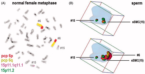
The 3D-interphase FISH was performed on sperm of two brothers with an inv dup(15)(q11.2) using probe sets comprised of partial chromosome painting (pcp) as well as commercially available centromeric probes (cep). The probe sets were tested on a normal metaphase spread, as shown in and applied in 3-D-FISH later (Figure 1B).
Figure 1. Example for a probeset used for fluorescence in situ hybridization (FISH) in the present study on a normal diploid female metaphase (A) and in a human sperm with a small supernumerary marker chromosome (sSMC) 15. (A) All probe sets used were designed as shown exemplarily for combination of chromosome-6- and -15-specific probeset. Here this probeset was hybridized on a normal diploid female, peripheral blood derived metaphase spread: partial chromosome paints (pcp) for short (p) and long (q) arm identify chromosome (= #) 6; a combination of an alphoid centromeric probe for 15p11.1 to 15q11.1 and a satellite III probe for 15p11.2 was applied to stain chromosome 15. (B) An example of how 3D-FISH analyses looked like using Cell-P-program (Olympus) is shown. The same sperm is shown twice. In the upper lane only chromosome 15 and sSMC(15) are depicted; as only probes covering overall 15p11.2 to 15q11.1 were applied the normal chromosome 15 shows a smaller staining than the sSMC(15) and both can be distinguished. In the lower lane all color channels used are depicted, i.e., the sSMC(15) and chromosomes 15 as well as 6 can be identified. Chromosomes 6, 15, and sSMC(15) were positioned in periphery. Chromosome 15 is located towards the sperm tail, chromosome 6 towards the head. Chromosome 6 and sSMC(15) are localized in close proximity, but also chromosome 15 and sSMC(15) are located together and not on opposite sides of the nucleus.
The results are summarized in and and depict the location of chromosomes 6, 15, 18, 19, 21, X, Y, and the sSMC (15) in the brothers (F and I). Also, in the same figures, these data are compared to the location of the corresponding chromosomes in a normal male without sSMC (data from [Manvelyan et al. Citation2008b]). Thus, as a result there was no change of the positioning of chromosomes 6, 15, and 18 in sperm of the brothers (I and F). Also the sSMC (15) was located at similar positions e.g., chromosome 15. Still, deviations from the normal localization were observed for chromosomes 21 and Y in both brothers and for chromosomes 19 and X only in brother I ( and ).
Figure 2. Positioning of the studied chromosomes, including small supernumerary marker chromosome (sSMC) 15, with respect to localization towards sperm center or periphery in both studied brothers and normal person. Positioning of chromosomes 6, 15, 18, 19, 21, X and Y, and the sSMC(15) towards sperm center and periphery in fertile brother (F) and infertile brother (I) studied, here compared to normal situation (N) according to [Manvelyan et al. Citation2008b]. Experiments were done using 3D-FISH (fluorescence in situ hybridization) as depicted in .
![Figure 2. Positioning of the studied chromosomes, including small supernumerary marker chromosome (sSMC) 15, with respect to localization towards sperm center or periphery in both studied brothers and normal person. Positioning of chromosomes 6, 15, 18, 19, 21, X and Y, and the sSMC(15) towards sperm center and periphery in fertile brother (F) and infertile brother (I) studied, here compared to normal situation (N) according to [Manvelyan et al. Citation2008b]. Experiments were done using 3D-FISH (fluorescence in situ hybridization) as depicted in Figure 1.](/cms/asset/299f2311-e053-4343-af17-d3df2866c3c0/iaan_a_979956_f0002_c.jpg)
Figure 3. Positioning of the studied chromosomes, including small supernumerary marker chromosome (sSMC) 15, with respect to localization towards sperm head, middle, or tail in both studied brothers and normal person. Depicted is the positioning of chromosomes as in towards sperm head, middle part, and tail in fertile brother (F) and infertile brother (I) studied here and compared to normal situation (N) according to [Manvelyan et al. Citation2008b]. Experiments were done using 3D-FISH (fluorescence in situ hybridization) as depicted in .
![Figure 3. Positioning of the studied chromosomes, including small supernumerary marker chromosome (sSMC) 15, with respect to localization towards sperm head, middle, or tail in both studied brothers and normal person. Depicted is the positioning of chromosomes as in Figure 2 towards sperm head, middle part, and tail in fertile brother (F) and infertile brother (I) studied here and compared to normal situation (N) according to [Manvelyan et al. Citation2008b]. Experiments were done using 3D-FISH (fluorescence in situ hybridization) as depicted in Figure 1.](/cms/asset/50069a5e-1bbe-466e-a43d-e2eb7da7f67b/iaan_a_979956_f0003_c.jpg)
Figure 4. Colocalization of the small supernumerary marker chromosome (sSMC) 15 and the corresponding tested chromosomes in both brothers. Percentage of sperm cells with colocalization of the sSMC and the corresponding tested chromosomes as in in fertile brother (F) and infertile brother (I). Experiments were done using 3D-FISH (fluorescence in situ hybridization) as depicted in .
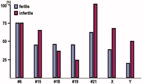
Figure 5. Monosomy and disomy for the studied chromosomes and the small supernumerary marker chromosome (sSMC) 15 in both brothers. Percentage of sperm with monosomy and disomy for the studied chromosomes and the sSMC(15) in fertile brother (F) and infertile brother (I). Experiments were done using 3D-FISH (fluorescence in situ hybridization) as depicted in .
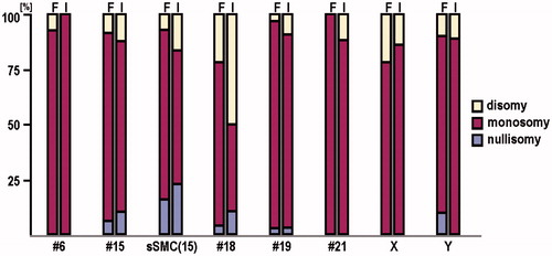
As highlighted in co-localization rates of sSMC (15) and the other tested chromosomes were overall high. Co-localization was highest with chromosomes 6, 21, 15, and X and less expressed with chromosomes 18, 19, and Y in both samples. In all cells studied here were included, e.g., aneuploid cells, which were excluded from evaluation before; while no exceptionally high rates of aneuploidy were detected for chromosomes 6, 19, 21, X, and Y, more than 25% up to ∼60% of the studied nuclei showed nullisomy and/or disomy for chromosomes 15, including sSMC (15), and 18. Aneuploid cells were more often observed in brother I as compared to brother F.
Discussion
To date, only a few sperm segregation analysis of sSMC carriers have been performed and reported for the sSMC derived from chromosomes 15 [Cotter et al. Citation2000; Guediche et al. Citation2012; Oracova et al. Citation2009; Paetzold et al. Citation2006], 20 [Wiland et al. Citation2005], and 22 [Perrin et al. Citation2012]. However, the nuclear architecture in sperm of sSMC carriers has never been done before. Based on our own previous study on sperm of a healthy male donor, the localization/positioning of all chromosomes, except acrocentric chromosomes which are dislocated in active cells due to their involvement in nucleolus organization, in sperm is similar to that known from peripheral blood lymphocytes [Manvelyan et al. Citation2008b].
In the present study, we observed that the positioning of chromosomes 6, 15, and 18 was not influenced and that sSMC (15) located at similar positions as chromosome 15, in sperm of two brothers with sSMC. The latter is in concordance with a previous observation of sSMC positioning in peripheral blood lymphocytes [Klein et al. Citation2012]. However, chromosomes 21 and Y were positioned more centrally in both brothers in comparison to the normal scenario (), with both chromosomes being displaced towards the central and/or tail part in the brother I (). For chromosomes 19 and X, a shift of the X-chromosome towards the central part and of chromosome 19 to the distal part of the nucleus were detected only in the infertile sibling (). Furthermore chromosome 19 was displaced from the head/middle to the middle/tail region (). This data supports the idea that sSMC influences the nuclear architecture [Klein et al. Citation2012]. Also it seems to be obvious that other factors besides sSMC presence can modulate the impact of the latter on the nuclear architecture, as the identical sSMC led to different effects in the two studied cases.
From a previous study it is known that sSMC show a preferential co-localization with their sister chromosome(s) in peripheral blood cells [Klein et al. Citation2012]. Also evidence was provided that this ‘attraction’ between an sSMC and one of its homologous sister chromosomes is transmitted by the euchromatic part of the sSMC rather than its heterochromatic one. In the present study chromosome 15 and sSMC (15) were co-localized in ∼45 to 65% of the sperm. However, the same or even higher co-localizations were detected for other chromosomes like 6, 21, and X. In peripheral blood cells sSMC (15) and one chromosome 15 were co-localized in 87% of the nuclei in fibroblasts in 58% of the studied cells [Klein et al. Citation2012]. A possible explanation for the deviations observed here might be that in the nucleus of a sperm such co-localization rates are influenced by the smaller size and higher compactness of the cell.
Previous studies on sperm of sSMC carriers have detected enhanced aneuploidy rates [Cotter et al. Citation2000; Guediche et al. Citation2012; Oracova et al. Citation2009; Paetzold et al. Citation2006; Perrin et al. Citation2012; Wiland et al. Citation2005]. The results obtained from the present study, confirmed and extended the results published by Guediche et al. [Citation2012] for both brothers (F and I). Nullisomy and/or disomy for chromosomes 15, including sSMC (15) and 18 were observed, but not for the other studied chromosomes. Interestingly higher aneuploidy rates were found in brother I than in brother F. This finding may be due to an adverse secondary effect of sSMC presence in sperm, which again is modulated by other genes, leading to infertility in the one and no influence on fertility in the other subject.
Overall this study provides further evidence that extra chromosomes, especially sSMC change the nuclear architecture of the cell. It is interesting that identical sSMC can be found in a fertile and an infertile male. Differences in nuclear sperm architecture were present in those siblings, still the major difference was the number of cells with aneuploidy. Enhanced aneuploidy-rates were also seen in other sSMC carriers studied due to infertility [Martin et al., 1986; Cotter et al. Citation2000; Oracova et al. Citation2009; Paetzold et al. Citation2006; Perrin et al. Citation2012; Wiland et al. Citation2005]. Further studies are necessary in sperm of fertile and infertile sSMC carriers to elaborate if the aneuploidy is due to sSMC presence and disturbance of nuclear architecture.
Material and Methods
Samples
Two brothers included in this study are carriers of an sSMC derived from chromosome 15 previously reported as inv dup(15)(q11.2) (see [Guediche et al. Citation2012]).
The younger brother was 39 years of age and presented with primary infertility; this infertile individual is referred as ‘brother I’; he had a severe oligozoospermia and the sperm was with low progressive motility and teratozoospermia. His 47-year-old brother had two healthy children (paternity test was not performed) and is referred as ‘brother F’ for fertile in this report; he had moderate oligozoospermia. Both males were otherwise phenotypically normal and there was no family history of infertility, congenital anomalies, or mental retardation (see as well [Guediche et al. Citation2012]). Sperm samples as used in Guediche et al. [Citation2012] were obtained from brothers I and F for research purposes with informed consent and institutional ethics committee approval.
Suspension FISH (S-FISH)
Whole chromosome painting (wcp), partial chromosome painting (pcp), and/or centromeric probes for chromosomes 6, 15, 18, 19, 21, X, and Y [Weise et al. Citation2008] were applied together in suspension-FISH (S-FISH) as previously reported [Iourov et al. Citation2007; Klein et al. Citation2012; Manvelyan et al. Citation2008b; Manvelyan et al. Citation2008c; Manvelyan et al. Citation2009; Steinhaeuser et al. Citation2002]. Images of 3D-preserved interphase nuclei were captured on a Zeiss Axioplan microscope (Car Zeiss, Jena, Germany) and analyzed by the Cell-P (Olympus, Hamburg, Germany) software. 30-50 interphase nuclei were acquired and processed per case. Between 20 and 30 nuclei were evaluated as reported previously [Iourov et al. Citation2007; Klein et al. Citation2012; Manvelyan et al. Citation2008b; Manvelyan et al. Citation2008c; Manvelyan et al. Citation2009; Steinhaeuser et al. Citation2002].
Evaluation
Only cells with one sSMC, one chromosome 15, and only one copy of the additionally tested chromosome were taken into account for the determination of localization. Sperm cells with nullisomy or disomy were recorded and are summarized in . Position and distance of chromosomes to the sSMC and/or each other were determined during the 3D-evaluation. An interphase nucleus was divided into two spheres, i.e., periphery (P) and center (C) and 50% of the nuclear radius was defined as ‘center'. Thus, analyzed chromosomes could either be allocated as C or P. Finally, the positioning of the chromosomes or the sSMC was recorded with respect to the sperm axis, i.e., if located in head (H), middle (M), or tail (T) [Klein et al. Citation2012; Manvelyan et al. Citation2008b] ().
Acknowledgment
This study was supported in part by the Deutsche Forschungsgemeinschaft (DFG) (LI 820/46-1).
Declaration of interest
The authors report no declarations of interest.
Author contributions
Performed the 3D-FISH experiments and evaluation: TK, NK; Provided the samples and did their cytogenetic workup: NG; Final evaluation and drafting of the manuscript: TL; All authors contributed to the final version of the manuscript.
References
- Cotter, P.D., Ko, E., Larabell, S.K., Rademaker, A.W. and Martin, R.H. (2000) Segregation of a supernumerary del(15) marker chromosome in sperm. Clin Genet 58:488–92
- Guediche, N., Tosca, L., Kara Terki, A., Bas, C., Lecerf, L., Young, J., et al. (2012) Array comparative genomic hybridization analysis of small supernumerary marker chromosomes in human infertility. Reprod Biomed Online 24:72–82
- Iourov, I.Y., Liehr, T., Vorsanova, S.G. and Yurov, Y.B. (2007) Interphase chromosome-specific multicolor banding (ICS-MCB): A new tool for analysis of interphase chromosomes in their integrity. Biomol Eng 24:415–7
- Klein, E., Manvelyan, M., Simonyan, I., Hamid, A.B., Guilherme, R.S., Liehr, T., et al. (2012) Centromeric association of small supernumerary marker chromosomes with their sister-chromosomes detected by three dimensional molecular cytogenetics. Mol Cytogenet 5:15
- Liehr, T. (2014) Homepage on small supernumerary marker chromosomes (sSMC). http://ssmc-tl.com/Start.html (accessed August 06, 2014)
- Liehr, T., Claussen, U. and Starke, H. (2004) Small supernumerary marker chromosomes (sSMC) in humans. Cytogenet Genome Res 107:55–67
- Liehr, T., Karamysheva, T., Merkas, M., Brecevic, L., Hamid, A., Ewers, E., et al. (2010) Somatic mosaicism in cases with small supernumerary marker chromosomes. Curr Genomics 11:432–9
- Liehr, T., Mrasek, K., Weise, A., Dufke, A., Rodríguez, L., Martínez Guardia, N., et al. (2006) Small supernumerary marker chromosomes–progress towards a genotype-phenotype correlation. Cytogenet Genome Res 112:23–34
- Liehr, T. and Weise, A. (2007) Frequency of small supernumerary marker chromosomes in prenatal, newborn, developmentally retarded and infertility diagnostics. Int J Mol Med 19:719–31
- Manvelyan, M., Hunstig, F., Bhatt, S., Mrasek, K., Pellestor, F., Weise, A., et al. (2008b) Chromosome distribution in human sperm – a 3D multicolor banding-study. Mol Cytogenet 1:25
- Manvelyan, M., Hunstig, F., Mrasek, K., Bhatt, S., Pellestor, F., Weise, A., et al. (2008c) Position of chromosomes 18, 19, 21 and 22 in 3D-preserved interphase nuclei of human and gorilla and white hand gibbon. Mol Cytogenet 1:9
- Manvelyan, M., Kempf, P., Weise, A., Mrasek, K., Heller, A., Lier, A., et al. (2009) Preferred co-localization of chromosome 8 and 21 in myeloid bone marrow cells detected by three dimensional molecular cytogenetics. Int J Mol Med 24:335–41
- Manvelyan, M., Riegel, M., Santos, M., Fuster, C., Pellestor, F., Mazaurik, M.L., et al. (2008a) Thirty-two new cases with small supernumerary marker chromosomes detected in connection with fertility problems: Detailed molecular cytogenetic characterization and review of the literature. Int J Mol Med 21:705–14
- Martin, R.H., Hildebrand, K.A., Yamamoto, J., Peterson, D., Rademaker, A.W., Taylor, P., et al. (1986) The meiotic segregation of human sperm chromosomes in two men with accessory marker chromosomes. Am J Med Genet 25:381–8
- Mateos-Langerak, J., Goetze, S., Leonhardt, H., Cremer, T., van Driel, R. and Lanctôt, C. (2007) Nuclear architecture: Is it important for genome function and can we prove it? J Cell Biochem 102:1067–75
- Oracova, E., Musilova, P., Kopecna, O., Rybar, R., Vozdova, M., Vesela, K., et al. (2009) Sperm and embryo analysis in a carrier of supernumerary inv dup(15) marker chromosome. J Androl 30:233–9
- Paetzold, U., Schwanitz, G., Schubert, R., van der Ven, K. and Montag, M. (2006) Sperm analyses, genetic counselling and therapy in an infertile carrier of a supernumerary marker chromosome 15. Adv Med Sci 51:31–5
- Perrin, A., Nguyen, M.H., Delobel, B., Gueganic, N., Basinko, A., Le Bris, M.J., et al. (2012) Characterization and meiotic segregation of a supernumerary marker chromosome in sperm of infertile males: Case report and literature review. Eur J Med Genet 55:743–6
- Steinhaeuser, U., Starke, H., Nietzel, A., Lindenau, J., Ullmann, P., Claussen, U., et al. (2002) Suspension (S)-FISH, a new technique for interphase nuclei. J Histochem Cytochem 50:1697–8
- Weise, A., Mrasek, K., Fickelscher, I., Claussen, U., Cheung, S.W., Cai, W.W., et al. (2008) Molecular definition of high-resolution multicolor banding probes: First within the human DNA sequence anchored FISH banding probe set. J Histochem Cytochem 56:487–93
- Wiland, E., Jarmuz, M. and Kurpisz, M. (2005) Segregation of the marker chromosome der(20) in the sperm of a male with karyotype 46,XY[96]/47,XY+mar[4]. Med Sci Monit 11:CS9–15