
Abstract
This study aimed to assess and compare global DNA methylation (GDM) between fertile men and infertile men with varicocele. In addition, we evaluated the correlations between DNA methylation with reactive oxygen species (ROS), protamine deficiency, and DNA integrity. Semen samples were collected from 44 men with grades II and III varicocele, and 15 fertile men for assessment of semen parameters, DNA methylation, DNA fragmentation, oxidative stress, and protamine deficiency. Samples were evaluated by the World Health Organization (WHO) criteria, immunostaining, the TUNEL assay, 2′, 7′-dichlorodihydrofluorescein diacetate (DCFH-DA) staining, and chromomycin A3 (CMA3) staining. Semen parameters were significantly lower in individuals with varicocele compared to fertile men. The percentage of GDM and intensity of DCFH were reduced and the percentages of DCFH, TUNEL, and CMA3 positive sperm significantly increased in individuals with varicocele compared to fertile men. Correlation analysis revealed a negative significant relation between DNA methylation and DNA fragmentation, but not with the degree of protamine deficiency and ROS production. The results have shown that individuals with varicocele show increased DNA susceptibility to damage when DNA is hypomethylated. This phenomenon appears to be independent of ROS production.
Introduction
Cellular functions are governed by epigenetic modifications which control gene expression. DNA methylation, as a key epigenetic modifier, plays a pivotal role in embryonic development, genomic imprinting, X-chromosome inactivation, and chromosome stability [Reik et al. Citation2001]. Analysis of total DNA methylation in primordial germ cells (PGCs), mature oocytes, and blastula show that these cells are relatively undermethylated. However, unlike mature oocytes and early embryos, mature sperm are highly methylated. The degree of DNA methylation in sperm is estimated to be twice that of oocytes [Kobayashi et al. Citation2012; Smallwood et al. Citation2011]. Unlike previous studies that suggest that only the male genome undergoes active demethylation after fertilization [Mayer et al. Citation2000; Reik et al. Citation2001; Santos et al. Citation2002], recent studies suggest that both the paternal and maternal genome undergo active and passive demethylation [Guo et al. Citation2014; Shen et al. Citation2014]. However the extent of active demethylation or the degree of demethylation is higher in the male pronucleus compared to the female pronucleus; this is likely due to a higher initial state of methylation in sperm [Smith et al. Citation2012, Citation2014]. A number of studies have shown that sperm from some infertile individuals are hypomethylated in comparison to normozoospermic men [Kobayashi et al. Citation2007; Tunc and Tremellen Citation2009]. Individuals with low levels of sperm methylation have a lower chance of pregnancy following assisted reproductive techniques [Benchaib et al. Citation2005]. Therefore, sperm epigenetic anomalies may have serious clinical consequences and warrant further investigation [Tunc and Tremellen Citation2009]. In addition to DNA methylation, in the past decade, researchers in the field of andrology have focused on sperm nuclear DNA integrity with a common consensus that male infertility is associated with sperm DNA damage [Schulte et al. Citation2010]. Sperm DNA damage can result from aberrant chromatin packaging during spermatogenesis that is associated with defects in histone/protamine exchange, abortive apoptosis, and/or excessive production of reactive oxygen species (ROS) [Shamsi et al. Citation2011]. The latter factors have been extensively studied and considered to be involved in the etiology of male infertility [Dada et al. Citation2010; Kiani-Esfahani et al. Citation2012].
The current literature supports the view that DNA base modifications, deletions, strand breakage, and chromosomal rearrangements are consequences of oxidative stress. Such alterations may affect efficiency of DNA methyltransferases (DNMTs) in performing DNA methylation and may account for observed global hypomethylation [Tunc and Tremellen Citation2009]. Varicocele is associated with chronic production of ROS and accounts for high lipid peroxidation [Agarwal et al. Citation2006] and increased DNA damage [Wang et al. Citation2012] in the sperm of these individuals.
It has been shown that increased ROS production in individuals with varicocele is associated with sperm immaturity [Zini et al. Citation2000]. Therefore, the aim of this study is, for the first time, to assess the association between global DNA methylation (GDM) with ROS production, sperm immaturity, and DNA integrity in individuals with varicocele.
Results
Comparison of sperm parameters between fertile men and individuals with varicocele
shows the semen parameters between fertile men and those with varicocele. There were significantly different sperm concentrations between fertile men (78.9 ± 11.5) and those with varicocele (35.8 ± 3.7; p ≤ 0.01), sperm motility in fertile men (57.3 ± 3.7) versus those with varicocele (43.08 ± 3.4; p ≤ 0.01), and percentage abnormal morphology between fertile men (92.13 ± 0.9) and those with varicocele (99.18 ± 0.1; p ≤ 0.01). The mean volumes of samples in individuals with varicocele (3.4 ± 0.2) and fertile men (4.1 ± 0.4) were not statistically different.
Table 1. Comparison of semen parameters and male age in fertile individuals and infertile men with varicocele.
Comparison of ROS activity, DNA integrity, protamine content and GDM in individuals with varicocele and fertile men
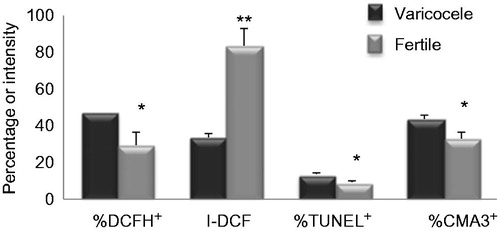
We assessed ROS with 2′, 7′-dichlorodihydrofluorescein (DCFH) staining. The percentage of positive DCFH was significantly higher in individuals with varicocele (46.8 ± 4.6) compared to fertile men (29.3 ± 7.03; p < 0.05 ). However,the intensity of sperm DCFH was significantly higher in fertile men (83.45 ± 9.1) compared to individuals with varicocele (33.4 ± 2.3, p < 0.01, ). The percentages of DNA fragmentation and protamine deficiency were assessed using TUNEL and chromomycin A3 (CMA3) staining (). There was a significantly lower percentage of TUNEL-positive sperm (p ≤ 0.01) in fertile men (8.9 ± 1.09) compared to individuals with varicocele (13.2 ± 0.8). Similarly, the percentage of CMA3-positive sperm was significantly (p ≤ 0.01) lower in fertile men (32.8 ± 3.2) compared to individuals with varicocele (43.5 ± 2.05).
Figure 1. Comparison of the mean percentages of 2′, 7′-dichlorodihydrofluorescein (DCFH+), TUNEL+, chromomycin A3 (), and intensity of DCFH (I-DCFH) between individuals with varicocele and fertile individuals. *p < 0.05; **p < 0.01.
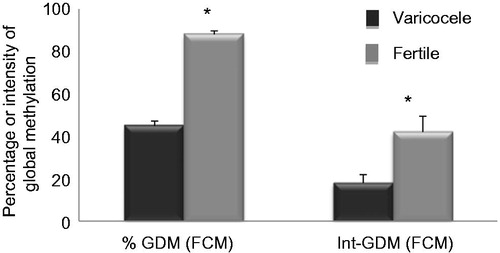
GDM was assessed by flow cytometry (). There was a significantly higher percentage of GDM (p ≤ 0.01) in fertile men (88.2 ± 1.6) compared to individuals with varicocele (45.03 ± 4.2). Intensity of GDM was also significantly higher (p ≤ 0.01) in fertile men (42.2 ± 7.6) compared to individuals with varicocele (18.2 ± 2.4).
Figure 2. Comparison of mean percentage of DNA methylation and intensity of DNA methylation between individuals with varicocele and fertile individuals. % GDM: % global DNA methylation; Int-GDM: intensity of DNA methylation; FCM: flow cytometry analyses. *p < 0.05.
Significant correlations were observed between the percentage of CMA3-positive sperm with sperm concentration (r = −0.2; p ≤ 0.05) and abnormal morphology (r = 0.3; p ≤ 0.01) (). Significant correlations were also observed between the percentage of TUNEL-positive sperm with sperm concentration (r = −0.3; p ≤ 0.01), sperm motility (r = −0.3; p ≤ 0.05), and abnormal morphology (r = 0.4; p ≤ 0.01). In addition, a significant correlation existed between the percentages of CMA3 and TUNEL positive sperm (r = 0.2; p ≤ 0.05). Examples are shown in .
Figure 3. Fluorescence images of sperm stained for DNA methylation (A), DNA fragmentation (B), and protamine deficiency (C). DNA methylation was assessed by antibody against 5-methylcytosine. DNA fragmentation was assessed by TUNEL staining (spermatozoa with fragmented DNA stained green (TUNEL positive, shown with arrow), whereas TUNEL negative sperm appear as red due to propidium iodide staining of nuclei). Protamine deficiency was assessed by chromomycin A3 (CMA3) staining, spermatozoa with bright yellow staining were considered as protamine deficient or CMA3 positive (arrow head), while spermatozoa with dull yellow staining were considered as having a normal amount of protamine or CMA3-negative.
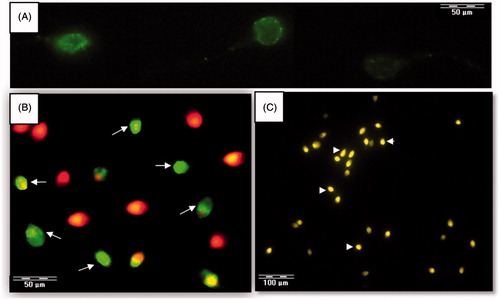
Table 2. Correlation between percentages of sperm positive for TUNEL, chromomycin A3 (CMA3), 2′, 7′-dichlorodihydrofluorescein (DCFH), and intensity of DCFH (I-DCFH) with sperm parameters in individuals with grades II and III varicocele and fertile individuals.
We also observed significant positive correlations between the intensity of DCFH with sperm concentration (r = 0.6; p ≤ 0.01), sperm motility (r = 0.2; p ≤ 0.05), and abnormal morphology (r = −0.5; p ≤ 0.01). This parameter also showed a significant correlation with TUNEL positive sperm (r = −0.5, p ≤ 0.01) but not with the percentage of CMA3 positive sperm. Unlike DCFH intensity, the percentage of DCFH positive sperm showed no correlation with any of these parameters.
We observed significant correlations between the percentage of GDM with sperm concentration (r = 0.4, p ≤ 0.01), abnormal morphology (r = −0.5, p ≤ 0.01), and intensity of DCFH (r = 0.4, p ≤ 0.01). Intensity of GDM did not shown any correlations with these parameters, except for abnormal sperm morphology (r = −0.4, p ≤ 0.01; ). In addition, we observed a significant correlation between percentage of GDM and TUNEL-positive sperm (r = −0.3, p ≤ 0.01; ).
Figure 4. Relationship between percentages of sperm global DNA methylation (GDM) with sperm DNA fragmentation in individuals with grades II and III varicocele and fertile men. N = 59.
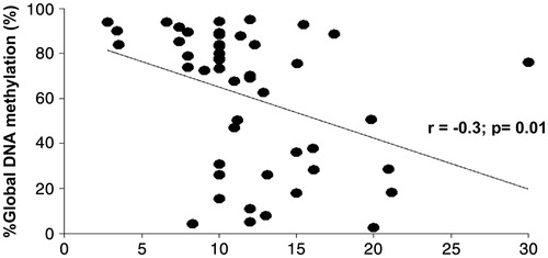
Table 3. Correlation between percentage and intensity of global DNA methylation (Int-GDM) by flow cytometry with sperm parameters, DNA fragmentation, and protamine deficiency in individuals with grades II and III varicocele and fertile individuals.
Discussion
Integrity of the sperm's genome is a prerequisite for the birth of healthy offspring [Borini et al. Citation2006]. Therefore, evaluation of DNA and epigenetic integrity of sperm, along with semen parameters may provide better diagnostic and prognostic value in infertility cases [Benchaib et al. Citation2005; Shamsi et al. Citation2011]. Sperm differs from the somatic cell due to a relative lack of cytoplasm and repair mechanisms. Spermatozoa are highly vulnerable to ROS damage due to the presence of polyunsaturated fatty acids in the sperm's plasma membrane, particularly when they are separated from seminal plasma which is rich in antioxidants [Henkel Citation2011]. Tunc and Tremellen [Citation2009] have reported that spermatozoa with normal DNA methylation are less susceptible to ROS induced DNA damage.
The results of this study showed that not only were semen parameters lower in individuals with varicocele, but also the percentages of ROS positive, DNA damaged, protamine deficient sperm were substantially higher in these individuals. Additionally, the percentage and intensity of DNA methylation were lower in individuals with varicocele compared to fertile men. This suggested that sperm from individuals with varicocele had a lower degree of maturity, higher degree of DNA fragmentation, and altered epigenetic state. These conclusions supported reports in the literature which suggested that individuals with varicocele had reduced efficiency of spermatogenesis which accounted for reduced semen parameters [Parikh et al. Citation1996; Pasqualotto et al. Citation2005]. Improper replacement of histone with protamine led to relatively decondensed sperm chromatin, which increased susceptibility to ROS mediated DNA damage [Sadek et al. Citation2011; Sakkas and Alvarez Citation2010].
In this study there was a significantly higher degree of DNA fragmentation in individuals with varicocele compared to fertile men. However, the overall range of DNA fragmentation was lower, with a mean of approximately 13% which was lower than the cut off value of 15%. One reason for this observation might be sample variation between different studies. The current study did not include individuals with very low sperm counts since substantial amounts of sperm were required to assess all the tests simultaneously. Other studies reported 10% to 12% as the cut-off value for achievement of a successful pregnancy [Duran et al. Citation2002; Robinson et al. Citation2012]. In a recent report, we stated that the percentage of DNA fragmentation significantly improved after varicocelectomy in infertile men with varicocele. Their partners became pregnant six months after varicocelectomy [Tavalaee et al. Citation2012].
In order to have further insight into the etiology of hypomethylation in individuals with varicocele, we assessed the correlation between percentage and intensity of DNA methylation with other studied parameters. There was a significant correlation between the percentage of DNA methylation and sperm concentration, which suggested that individuals with low sperm concentration had reduced degrees of DNA methylation. This result supported our recent study which showed that improvement in GDM post-surgery mainly occurred in varicocele individuals with substantially reduced sperm counts [Tavalaee et al. Citation2014]. In conjunction, these results emphasize that when spermatogenesis is severely affected, the degree of methylation is also reduced. However, it remains to be determined whether reduced spermiogenesis leads to hypomethylation or vice versa. The majority of reports have observed no correlation between the intensity of GDM and sperm concentration. In these reports the percentage of positive DNA methylation was not reported, rather the degree of DNA methylation as an arbitrary unit was reported according to fluorescent microscopy results. In the current study we have used flow cytometry which assesses approximately 10,000 sperm per sample and reports both percentage and intensity of GDM. We found no correlation between fluorescence intensity for GDM and sperm concentration which confirmed results from other studies [Benchaib et al. Citation2005]. In contrast, only one study showed a negative correlation between sperm concentration and intensity of GDM [Tunc and Tremellen Citation2009]. Although our results were mostly consistent with the literature, variance may reflect the etiology of infertility. The current study assessed infertile individuals with only varicocele, whereas other studies have assessed infertility due to other reasons. The presence of heat in varicocele conditions can affect enzyme kinetics and enzymes involved in epigenetic modifications. The percentage of abnormal morphology showed a negative correlation with both percentage and intensity of GDM. This suggested that abnormal spermiogenesis was associated with reduced DNA methylation. A similar relationship between GDM and sperm concentration and sperm morphology was observed.
One of the major events during spermiogenesis is the replacement of histone with protamine which has an important role in chromatin packaging [Schulte et al. Citation2010]. We found no significant correlation between the percentage of protamine deficient sperm with percentage or intensity of DNA methylation. This suggested that the two events were not likely related. This agrees with previous reports that assessed the relation between degrees of DNA methylation with ratio of protamine1/protamine2 in infertile individuals [Aoki et al. Citation2006]. These results might suggest that sperm DNA methylation occurrs before and after spermatid differentiation and was likely independent of histone protamine replacement.
In this study, no relation was observed between DNA methylation and sperm motility. Sperm acquire motility during epididymal maturation therefore this might suggest that the observed hypomethylation was unrelated to epididymal maturation. Additional studies should be conducted to define at which stage of spermatogenesis DNA methylation takes place, and determine the relationship of varicocele to these events.
No correlation was observed between the parameters studied and the intensity of DNA methylation, except for sperm with abnormal morphology. A highly significant correlation between the percentage of DNA methylation with intensity of DNA methylation (data not shown; r = 0.6; p = 0.00) suggests that DNA methylation is an ‘all or none’ event. In other words, if DNA methylation occurs the intensity is high and vice versa. However, the correlation between sperm morphology and intensity of DNA methylation suggest that anomalies which occur during spermiogenesis affect the degree of DNA methylation. This may be related to the severity of varicocele as it impacts spermiogenesis.
In this study there was a negative correlation between percentage of DNA methylation and percentage of DNA fragmentation which suggested that sperm with high DNA damage had a low percentage of DNA methylation. Two hypotheses could explain this phenomenon. First, DNA methylated regions were less prone to DNA damage or alternatively, damaged regions were less capable of methylation. Tunc and Tremellen [Citation2009] stated that the latter hypothesis might account for hypomethylation in infertile individuals. These researchers supported their hypothesis by the correlation between oxidative DNA adducts in somatic cells and impaired DNA methyltransferase activity [Turk et al. Citation1995]. They reported incorporation of 8-OHdG in the methyl CpG binding protein (MBP) recognition sequence. This resulted in significant inhibition of MBP binding and hindered the process of DNA methylation [Valinluck et al. Citation2004; Tunc and Tremellen Citation2009]. They did not rule out the former hypothesis. There is no evidence in the literature that normal DNA methylation protects sperm or somatic cells from apoptosis or oxidative stress, which are the major causes of DNA fragmentation. These authors have also reported that anomalies in the methyl donor pathway (the folate/homocysteine pathway) do not account for the hypomethylation state in an infertile individual [Tunc and Tremellen Citation2009]. Anomalies in this pathway in individuals with varicocele need additional research. In support of the latter hypothesis, Tunc and Tremellen [Citation2009] have shown that antioxidant therapy reduces DNA fragmentation and overcomes the hypomethylation status.
Taking into consideration the relationship between DNA methylation and DNA damage, we have expected to obtain a correlation between DNA methylation with the percentage of ROS positive sperm as it is believed that DNA damage is induced by ROS. However, we did not observe this correlation. There was a significant negative correlation between the percentage of DNA methylation and ROS intensity. Although individuals with varicocele have a higher percentage of ROS positive sperm compared to fertile men, [Singh and Agarwal Citation2011] their ROS intensity is lower. Most likely this is attributed to leakage of ROS or inability to produce ROS due to loss of enzyme function in a portion of these sperm that are in the final stage of apoptosis. Such a hypothesis has been previously proposed by Aitken et al. [Citation2010] who have shown that sperm which are initially ROS positive gradually become TUNEL positive [Aitken et al. Citation2010]. Additionally, the association between DNA hypomethylation and DNA fragmentation in individuals with varicocele might be independent of ROS production and related to other phenomenon such as hyperthermia which can affect enzyme efficiency. The correlations observed between the semen parameters with percentage of DNA damage and protamine deficiency were consistent with the literature.
In our recent study, we classified individuals with varicocele according to World Health Organization (WHO) [Citation2010] criteria. We reported that although the degree of DNA fragmentation, oxidative stress, and protamine deficiency significantly reduced after varicocelectomy, the percentage of GDM only significantly improved in oligozoospermic individuals. According to the literature, we believe that DNA methylation was affected in chronic varicocele which severely affected sperm concentration [Tavalaee et al. Citation2014]. A limitation of this research was the significantly higher age of fertile men compared to individuals with varicocele. Although it was unlikely that this difference affected our conclusion. However, we have proposed that this point should be taken into considered in future experiments by investigating effect of age on DNA methylation.
The assessment of DNA methylation by immunostaining is a relative approach and few studies have used flow cytometry to assess DNA methylation [this study and Benchaib et al. Citation2005]. This approach cannot simultaneously assess 5-methylcytosine (5mC), 5-hydroxy-methylcytosine, etc. Therefore, we believe new and more robust and molecular approachs should be implemented for assessment of methylation status not only to define the global DNA methylation but also to assess DNA methylation at the specific gene level, like microarray analysis for DNA methylation [Krausz et al. Citation2012] or direct sequencing of the methylated genome [Smith et al. Citation2012, Citation2014].
Reduced pregnancies are associated with sperm DNA fragmentation and hypomethylation. Further evidence also suggests an association between epigenetic disorders and assisted reproductive techniques [Tavalaee et al. Citation2009]. Therefore, there is a strong interest in epigenetic processes involved in reproduction and how they might affect assisted reproductive outcomes. In this study, for the first time, we have provided evidence that varicocele affects the epigenetic status of sperm. We provided associative evidence that the state of hypomethylation observed in individuals with varicocele might be related to increased DNA damage.
Materials and Methods
This study received the approval of the Institutional Review Board of Isfahan Fertility and Infertility Center and Royan Institute from 2012 to 2013. Semen samples were obtained from 44 individuals diagnosed with grades II and III varicocele. The control group was comprised of 15 fertile individuals who had no clinical presentations of infertility and were participants of the embryo donation program. Fertile men and individuals with varicocele were concomitantly included in the study in order to perform the experiment simultaneously on both groups. The mean age of fertile men (37.8 ± 2.0 years) was significantly higher than individuals with varicocele (31.1 ± 0.6 years).
The semen samples were collected by masturbation after 3 to 4 days of abstinence. Informed consent were signed by the study participants who provided the samples. The samples were examined for concentration, morphology, and motility according to WHO [Citation2010] guidelines. The number of white blood cells in individuals with varicocele were less than 1 × 10⁶.
Inclusion criteria
Inclusion criteria consisted of the following: male gender, ages 24 to 46, diagnosed with primary infertility, and the presence of left-sided varicocele (grades II and III) as diagnosed by palpation and Doppler duplex ultrasound.
Exclusion criteria
Individuals were excluded if they had grade I varicocele, azoospermia, recurrent varicocele, leukocytospermia, urogenital infections, abnormal hormonal profiles, anatomical disorders, Klinefelter’s syndrome, cancer, fever approximately 90 days prior to the seminal analysis, seminal sperm antibodies, excessive alcohol and drug use, previous history of scrotal trauma or surgery, and occupational exposure to heat.
Evaluation of sperm protamine deficiency by CMA3 staining
CMA3 staining was carried out according to Nasr-Esfahani et al. [Citation2001]. Briefly, semen samples from fertile and infertile men were washed and fixed in Carnoy’s solution (methanol:glacial acetic acid 3:1; Merck, Germany) at 4°C for 5 min. Then, smears were prepared and each slide was incubated for 20 min with 100 µL of CMA3 solution (Sigma, St. Louis, MO, USA) that consisted of 0.25 mg/mL in McIlvaine buffer (7 mL citric acid (0.1 M), 32.9 mL Na2HPO4 .7H2O (0.2 M), pH 7.0, that contained 10 mM MgCl2). The slides were subsequently washed in buffer and mounted. Microscopic analysis of the slides was performed on an Olympus fluorescent microscope (BX51, Tokyo, Japan) with the appropriate filters (460–470 nm). We evaluated 500 sperm cells per slide. Spermatozoa that stained bright yellow were considered protamine deficient (CMA3 positive) whereas those with dull yellow stain contained a normal amount of protamine (CMA3-negative; ) [Nasr-Esfahani et al. Citation2001].
Evaluation of GDM by immunostaining
Semen samples from fertile and infertile men were washed in phosphate-buffered saline (PBS) and fixed in ethanol (70%) at 4°C for 60 min. Then, cell pellets were washed in PBS-ethylene diamine tetra acetic acid (EDTA) for 5 min at 3,000 rpm. For permeabilization and DNA decondensation, we treated the sperm with 0.5% triton x-100 for 15 min and incubated them in 1 mol/l hydrochloric acid HCL-tris buffer (pH 9.5) that contained 25 mmol/l dithiothreitol for 20 min at room temperature, respectively. Then, sperm were washed twice in PBS-EDTA and allowed to denature with 4N HCl for 30 min. The sperm pellets were washed with Tris (1 mol/l, pH 9), then washed with PBS-EDTA.
The sperm pellets were blocked with 1% BSA + 10% goat serum blocking solution for 60 min at room temperature. Pellets were then incubated with mouse anti-5mc antibody (Eurogentec, Ougree, Belgium) diluted 1:400 in 1% BSA + 3% goat serum overnight at 4°C and washed with PBS-EDTA. Goat anti-mouse IgG secondary antibody FITC conjugated diluted 1:50 incubated with the pellet for 60 min at 37°C. The isotype control (CBL 600 mouse IgG1) was used as a negative control. The cells were then washed in PBS-EDTA and maintained at 4°C in a dark chamber until quantification by flow cytometry. GDM was assessed by a FACSCalibur flow cytometer (Becton Dickinson, San Jose, CA, USA) using an argon laser with an excitation wave length of 488 nm. The results were defined as percentage and intensity of DNA methylation. A minimum of 10,000 sperm were examined for each assay and analyzed using BD CellQuest Pro software according to a modified protocol by Benchaib et al. [Citation2005].
Evaluation of DNA fragmentation by TUNEL
Evaluation of DNA fragmentation was carried out according to Kheirollahi-Kouhestani et al. [Citation2009]. Briefly, semen samples were washed with PBS and fixed in 4% methanol-free formaldehyde. A commercial detection kit was used for the detection of DNA fragmentation (Apoptosis Detection System Fluorescein, Promega, Mannheim, Germany). Microscopic analysis of the slides was performed on an Olympus fluorescent microscope (BX51, Tokyo, Japan) with the appropriate filters (460–470 nm). Nuclei from the sperm with fragmented DNA stained green (TUNEL positive) whereas the other cell nuclei stained red (TUNEL negative). We counted 500 cells per slide and determined the percentages of sperm with fragmented DNA ().
Evaluation of ROS production by DCFH-DA staining
We performed 2′, 7′-dichlorodihydrofluorescein diacetate (DCFH-DA) staining according to Kiani-Esfahani et al. [Citation2012] with minor modifications. For DCFH-DA staining, 1 × 10⁶ sperm/ml in PBS were exposed to 5 μM of DCFH-DA at room temperature for 30 min after which the percentage and intensity of ROS positive sperm were assessed by a FACSCalibur flow cytometer (Becton Dickinson, San Jose, CA, USA). Unstained samples were used as the negative control via the application of PBS without DCFH-DA [Kiani-Esfahani et al. Citation2012].
Statistical analysis
Results were expressed as mean ± SEM. For comparison of mean values for the studied parameters between fertile and infertile couples, we used the independent student’s t test. Pearson correlation coefficients were used to assess correlations between different parameters. Statistical Package for the Social Sciences software (SPSS 11.5; SPSS, Chicago, IL, USA) was utilized. p Values of <0.05 were considered significant. Flow cytometry data were analyzed with WIN MDI 2.9 software.
| Abbreviations | ||
| CMA3 | = | chromomycin A3 |
| DCFH-DA | = | 2′, 7′-dichlorodihydrofluorescein diacetate |
| DNMTs | = | DNA methyltransferases |
| GDM | = | global DNA methylation |
| PBS | = | phosphate-buffered saline |
| PBS-EDTA | = | ethylene diamine tetra acetic acid |
| PGCs | = | primordial germ cells |
| ROS | = | reactive oxygen species |
| WHO | = | World Health Organization |
Acknowledgments
This study was supported by Royan Institute. We would like to express our appreciation to the staff of Isfahan Fertility and Infertility Center for their full support.
Declaration of interest
The authors have no financial or commercial conflicts with this project. Royan Institute is a nongovernment organization (NGO) that belongs to the Iranian Academic Center for Education, Culture & Research (ACECR). None of the authors are employed in any respect by the Government of Iran.
Author contributions
Collection and/or assembly of data, and manuscript writing; MB; Collection and/or assembly of data, interpretation, manuscript writing, and final approval of the manuscript: MT; Urologist: HA; Flow cytometry analyses: AK; Student supervisor: AHS; Experimental design, student supervisor, interpretation, manuscript writing, and final approval of the manuscript: MHN-E.
References
- Agarwal, A., Prabakaran, S. and Allamaneni, S.S. (2006) Relationship between oxidative stress, varicocele and infertility: A meta-analysis. Reprod Biomed Online 12:630–633
- Aitken, R.J., De Iuliis, G.N., Finnie, J.M., Hedges, A. and McLachlan, R.I. (2010) Analysis of the relationships between oxidative stresses, DNA damage and sperm vitality in a patient population: Development of diagnostic criteria. Hum Reprod 25:2415–2426
- Aoki, V.W., Emery, B.R. and Carrell, D.T. (2006) Global sperm deoxyribonucleic acid methylation is unaffected in protamine-deficient infertile males. Fertil Steril 86:1541–1543
- Benchaib, M., Braun, V., Ressnikof, D., Lornage, J., Durand, P., Niveleau, A., et al. (2005) Influence of global sperm DNA methylation on IVF results. Hum Reprod 20:768–773
- Borini, A., Tarozzi, N., Bizzaro, D., Bonu, M.A., Fava, L., Flamigni, C., et al. (2006) Sperm DNA fragmentation: Paternal effect on early post-implantation embryo development in ART. Hum Reprod 21:2876–2881
- Dada, R., Shamsi, M.B., Venkatesh, S., Gupta, N.P. and Kumar, R. (2010) Attenuation of oxidative stress and DNA damage in varicocelectomy: Implications in infertility management. Indian J Med Res 132:728–730
- Duran, E.H., Morshedi, M., Taylor, S. and Oehninger S. (2002) Sperm DNA quality predicts intrauterine insemination outcome: A prospective cohort study. Hum Reprod 17:3122–3128
- Guo, F., Li, X., Liang, D., Li, T., Zhu, P., Guo, H., et al. (2014) Active and passive demethylation of male and female pronuclear DNA in the Mammalian zygote. Cell Stem Cell 15:447–458
- Henkel, R.R. (2011) Leukocytes and oxidative stress: Dilemma for sperm function and male fertility. Asian J Androl 13:43–52
- Kheirollahi-Kouhestani, M., Razavi, S., Tavalaee, M., Deemeh, M.R., Mardani, M., Moshtaghian, J., et al. (2009) Selection of sperm based on combined density gradient and Zeta method may improve ICSI outcome. Hum Reprod 24:2409–2416
- Kiani-Esfahani, A., Tavalaee, M., Deemeh, M.R., Hamiditabar, M. and Nasr-Esfahani, M.H. (2012) DHR123: An alternative probe for assessment of ROS in human spermatozoa. Syst Biol Reprod Med 58:168–174
- Kobayashi, H., Sato, A., Otsu, E., Hiura, H., Tomatsu, C., Utsunomiya, T., et al. (2007) Aberrant DNA methylation of imprinted loci in sperm from oligospermic patients. Hum Mol Genet 1:2542–2551
- Kobayashi, H., Sakurai, T., Imai, M., Takahashi, N., Fukuda, A., Yayoi, O., et al. (2012) Contribution of intragenic DNA methylation in mouse gametic DNA methylomes to establish oocyte-specific heritable marks. PLoS Genet 8:e1002440
- Krausz, C., Sandoval, J., Sayols, S., Chianese, C., Giachini, C., Heyn, H., et al (2012) Novel insights into DNA methylation features in spermatozoa: Stability and peculiarities. PLoS One 7(10):e44479
- Mayer, W., Niveleau, A., Walter, J., Fundele, R. and Haaf, T. (2000) Demethylation of the zygotic paternal genome. Nature 403:501–502
- Nasr-Esfahani, M.H., Razavi, S. and Mardani, M. (2001) Relation between different human sperm nuclear maturity tests and in vitro fertilization. J Assist Reprod Genet 18:219–225
- Parikh, F.R., Kamat, S.A., Kodwaney, G.G. and Balaiah, D. (1996) Computer-assisted semen analysis parameters in men with varicocele: Is surgery helpful? Fertil Steril 66:440–445
- Pasqualotto, F.F., Lucon, A.M., de Góes, P.M., Sobreiro, B.P., Hallak, J., Pasqualotto, E.B., et al. (2005) Semen profile, testicular volume, and hormonal levels in infertile patients with varicoceles compared with fertile men with and without varicocele. Fertil Steril 83:74–77
- Reik, W., Dean, W. and Walter, J. (2001) Epigenetic reprogramming in mammalian development. Science 293:1089–1093
- Robinson, L., Gallos, I.D., Conner, S.J., Rajkhowa, M., Miller, D., Lewis, S., et al. (2012) The effect of sperm DNA fragmentation on miscarriage rates: A systematic review and meta-analysis. Hum Reprod 27:2908–2917
- Sadek, A., Almohamdy, A.S., Zaki, A., Aref, M., Ibrahim, S.M. and Mostafa, T. (2011) Sperm chromatin condensation in infertile men with varicocele before and after surgical repair. Fertil Steril 95:1705–1708
- Sakkas, D. and Alvarez, J.G. (2010) Sperm DNA fragmentation: Mechanisms of origin, impact on reproductive outcome, and analysis. Fertil Steril 93:1027–1036. Review
- Santos, F., Hendrich, B., Reik, W. and Dean, W. (2002) Dynamic reprogramming of DNA methylation in the early mouse embryo. Dev Biol 241:172–182
- Schulte, R.T., Ohl, D.A., Sigman, M. and Smith, G.D. (2010) Sperm DNA damage in male infertility: Etiologies, assays, and outcomes. J Assist Report Genet 27:3–12
- Shamsi, M.B., Imam, S.N. and Dada, R. (2011) Sperm DNA integrity assays: Diagnostic and prognostic challenges and implications in management of infertility. J Assist Reprod Genet 28:1073–1085
- Shen, L., Inoue, A., He, J., Liu, Y., Lu, F. and Zhang, Y. (2014) Tet3 and DNA replication mediate demethylation of both the maternal and paternal genomes in mouse zygotes. Cell Stem Cell 15:459–470
- Singh, A. and Agarwal, A. (2011) The Role of Sperm Chromatin Integrity and DNA Damage on Male Infertility. Open Reprod Sci J 3:65–7165
- Smallwood, S.A., Tomizawa, S., Krueger, F., Ruf, N., Carli, N., Segonds-Pichon, A., et al. (2011) Dynamic CpG island methylation landscape in oocytes and preimplantation embryos. Nat Genet 43:811–814
- Smith, Z.D., Chan, M.M., Mikkelsen, T.S., Gu, H., Gnirke, A., Regev, A., et al. (2012) A unique regulatory phase of DNA methylation in the early mammalian embryo. Nature 484:339–344
- Smith, Z.D., Chan, M.M., Humm, K.C., Karnik, R., Mekhoubad, S., Regev, A., et al. (2014) DNA methylation dynamics of the human preimplantation embryo. Nature 511:611–615
- Tavalaee, M., Razavi, S. and Nasr-Esfahani, M.H. (2009) Influence of sperm chromatin anomalies on assisted reproductive technology outcome. Fertil Steril 91:1119–1126
- Tavalaee, M., Abbasi, H., Deemeh, M.R., Fotohi, F., Sadoughi Gilani, M.A. and Nasr Esfahani, M.H. (2012) Semen parameters and chromatin packaging in microsurgical varicocelectomy patients. Int J Fertil Steril 6:165–174
- Tavalaee, M., Bahreinian, M., Barekat, F., Abbasi, H. and Nasr-Esfahani, M.H. (2014) Effect of varicocelectomy on sperm functional characteristics and DNA methylation. Andrologia J. 1–6. doi: 10.1111/and.12345. [Epub ahead of print]
- Tunc, O. and Tremellen, K. (2009) Oxidative DNA damage impairs global sperm DNA methylation in infertile men. J Assist Reprod Genet 26:537–544
- Turk, P.W., Laayoun, A., Smith, S.S. and Weitzman, S.A. (1995) DNA adducts 8-hydroxyl-2′-deoxyguanosine (8hydroxyguanine) affects function of human DNA methyltransferase. Carcinogenesis 16:1253–1255
- Valinluck, V., Tsai, H.H., Rogstad, D.K., Burdzy, A., Bird, A. and Sowers, L.C. (2004) Oxidative damage to methyl-CpG sequences inhibits the binding of the methyl-CpG binding domain (MBD) of methyl-CpG binding protein 2 (MeCP2). Nucleic Acids Res 32:4100–4108
- Wang, Y.J., Zhang, R.Q., Lin, Y.J., Zhang, R.G. and Zhang, W.L. (2012) Relationship between varicocele and sperm DNA damage and the effect of varicocele repair: A meta-analysis. Reprod Biomed Online 25:307–314
- WHO (2010) WHO laboratory manual for the examination and processing of human semen. Fifth edition, World Health Organization. WHO Press, Geneva, Switzerland
- Zini, A., Defreitas, G., Freeman, M., Hechter, S. and Jarvi, K. (2000) Varicocele is associated with abnormal retention of cytoplasmic droplets by human spermatozoa. Fertil Steril 74:461–464