
Abstract
Testicular apoptosis is activated by stress, but it is not clear which signaling pathway is activated in response to stress. The aim of this study was to investigate whether intrinsic, extrinsic, or both apoptotic signaling pathways are activated by acute and chronic stress. Adult male rats were subjected to cold water immersion-induced stress for 1, 20, 40, and 50 consecutive days. The seminiferous tubules:apoptotic cell ratio was assayed on acute (1 day) and chronic (20, 40, 50 days) stress. Apoptotic markers, including cleaved-caspase 3 and 8, the pro-apoptotic Bax and anti-apoptotic Bcl-2 proteins were also determined after acute and chronic stress induction. Additionally, epididymal sperm quality was evaluated, as well as corticosterone and testosterone levels. An increase in tubule apoptotic cell count percentage after an hour of acute stress and during chronic stress induction was observed. The apoptotic cells rate per tubule increment was only detected one hour after acute stress, but not with chronic stress. Accordingly, there was an increase in Bax, cleaved caspase-8 and caspase-3 pro-apoptotic proteins with a decrease of anti-apoptotic Bcl-2 in both acutely and chronically stressed male testes. In addition, sperm count, viability, as well as total and progressive motility were low in chronically stressed males. Finally, the levels of corticosterone increased whereas testosterone levels decreased in chronically stressed males. Activation of the extrinsic apoptotic pathway was shown by cleaved caspase-8 increase whereas the intrinsic apoptotic pathway activation was determined by the increase of Bax, along with Bcl-2 decrease, making evident a cross-talk between these two pathways with the activation of caspase-3. These results suggest that both acute and chronic stress can potentially activate the intrinsic/extrinsic apoptosis pathways in testes. Chronic stress also reduces the quality of epididymal spermatozoa, possibly due to a decrease in testosterone.
Introduction
Apoptosis is an innate and evolutionarily conserved process of cellular death that can occur in tissue under both normal and pathological conditions. Apoptosis plays an essential role in the control of germ cell rate in the testes [Stiblar-Martinic Citation2009]. Testicular apoptosis is a common event that takes place during spermatogenesis in a variety of mammalian species, including humans [Allan et al. Citation1987; Stiblar-Martinic Citation2009], and can occur in spermatogenic cells at various stages of differentiation. During early development, particularly in the germ line ontogeny, apoptosis represents a pivotal event regulating of the germ cell:Sertoli cell ratio [Aitken and Baker Citation2013]. The progression to the first wave of spermatogenesis relies on the coordinated Fas/Fasl activation and Bax-dependent spermatogonia and spermatocytes physiological apoptosis [Koji Citation2001; Nagata and Golstein Citation1995; Print and Loveland Citation2000; Shaha et al. Citation2010]. In a healthy male adult, apoptosis is required as a cell production regulating process or when spermatogenesis cannot be supported by the testicular environment [Lee et al. Citation1999]. Thus, germ cell apoptosis may help in maintaining a viable number of germ cells that can be sustained by Sertoli cells [Braun Citation1998; Lee et al. Citation1997, Citation1999; Nakanishi and Shiratsuchi Citation2004].
Germ cell removal is another role for apoptosis when damage is induced in the seminiferous epithelium, this damage could be initiated by a broad range of physiological and environmental factors [Aitken and Baker Citation2013; Blanco-Rodríguez Citation1998; Francavilla et al. Citation2002] such as when serum testosterone [Shaha Citation2008] or gonadotrophin levels decrease [Pareek et al. Citation2007; Sinha Hikim et al. Citation1997], or by external factors such as UV or ionizing radiation [Otala et al. Citation2005], endocrine disruptors [Jin et al. Citation2013], and stress [Ozawa et al. Citation2002]. Within this context, testicular heat stress (43°C for 15 minutes) causes an increase in rat testicular seminiferous epithelium germ cell apoptosis at stages l–Vl and Xll–XIV [Banks et al. Citation2005; Lue et al. Citation1999; Rockett et al. Citation2001]. In addition, immobilization stress for 7 consecutive days induces testicular germ cell apoptosis [Yazawa et al. Citation1999]. In a similar way, exogenous dexamethasone administration induces male germ cell apoptosis, affecting mainly the spermatogonia and primary spermatocytes [Mahmoud et al. Citation2009; Yazawa et al. Citation1999; Yazawa et al. Citation2000]. Testes exposed to mild hypothermia (30 minutes at 10°C) induces seminiferous epithelium germ cell apoptosis at specific stages (XII and XIV), displaying maximum damage after 2 hours of hypothermic exposure. Testes direct cooling achieved by using Ringer solution at 0°–1°C for 60 minutes, causes apoptosis-mediated germ cell loss within 8 hours. Stages XII–I pachytene spermatocytes are hypersensitive to cooling, thus, germ cell depletion is usually seen 2–24 hours after treatment [Zhang et al. Citation2004]. In addition, acute stress by heat shock (40°C for 2 hours) or cold-induced stress (4°C for 12 hours) leads to apoptosis in spermatids and sperm. Furthermore, these stressors can cause an increase in caspase-3 gene expression, as well as an induction of caspase-3, -8, and -9 activities, supporting a double apoptosis pathway activation (extrinsic/intrinsic) [Wang et al. Citation2012]. One hour immobilization-induced stress, followed by forced swimming (15 minutes) daily for 60 days in rats, causes an increase in testicular germ cell apoptosis and a decrease in epididymal sperm count [Nirupama et al. Citation2012]. Although in all these conditions germ cells undergo apoptosis, and it has been speculated that an apoptotic pathway (intrinsic, extrinsic) is activated, it is not clear which one of the apoptotic signaling pathways (intrinsic/extrinsic) is involved in the testes stress response. Therefore, the aim of this work was to investigate whether intrinsic, extrinsic, or both apoptotic signaling pathways activate during acute and chronic stress. Considering that an increase in germ cell apoptosis might cause an imbalance between cell proliferation and cell death, lowering sperm daily production and male fertility, count, and quality of epididymal spermatozoa was also assessed. Additionally, serum corticosterone and testosterone concentration changes were evaluated.
Results
Total body and testicular weight of rats under acute stress were determined and data showed no significant variation (). In contrast, total body and testicular weight of rats under chronic stress was significantly lower than controls (p < 0.01) from day 20 to 50 days of chronic stress induction (). Testes from rats exposed to acute stress showed a significant increase in tubules apoptotic germ cell percentage at 1 and 24 hours after the stressor exposure (). Tubules apoptotic germ cell percentage was significantly increased when compared with the control group (p < 0.01). Testes from rats exposed to chronic stress showed higher percentages of tubules with apoptotic germ cells starting at day 20 of stress induction. The percentages of tubules at 20, 40, and 50 days under chronic stress, were significantly higher than in the control group (p < 0.01) ().
Figure 1. Percentage of seminiferous tubules with TUNEL-positive cells at different experimental times after a single stressor exposure. Percentages were significantly higher than those observed in control groups from the first hour until reaching a peak 24 hours after acute stress induction. Data shown as Mean ± SEM and subjected to one-way ANOVA and Newman-Keulls analysis. *p < 0.01 vs. control group.
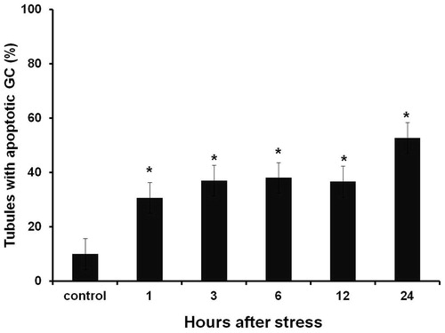
Figure 2. Seminiferous tubules TUNEL-positive cells at 20, 40, and 50 days after chronic stress induction. Percentages were significantly higher than those observed on control groups. Data shown as Mean ± SEM and subjected to one-way ANOVA and Newman-Keulls analysis. *p < 0.01 vs. control group.
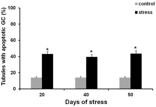
Table 1. Effect of acute stress on body and testes weights of male rats.
Table 2. Effect of chronic stress on body and testes weights of male rats.
The number of apoptotic testes male germ cells exposed to acute stress were higher than in the control group (). This increase was significant only one hour after stress induction (p < 0.05) ( and ). No significant increase of the seminiferous tubules apoptotic germ cell rate was found in any of the chronic stress groups (, ). Of note, Leydig apoptotic cells were observed in the testes of acutely stressed males ().
Figure 3. Control and stressed rat testicular sections. (A) Control testis showed non-significant TUNEL-positive cells. (B) Seminiferous tubules TUNEL-positive cells increase (arrows) observed only at 1 hour after acute stress induction. (C) Chronic stressed (50 days) seminiferous tubules displayed scattered TUNEL-positive cells. Some apoptotic Leydig cells were also observed (arrowhead). Bar = 50 µm; magnification: 400×.
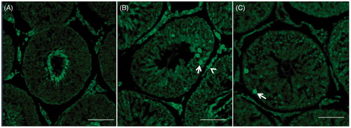
Figure 4. Number of apoptotic germ cells contained in seminiferous tubules in testes of males exposed to acute stress. The number of apoptotic germ cells was significantly higher than in testes of the control group one hour after exposure to the stressor. Data shown as mean ± SEM. One-way ANOVA followed by Newman-Keulls test; *p < 0.01 compared with control.
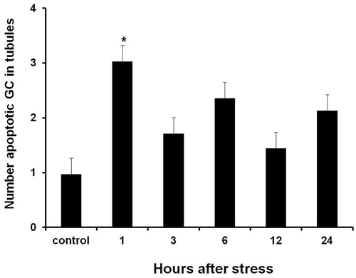
Table 3. Number of apoptotic germ cells (GC) contained in seminiferous tubules (ST) at days 20, 40, and 50 of chronic stress.
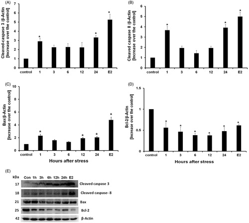
In males exposed to acute stress, the level of cleaved-caspase-3 protein increased significantly (p < 0.05) at one and twenty-four hours after stressor exposure (). The level of cleaved caspase-8 protein in total testes lysates also increased significantly (p < 0.05) at one and twenty-four hours after stress induction (). Bax protein content increased significantly (p < 0.05) at one, twelve, and twenty-four hours after stress exposure (). Bcl-2 expression decreased significantly (p < 0.05), when compared with the control group, at all the experimental times (1, 3, 6, 12, and 24 hours) after acute stress induction ().
Figure 5. Western blot cleaved caspase-8, Bax, Bcl-2, cleaved-caspase-3 proteins detection, and estradiol benzoate (E2) treatment as apoptosis positive control. (A) Active caspase-3 content increased from one until 24 hours after acute stress induction. (B) Cleaved-caspase-8 content increased from one until 24 hours after acute stress induction. (C) Bax content increased from 1, 12, and 24 hours after acute stress induction. (D) Bcl-2 expression decreased at all experimental times. Data is presented as relative protein expression (optical density) normalized β-actin OD quantities from identical blot. (E) Gels from five protein expression analysis for all experimental times. Each data point represents the mean ± SEM; n = 5. *p < 0.05 vs. control groups.
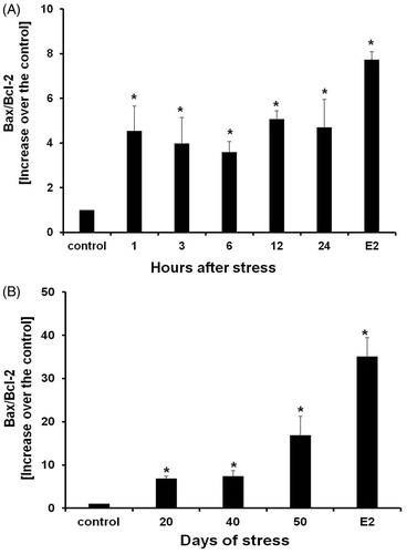
The level of cleaved-caspase-3 protein increased significantly (p < 0.05) every day that the rats were exposed to chronic stress (). Cleaved-caspase-8 levels also increased significantly (p < 0.05) in the testes of chronically stressed males (). Similarly, Bax protein levels increased significantly (p < 0.05) for chronic stress induction () whereas Bcl-2 protein decreased significantly (p < 0.05) (). The Bax/Bcl-2 ratio increased significantly (p < 0.05) in both acute and chronic stressed males ().
Figure 6. Western blot cleaved caspase-8, Bax, Bcl-2, cleaved-caspase-3 proteins detection, and estradiol benzoate (E2) treatment as apoptosis positive control. (A) Cleaved-caspase-3 content increased at all experimental times. (B) Cleaved-caspase-8 content increased at all experimental times in all days of stress. (C) Bax content also increased during chronic stress at all experimental times. (D) Bcl-2 content decreased in all experimental times. Data is presented as relative protein expression (optical density) normalized with β-actin OD quantities from identical blot. (E) Gels from five protein expression analysis for all experimental times. Each data point represents the mean ± SEM; n = 5. *p < 0.05 vs. control groups.
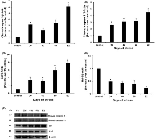
Figure 7. Bax/Bcl-2 relative ratios of acute stress (A) and chronic stress (B). Bax content was significantly increased. Data is presented as Bax and Bcl-2 relative expression. Each data point represents the mean ± SEM; n = 5. *p < 0.05 vs. control groups.
Physiological sperm analysis
Acute stress induction caused no changes in any of the analyzed parameters (). However, chronically stressed males showed a significantly lower percentage on sperm viability at 20, 40, and 50 days after stress induction in comparison with the control group (p < 0.01) (). Chronically stressed males also displayed a lower sperm count at days 20, 40, and 50 after chronic stress induction when compared with the control group (p < 0.01) (). Total sperm motility decreased significantly at 20, 40, and 50 days after stress induction. These percentages were significantly lower than those observed in the control groups (p < 0.01) (). Interestingly, the percentage of progressively motile sperm from males exposed to 20 days of chronic stress was not different from the control group. Nevertheless, at 40 and 50 days after stress induction progressive motility percentage decreased significantly when compared with the control group (p < 0.01) ().
Figure 8. Sperm viability (A), concentration (B), total sperm motility (C), and progressive sperm motility (D). All these parameters appeared lower than when compared to control groups. Data shown as mean ± SEM. Two-way ANOVA and Newman-Keulls analysis; *p < 0.01 vs. control groups.
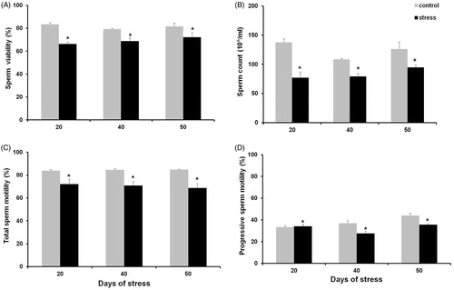
Table 4. Effect of acute stress on viability, concentration, motility, and total and progressive motility of spermatozoa obtained from the cauda epididymis of the rat.
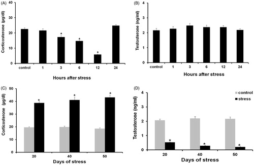
Serum corticosterone from males exposed to acute stress did not show an increase at any of the evaluated experimental times. Instead, there was a decrease on this steroid level from 3 to 12 hours after stressor exposure, followed by an increase at 24 hours (). Rats exposed to chronic stress, showed a significant increase in corticosterone levels at all experimental times (p < 0.01) (). The level of testosterone showed no differences when compared with the control group after acute stress induction (), but decreased significantly in rats exposed to chronic stress, at all experimental times (p < 0.01) ().
Figure 9. Corticosterone (A) and testosterone levels (B) in males exposed to acute stress. No changes were detected in any of these hormones. (C) Corticosterone levels in males exposed to chronic stress showed a significant increase when compared with the control group. (D) Testosterone levels were significantly lower when compared with the control group. Data shown as mean ± SEM. Two-way ANOVA and Newman-Keulls analysis; *p < 0.01 vs. control groups.
Discussion
Our data show that acute and chronic stress by cold water immersion eventually leads to apoptosis in testicular germ cells, and that both intrinsic and extrinsic pathways are involved in this process. In the acutely stressed males, the percentage of apoptotic germ cells and tubules increased during the first hour after stress exposure, reaching a peak at 24 hours. Cleaved caspase-3 increased one hour after stressor exposure, reaching a peak at 24 hours. Cleaved caspase-3 is an important effector caspase because activation marks the point of no return of apoptotic signaling [Earnshaw et al. Citation1999]. Cleaved-caspase 8 also increased at one and 24 hours after acute stress induction, indicative of the activation of the extrinsic apoptotic pathway, as early as one hour after stressor exposure, suggesting that acute stress can activate testes germ cell apoptosis through the extrinsic pathway. Moreover, it is known that the testes extrinsic apoptotic pathway involves the activation of the Fas receptor and its ligand (FasL), expressed by Sertoli, spermatocyte, and Leyding cells. Once bound to its ligand, Fas can trigger the apoptotic event [Pentikainen et al. Citation1999; Porcelli et al. Citation2006], by a caspase-8 dependent mechanism [Maheshwari et al. Citation2009; Said et al. Citation2004]. Caspase-8 is required for caspase-3 cleavage [Said et al. Citation2004] that is necessary to trigger apoptosis. However, the role of Fas/FasL in activating acute stress-induced testicular apoptosis remains to be elucidated.
The level of Bax increased in contrast with Bcl-2 that decreased (both markers of the intrinsic apoptotic pathway) at all experimental times after stressor exposure. This, indicates that the intrinsic pathway is also activated by acute stress. It has been proposed that the Bcl-2 family proteins are either germ cell survival or death modulators [Knudson et al. Citation1995; Mahmoud et al. Citation2009; Yan et al. Citation2000]. Functions of the Bcl-2 related proteins comprise pro-apoptotic (Bax, Bad, and Bid) and anti-apoptotic (Bcl-2, Bcl-xL) proteins [Knudson et al. Citation1995; Maheshwari et al. Citation2009; Yamamoto et al. Citation2000]. Induction of Bax acute stress may promote changes in mitochondrial membrane permeability and release cytochrome C into the cytosol [Blatt and Glick Citation2001]. Once released, cytochrome C binds to apoptotic protease activating factor-1 (Apaf-1) and pro-caspase-9 [Nuñez et al. Citation1998; Wang et al. Citation2012], activating the initiator caspase-9, that in turn activates caspase-3 [Maheshwari et al. Citation2009]. Therefore, it is possible that rapid stress-induced testes germ cell apoptosis involves both extrinsic and intrinsic apoptotic signaling pathways.
During chronic stress, a significant increase in the percentage of apoptotic germ cell tubules was observed at all experimental times (20, 40, and 50 days). Although the apoptotic germ cell rate did not increase with chronic stress, the seminiferous tubule apoptotic germ cells rate did, and remained high at all experimental times. In contrast, the number of tubules with apoptotic cells increased greatly after seven days of immobilization-induced stress [Yazawa et al. Citation1999]. The differences may reflect the experimental conditions. The steady apoptotic germ cells rate during chronic stress in the present study may reflect the efficient phagocytic removal of seminiferous epithelium apoptotic cells. Moreover, Sertoli cells are capable of recognizing and then engulfing germ cells initiating apoptosis through scavenger receptor type l (SR-BI) binding, present on the Sertoli cell membrane [Nakanishi and Shiratsuchi Citation2004]. The increase in the number of tubules with apoptotic germ cells likely yields the reduction in testicular weight during chronic stress.
The present work showed that chronic stress paralled increased levels of testicular caspase-3, caspase-8, and Bax. Accordingly, Bcl-2 content decreased at all experimental times. Taken together, these results indicate that both intrinsic and extrinsic signaling pathways are involved in testicular germ cell apoptosis during chronic stress, converging at caspase-3 activation [Kaufmann et al. Citation2012] that is tightly associated with DNA fragmentation [Maheshwari et al. Citation2009]. Increased apoptosis activation is demonstrated with the elevation of the Bax/Bcl2 ratio, which indicates an up-regulation of caspase-3 activation. The activation of testicular extrinsic and intrinsic apoptotic signaling pathways may also reflect the cross-talk between both pathways [Said et al. Citation2004] with caspase-8-mediated proteolytic activation of the pro-apoptotic protein Bid [Kaufmann et al. Citation2012; Said et al. Citation2004]. In this mechanism, truncated Bid (tBid) is then translocated to the mitochondria where it promotes cytochrome C Bax-dependent release and Apaf1-caspase-9 complex activation, followed by caspase-3 cleavage and activation that leads to apoptosis. The cross-talk between these pathway allows caspase-driven signal amplification to ensure efficient removal of damaged germ cells [Kaufmann et al. Citation2012].
Testicular apoptosis observed in chronically stressed males is consistent with the decrease in testosterone levels, since it has been shown that decreased intratesticular testosterone is sufficient to activate caspase-3 in primary spermatocytes [Kim et al. Citation2001]. The decrese in testosterone likely reflects the increase in corticosterone during chronic stress. This may be mediated by non-genomic mechanisms [Dong et al. Citation2004] or genetic mechanisms through the inhibition of genes encoding testosterone biosynthetic enzymes [Payne and Sha Citation1991], such as 3β-hydroxysteroid dehydrogenase and 17α-hydroxylase/17-20-lyase [Hales and Payne Citation1989; Orr et al. Citation1994]. Corticosterone also induces Leydig cell apoptosis [Chen et al. Citation2012], resulting in a significant reduction in the secretion of testosterone [Hardy et al. Citation2005] as confirmed by TUNEL. It appears that the number of apoptotic Leydig cells observed were not sufficient to cause a decrease in the level of testosterone after acute stress induction. As above, germ cell apoptosis might also be induced by corticosterone, since glucocorticoid receptors (GR) have been identified in the cytoplasm of primary spermatocytes [Schultz et al. Citation1993]. Therefore, corticosterone may be inhibiting, through a genomic mechanism, the transcription of certain transcription factors, such as c-jun/c-fos and NF-kB (nuclear factor kB) whose target genes are related to cell survival [Greenstein et al. Citation2002; Sionov et al. Citation2006]. Moreover, corticosterone could potentially lead germ cells to apoptosis through the intrinsic apoptotic signaling pathway, as observed during dexamethasone treatment [Yazawa et al. Citation2000], where Bax expression increases at specific androgen-dependent stages (Vll–Vlll) of the seminiferous epithelium cycle [Mahmoud et al. Citation2009]. Testicular corticosterone-induced apoptosis may also be affected by the level of oxidative stress, since increases in hydrogen peroxide (H2O2) and lipid peroxidation were observed along with attenuated superoxide dismutase (SOD) and catalase (CAT) activity [Dhanabalan et al. Citation2010]. H2O2 is known to induce germ cell apoptosis in the testes, through the Fas/FasL complex and other pro-apoptotic proteins, such as Bax, Bid, and Bad [Maheshwari et al. Citation2009]. Therefore, it is possible that the decrease in testosterone paralling the increase in corticosterone from chronic stressed males can induce testicular germ cell apoptosis through ROS overproduction and generation of oxidative stress, thus directly activating both apoptotic signaling pathways.
Chronic stress also caused a reduction of epididymal spermatozoa and sperm count as well as total and progressive motility. Similar results have been reported in sperm quality and sperm fertilization ability [Saki et al. Citation2010]. In the present work, epididymal quality and count changes were assayed as a function of stressor exposure. Epididymal sperm mutation is characterized by nuclear condensation, the acquisition of motility, and capacitation capabilities. This is an androgen-dependent process, mainly orchestrated by the conjugated action of testosterone and dihydrotestosterone. Androgen deprivation causes loss of sperm motility, fertilization ability, and eventually leads to cell death [Robaire et al. Citation2006]. A decrease in the level of testosterone affects conversion of round spermatids during spermiogenesis at stages 7 to 8 causing premature detachment that prevents elongation [O'Donnell et al. Citation1996; Sofikitis et al. Citation1999], thereby decreasing daily sperm production which may reflect our previous observation of decreased testosterone. Conversely, stress-induced corticosterone release might affect the epididymal microenvironment through the epithelial cytoplasmic and nuclear glucocorticoid receptor [Schultz et al. Citation1993; Silva et al. Citation2010]. The loss of total progressive motility could be owed to epididyimal corticosterone-induced ROS overproduction and lipid peroxidation [Dhanabalan et al. Citation2010]. ROS-induced sperm lipid peroxidation may alter sperm plasma membrane fluidity [Agarwal et al. Citation2014], causing a defect in the middle piece [Rao et al. Citation1989], decreasing motility [Agarwal et al. Citation2003; Alvarez and Storey Citation1982] due to defects in the axoneme [De Lamirande and Gagnon Citation1992a] and ATP depletion [De Lamirande and Gagnon Citation1992b]. In turn this would also reconcile the loss of sperm viability.
In conclusion, germ cell death after acute or chronic stress by cold water immersion leads to an increase in the number of apoptotic germ cell tubules. This involves both of the extrinsic and intrinsic apopototic pathways. Immediately after acute stress this is characterized by an elevated apoptotic germ cell rate. This increase is also reflected as a function of sperm count, motility, and viability that may be owed to the opposite response of corticosterone and testosterone levels.
Materials and Methods
Animals
Animal handling and experimentation was performed in accordance to Mexican Official Rules (NOM-062-ZOO-1999). Wistar adult male rats, (90 d old, 250–300 g/body weight), housed under the Universidad Autónoma Metropolitana-Iztapalapa facilities were used. Five animals per cage were placed (50X30X20 cm) under standard conditions. The room was maintained on a 12:12 reverse light/dark cycle (lights off: 0900) and controlled temperature (22 ± 1°C). Food and water were available ad libitum throughout the experimental times. Animals were kept in these conditions for one week before being randomly assigned to one of the following groups: control group: 1, 20, 40, and 50 d (n = 5 per group); acute stress group (a single exposure): 1, 3, 6, 12, and 24 h (n = 5 per experimental time) after stressor exposure; chronic stress group: 20, 40, and 50 consecutive days (n = 5 per experimental time).
Cold water immersion-induced stress
Stressor exposure took place during the dark phase (0900) of the light/dark cycle. Stressor exposure was performed in a separated room. Rats were placed individually in a cold water covered tank (15.5 cm deep, at 15°C) where they could either swim or remain in an upright position, keeping their heads above water level for 15 min. After stress induction, animals were dried with a cloth and immediately returned to their cages [Retana-Márquez et al. Citation1996]. This procedure was done once/d for one (acute stress) or 20, 40, and 50 consecutive d (chronic stress). Experimental times were chosen for chronic stress effect evaluation along with spermatogenesis that is known to last 48–52 d in rats.
Control animals were kept in their home cages without any disturbance. Rats under acute stress were anesthetized with sodium pentobarbital (25 mg/kg body weight; Pfizer, SA de CV, Toluca, Mexico, ip) at 1, 3, 6, 12, and 24 h after stressor exposure. Rats under chronic stress were anesthetized 1 h after the last stress induction session. Testes were immediately removed and weighed. Apoptosis was performed using the left testis. The right testis was stored at −70°C for protein extraction and Western blot analysis. Cauda epididymis was removed for determination of sperm motility, viability, and count. Blood samples from control and stressed rats were collected by cardiac puncture (3–4 ml) in test tubes, immediately after the cauda epididymis and testis were obtained (∼3 min) followed by centrifugation (2500 rpm/10 min). Serum samples were stored at −20°C for hormone content determination.
Testicular tissue sampling
Left testis was fixed by using 4% paraformaldehyde testicular artery perfusion (Sigma-Aldrich, USA) and paraffin embedding (Paraplast-plus). Transverse sections (5 µm) were obtained by using a Leica microtome mounted on poly-l-lysine hydrobromide (Sigma Aldrich) treated slides.
Deoxyuridine nick-end labeling (TUNEL) assay
Testis tissue sections (5µm) were deparaffinized in xylol and sequentially rehydrated in ethanol dilutions: 100, 96, 80, and 70%, washed, and maintained in PBS (137 mM NaCl, 2.68 mM KCl, 8.03 mM Na2HPO4, 1.47 mM K2HPO4, pH 7.4). The in-situ cell detection kit (POD kit, Cat. No. 11684817910 Roche, Germany) was used for apoptosis detection by TUNEL assay according to the manufacturer’s instruction. Briefly, tissue sections were dewaxed and treated with 20 µg/ml Proteinase K (Proteinase K recombinant PCR Grade, Cat. No. 03 115 887 001 Roche, Germany) for 30 min at 37°C in a dark humidity chamber.
Permeabilization was achieved by using 0.1% Triton X-100 in 0.1% sodium citrate for 5 min on ice (4°C); sections were incubated with the TUNEL reaction mixture (terminal deoxynucleotidyl transferase (TdT) 25 µl) and fluorescein-dUTP for 60 min at 37°C in a dark humidity chamber. At least 50 tubules were randomly analyzed per slide in different fields. Cells were first identified by using fluorescent microscopy (Axiostar Plus, HBO 50/Ac, Carl Zeiss) and then analyzed using confocal microscopy (LSM 780 multiphoton confocal microscope, Carl Zeiss) at a 450–500 nm wavelength excitation and a 515–565 nm (green light) detection range. Apoptotic cells (intense green nuclear fluorescence) or normal (no fluorescence) were identified. Fifty randomly selected tubules were counted for each individual and the tubules apoptotic germ cell percentage was estimated as the other apoptotic cell type contained in these tubules.
Western blot analysis
Frozen testes tissue (0.2 g) was minced in 100 µl ice-cold PBS (pH 7.4) (1% Igepal, 20 mM Tris-HCl, 120 mM NaCl, protease inhibitors: Complete Mini Protease Inhibitor Cocktail (Roche Molecular Biochemical, Indianapolis, IN, USA). Tissue was homogenized using a dunce homogenizer. After incubation at 4°C for 15 min, samples were centrifuged at 12,000 X g for 10 min at 4°C. Supernatants (total protein extract) were aliquoted and stored at −70°C for further use. Protein concentration was determined using the Thermo Scientific NanoDrop 2000c spectrophotometer (spectral range of 280 nm).
For Western blot analysis, 300 µg of protein was loaded on a 10% SDS-polyacrylamide gel at 120 V in a Mini-Protean ll Cell (Bio-Rad, CA, USA) along with a low-range marker standard (NuPAGE Novex, Invitogen, CA, USA). Proteins were transferred to a 0.45 µm nitrocellulose membranes (PVDF Transfer Membrane; GE Healthcare, Buckinghamshire, UK) embed in transfer buffer (25 mM Tris base, 190 mM glycine, 20% methanol) at 120 V for 30 min by using the Trans-Blot Turbo (Bio-Rad) protein transfer system. Membranes were blocked with nonfat dried milk 5% in TTBS (137 mM NaCl, 20 mM Tris- HCl and 0.1% Tween 20, pH 7.6) for 30 min at room temperature. After three TTBS washes (5 min/each), membranes were incubated with primary antibodies (diluted 1:500) against Bax (Cat No. 3331-100, BioVision), or Bcl-2 (Cat. No. 3033-100, BioVision, CA, USA), or anti- cleaved caspase-3 (Cat No. 9665, Cell Signaling Technology) or anti-caspase-8 (Cat No. Sc-5263, Santa Cruz Biotechnology, TX, USA) overnight at 4°C. After three TTBS washes (10 min/each), membranes were incubated with the appropriate horseradish peroxidase-conjugated goat antirabbit or horse antimouse (BA-1000 or BA-9200, Vector Laboratories, CA, USA) secondary antibodies at a 1:10,000 dilution for 2 h. Membranes were washed again with TTBS three times (10 min/each). Results were visualized by chemiluminescent detection using luminol (5-amino-2,3 dihydro-1,4 phalazinedione; Sigma Chemical, St. Louis, MO, USA) and H2O2. Results were analyzed by using Kodak Molecular Imaging Software for densitometric data. Stripping buffer was used on the membranes for later incubation with β-actin monoclonal antibody (1:500) (Sigma) as protein loading control. Signal protein specific ratio was compared with actin, the internal standard. Rat testes protein extracts were treated with estradiol benzoate (E2, 10 µg/0.2 ml oil for 50 d, sc) as apoptosis-induced positive control.
Sperm analysis
Cauda epididymis was stored with 1 ml of saline phosphate buffer (PBS: 137 mM NaCl, 2.68 mM KCl, 8.03 mM Na2HPO4, 1.47 mM K2HPO4, pH 7.4) at 37°C. Each cauda epididymis was cut using fine tip dissection scissors to release the sperm stored in the cauda. Spermatozoa were evenly distributed in the PBS, for the following test.
Sperm viability
Sperm viability was determined using a colorant (5% nigrosin, 1% eosin, and sodium citrate), dissolved in distilled water [Lucio et al. Citation2009]. Epididymal sperm suspension (10 µl) was mixed with the colorant solution (10 µl) and analyzed on a slide with a cover slip. Slides were observed under an optical microscope (Olympus Light Microscope CX 41) 40× objective lens. Undyed sperm were counted as viable whereas dyed sperm were counted as dead. Different fields were analyzed randomly until two hundred sperm were counted. Sperm viability was reported as viable spermatozoa percentage out of the total counting.
Sperm count
Epididymal sperm count was performed by using a Neubauer haemocytometer. Epididymal sperm suspension (25 µl) was taken to a 500 µl final volume with distilled water. Sperm suspension (10 µl) was placed on the Neubauer haemocytometer and counted on eight different spots. Count discrimination criteria were settled using the spermatozoa head position inside of the analyzed chamber squares. Formula count was resolved as follows:
Final sperm count was expressed in millions per milliliter.
Sperm motility
Spermatozoa suspension was placed into flat deep cannula (100 µm) for CASA analysis by using HTM-IVOS motility analyzer (Hamilton Thorne IVOS CASA instrument, MA, USA), software version 10.6. Trails (1 s/each) were captured at 60 Hz under X4 dark-field illumination (37°C, video frequency = 60, magnification = 1.95). Five experimental individuals were used for each of the reported experimental times. Total and progressive sperm motility percentage was assayed.
Steroid purification
Blood serum was stored in test tubes at −20°C for further evaluation. Corticosterone and testosterone levels were evaluated by using high resolution liquid chromatographic (HPLC). Steroids were isolated from blood serum using the Woodward and Emery modified method [Woodward and Emery Citation1987]. Isolation was performed using diethyl ether-dicloromethane (5 ml, 60:40 v/v), vortexed, and immediately centrifuged (2 500 rpm/5 min). Supernatants were vortexed with 1 ml of HPLC-water. After a second centrifugation, the organic phase (3 ml) was separated and evaporated at room temperature. The resulting precipitate was suspended in a methanol-water solution (100 ml, 60:35 v/v).
Columns were equilibrated using a acetonitrile-water solution (65:35 v/v) flow rate of 0.4 ml/min. Separations were performed at 40°C in a Waters symmetry C18 column (particle size 5 µl; column size 2.0 X 150 mm; Waters Corp., Milford, MA, USA). Waters 600-MS system controller was used to elute the mobile phase and steroid levels were determined by a UV absorbance detector (486 Waters) at 250 nm.
HPLC system calibration was performed using different nortestosterone, testosterone (0–34 ng/ml), and corticosterone (0–50 µg/dl). Sample corticosterone and testosterone levels slopes were calculated. Testosterone regression trend was: y = −0.774 + 0.2181x (r2 = 0.99896), and y = −0.6464 + 0.01655x (r2 = 0.99986) for corticosterone. Corticosterone and testosterone assay detection limits were 1.5–50 µg/dl and 0.2–8 ng/ml, respectively.
Statistical analysis
Data was reported as mean ± S.E.M and spermatic parameters as percentages. Tubules positive for apoptotic germ cells, the apoptotic germ cells rate/per tubule, and hormone concentrations for acute stressed experimental individuals were subjected to a one-way ANOVA analysis. Hormone concentrations and sperm parameters for chronic stressed experimental individuals were subjected to a two-way ANOVA analysis, with condition and days as factors. When significant, ANOVA analysis was subjected to Newman-Keuls test for significance determination. Western blot data was subjected to Kruskal-Wallis one way ANOVA analysis. The level of significance was fixed at p < 0.05.
Acknowledgments
The authors want to express their gratitude to Dr. Miguel Betancourt for his support in the use of the fluorescence microscope.
Declaration of interest
This study was partially supported by CONACyTPIFOP-CONACYT-SEP 309-0 (C/PFPN-2002-35-32). PROMEP, grant 1035/09/1247. The authors report no conflict of interest.
Author contributions
Study design: Conceived, designed, and coordinated the study, evaluated steroid hormones, analysis and interpretation of data, and participated in the critical review of the manuscript: MSIRM; Carried out TUNEL, western blot, sperm analysis, statistical data analysis, and drafted the manuscript: ALJR; Participated in the critical review of the manuscript: MGL, RMVV, AAM, LEGQ. All authors read and approved the final version of the manuscript.
References
- Agarwal, A., Saleh, R.A. and Bedaiwy, M.A. (2003) Role of reactive oxygen species in the pathophysiology of human reproduction. Fertil Steril 79:829–843
- Agarwal, A., Virk, G., Ong, C. and du Plessis, S.S. (2014) Effect of Oxidative Stress on Male Reproduction. World J Mens Health 32:1–17
- Aitken, R.J. and Baker, R.A. (2013) Causes and consequences of apoptosis in spermatozoa; contributions to infertility and impacts on development. Int J Dev Biol 57:265–272
- Allan, D.J., Harmon, B.V. and Kerr, J.F.R. (1987) Cell death in spermatogenesis. In: Perspectives on mammalian cell death. Potten CS, ed. Oxford University Press, UK, pp. 229–258
- Alvarez, J.G. and Storey, B.T. (1982) Spontaneous lipid peroxidation in rabbit epididymal spermatozoa: its effect on sperm motility. Biol Reprod 27:1102–1108
- Banks, S., King, S.A., Irvine, D.S. and Saunders, P.T.K. (2005) Impact of a mild scrotal heat stress on DNA integrity in murine spermatozoa. Reproduction 129:505–514
- Blanco-Rodríguez, J. (1998) A matter of death and life: the significance of germ cell death during spermatogenesis. Int J Androl 21:236–248
- Blatt, N.B. and Glick, G.C. (2001) Signaling pathways and effector mechanisms pre-programmed cell death. Bio Org Med Chem 9:1371–1384
- Braun, R.E. (1998) Early sperm is sacred or is it not? Nat Genet 18:202–204
- Chen, Y., Wang, Q., Wang, F.-F., Gao, H.-B. and Zhang, P. (2012) Stress induces glucocorticoid-mediated apoptosis of rat Leydig cells in vivo. Stress 15:74–84
- De Lamirande, E. and Gagnon, C. (1992a) Reactive oxygen species and human spermatozoa. l. Effects on the motility of intact spermatozoa and on sperm axonemes. J Androl 13:368–378
- De Lamirande, E. and Gagnon, C. (1992b) Reactive oxygen species and human spermatozoa. ll. Depletion of adenosine triphosphate plays an important role in the inhibition of sperm motility. J Androl 13:379–386
- Dhanabalan, S., Jubendradass, R., Latha, P. and Mathur, P.P. (2010) Effect of restraint stress on 2,3,7,8 tetrachlorodibenzo-p-dioxin induced testicular and epididymal toxicity in rats. Human Exp Tox 30:567–578
- Dong, Q., Salva, A., Sottas, C.M., Niu, E., Holmes, M. and Hardy, M.M. (2004) Rapid glucocorticoid mediation of suppressed testosterone biosynthesis in male mice subjected to immobilization stress. J Androl 25:972–981
- Earnshaw, W.C., Martins, L.M. and Kaufman, S.H. (1999) Mammalian caspases: structure, activation, substrates, and functions during apoptosis. Annu Rev Biochem 68:383–424
- Francavilla, S., D’Abrizio, P., Cordeschi, J., Pelliccione, F., Necozione, S., Ulisse, S., et al. (2002) Fas expression correlates with human germ cell degeneration in meiotic and post-meiotic arrest of spermatogenesis. Mol Hum Reprod 8:213–220
- Greenstein, S., Ghias, K., Krett, N.L. and Rosen, S.T. (2002) Mechanisms of glucocorticoid-mediated apoptosis in hematological malignancies. Clin Cancer Res 8:1681–1694
- Hales, D.B. and Payne, A.H. (1989) Glucocorticoid-mediated repression of P450scc mRNA and de novo synthesis in cultured Leydig cells. Endocrinol 124:2099–2104
- Hardy, M.P., Hui-Bao, G., Qiang, D., Renshan, G., Qian, W., Wei, R.C., et al. (2005) Stress hormone and male reproductive function. Cell Tissue Res 322:147–153
- Jin, P., Wang, X., Chang, F., Bai, Y., Li, Y., Zhou, R., et al. (2013) Low dose bisphenol A impairs spermatogenesis by suppressing reproductive hormone production and promoting germ cell apoptosis in adult rats. J Biomed Res 27:135–144
- Kaufmann, T., Strasser, A. and Jost, P.J. (2012) Fas death receptor signaling: roles of Bid and XIAP. Cell Death Differ 19:42–50
- Kim, J-M., Ghosh, S.R., Weill, A.C.P. and Zirkin, B.R. (2001) Caspase-3 and Caspase-Activated Deoxyribonuclease are associated with testicular germ cell apoptosis resulting from reduced intratesticular testosterone. Endocrinol 142:3809–3816
- Knudson, C.M., Tung, K.S.K., Toutellotte, W.G., Brown, G.A.J. and Korsmeyer, S.J. (1995) Bax-deficient mice with lymphoid hyperplasia and male germ cell death. Science 270:96–99
- Koji, T. (2001) Male germ cell death in mouse testes: possible involvement of Fas and Fas ligand. Med Electron Microsc 34:213–222
- Lee, J., Richburg, J.H., Younkin, S.C. and Boekelheide, K. (1997) The Fas system is a key regulator of germ cell apoptosis in the testis. Endocrinol 138:2081–2088
- Lee, J., Richburg, J.H., Shipp, E.B., Meistrich, M.L. and Boekelheide, K. (1999) The Fas system, a regulator of testicular germ cell apoptosis, is differentially up-regulated in Sertoli cell versus germ cell injury of the testis. Endocrinol 140:852–858
- Lucio, R.A., Tlachi, J.L., López, A.A., Zempoalteca, R. and Velázquez-Moctezuma, J. (2009) Análisis de los parámetros del eyaculado en la rata Wistar de laboratorio: descripción de la técnica. Vet Méx 40:405–415
- Lue, Y.H., Amiya, P., Sinha-Hikim, R.S., Swerdloff, P.I., Khay, S.T., Tan, B., et al. (1999) Single exposure to heat induces stage-specific germ cell apoptosis in rats: role of intratesticular testosterone on stage specificity. Endocrinol 140:1709–1717
- Maheshwari, A., Misro, M.M., Aggarwal, A., Sharma, R.K. and Nandan, D. (2009) Pathways involved in testicular germ cell apoptosis induced by H2O2 in vitro. FEBS J 276:870–881
- Mahmoud, H., Mahmoud, O., Layasadat, K. and Naeim, A. (2009) Dexamethasone effects on Bax expression in the mouse testicular germ cells. Fol Histochem Cytobiol 47:237–241
- Nagata, S. and Golstein, P. (1995) The Fas death factor. Science 267:1449–1456
- Nakanishi, Y. and Shiratsuchi, A. (2004) Phagocytic removal of apoptotic spermatogenic cells by Sertoli cells: mechanisms and consequences. Biol Pharm Bull 27:13–16
- Nirupama, M., Devaki, M., Nirupama, R. and Yajurvedi, H.N. (2012) Chronic intermittent stress-induced alterations in the spermatogenesis and antioxidant status of the testis are irreversible in albino rat. J Physiol Biochem 69:59–68
- Nuñez, G., Benedict, M.A., Hu, Y. and Inohara, N. (1998) Caspases: the proteases of the apoptotic pathway. Oncogene 17:3237–3245
- O'Donnell, L., McLachlan, R.I., Wreford, N.G., de Kretser, D.M. and Robertson, D.M. (1996) Testosterone withdrawal promotes stage-specific detachment of round spermatids from the rat seminiferous epithelium. Biol Reprod 55:895–901
- Otala, M., Suomalainen, L., Pentikäinen, M.O., Kovanen, P., Tenhunen, M., Erkkilä, K., et al. (2005) Protection from radiation-induced male germ cell loss by sphingosine-1-phosphate. Biol Reprod 70:759–767
- Orr, T.E., Taylor, M.F., Bhattacharyya, A.K., Collins, D.C. and Mann, D.R. (1994) Acute immobilization stress disrupts testicular steroidogenesis in adult male rats by inhibiting the activities of 17α-hydroxylase and 17,20-lyase without affecting the binding of LH/hCG receptors. J Androl 15:302–308
- Ozawa, N., Goda, N., Makino, N., Yamaguchi, T., Yoshimura, Y. and Suematsu, M. (2002) Leydig cell-derived heme oxygenase-1 regulates apoptosis of premeiotic germ cells in response to stress. J Clin Invest 109:457–467
- Pareek, T.K., Joshi, A.R., Sanyal, A. and Dighe, R.R. (2007) Insights into male germ cell apoptosis due to depletion of gonadotropins caused by GnRH antagonists. Apoptosis 12:1085–1100
- Payne, A.H. and Sha, L.L. (1991) Multiple mechanism for regulation of 3β-hydroxysteroid dehydrogenase/Δ5-Δ4isomerase, 17α-hydroxylase/C17-20 lyase cytochrome P450, and cholesterol side-chain cleavage cytochrome P450 messenger ribonucleic acid levels in primary cultures of mouse Leydig cells. Endocrinol 129:1429–1435
- Pentikainen, V., Erkkila, K. and Dunkel, L. (1999) Fas regulates germ cell apoptosis in the human testis in vitro. Am J Physiol 276:310–316
- Porcelli, F., Megiollaro, D., Carnevali, A. and Ferrandi, B. (2006) Fas ligand in bull ejaculated spermatozoa: A quantitative immunohistochemical study. Acta Histochem 13:287–292
- Print, C.G. and Loveland, K.L. (2000) Germ cell suicide: new insights into apoptosis during spermatogenesis. Bioessays 22:423–430
- Rao, B., Sourfir, J.C., Martin, M. and David, G. (1989) Lipid peroxidation in human spermatozoa as related to midpiece abnormalities and motility. Gamete Res 24:127–134
- Retana-Márquez, S., Domínguez-Salazar, E. and Velázquez-Moctezuma, J. (1996) Effect of acute and chronic stress on masculine sexual behavior in the rat. Psychoneuroendocrinology 21:39–50
- Robaire, B., Hinton, B.T. and Orgebin-Crist, M.C. (2006) The epididymis In: Knobil and Neill's Physiology of Reproduction. Neill JD, Plant MT, Donald WP, Challis JGC, De Kretser DM, Richards JS, et al., eds. St. Louis (MO): Academic Press, pp. 1081–1092
- Rockett, J.C., Mapp, F.L., Garges, J.B., Luft, J.C. and Dix, D.J. (2001) Effects of hyperthermia on spermatogenesis, apoptosis, gene expression, and fertility in adult male mice. Biol Reprod 65:229–239
- Said, T.M., Paasch, U., Glander, H.J. and Agarwal, A. (2004) Role of caspases in male infertility. Hum Reprod 10:39–51
- Saki, G., Fakher, R. and Alizadeh, K. (2010) Effect of forced swimming stress on count, motility and fertilization capacity of the sperm in adult rats. Hum Reprod Sci 2:72–75
- Schultz, R., Isola, J., Parvinen, M., Honkaniemi, J., Wikström, A.C., Gustafsson, J.A., et al. (1993) Localization of the glucocorticoid receptor in testis an accessory sexual organs of male rat. Mol Cell Endocrinol 95:115–120
- Shaha, C. (2008) Germ cell apoptosis: relevance to infertility and contraception. Immunol Endocr Metab Agents Med Chem 8:66–78
- Shaha, C., Tripathi, R. and Mishra, D.P. (2010) Male germ cell apoptosis: regulation and biology. Phil Trans R Soc B 365:1501–1515
- Silva, E.J., Queiróz, D.B.C., Honda, L. and Avellar, M.C.W. (2010) Glucocorticoid receptor in the rat epididymis: expression, cellular distribution and regulation by steroid hormones. Mol Cell Endocrinol 325:64–77
- Sinha Hikim, A.P., Rajavashisth, T.B., Sinha Hikim, I., Lue, Y., Bonavera, J.J., Leung, A., et al. (1997) Significance of apoptosis in the temporal and stage-specific loss of germ cells in the adult rat after gonadotropin deprivation. Biol Reprod 57:1193–1201
- Sionov, R.V., Cohen, O., Kfir, S., Zilberman, Y. and Yefenof, E. (2006) Role of mitochondrial glucocorticoid-induced apoptosis. J Exp Med 203:189–201
- Sofikitis, N., Ono, K., Yamamoto, Y., Papadopoulos, H. and Miyagawa, I. (1999) Influence of the male reproductive tract on the reproductive potential of round spermatids abnormally released from the seminiferous epithelium. Horm Reprod 14:1998–2006
- Stiblar-Martinic, D. (2009) Morphometrical evaluation of germ cell apoptosis in infertile men. Folia Biol (Praha) 55:233–237
- Wang, D.-H., Hu, J.-R., Wang, L.-Y., Hu, Y.-J., Tan, F.-Q., Zhou, H., et al. (2012) The Apoptotic Function Analysis of p53, Apaf1, Caspase3 and Caspase7 during the Spermatogenesis of the Chinese Fire-Bellied Newt Cynops orientalis. PLoS One 7:e39920
- Woodward, C. and Emery, P.W. (1987) Determination of plasma corticosterone using high-performance liquid chromatography. J Chrom 419:280–284
- Yan, W., Suominen, J., Samson, M., Jegou, B. and Toppari, J. (2000) Involvement of Bcl-2 family proteins in germ cell apoptosis during testicular development in the rat and pro-survival effect of stem cell factor on germ cell in vitro. Mol and Cell Endocrinol 165:115–129
- Yamamoto, C.-M., Sinha, H.A.P., Huynh, P.N., Shapiro, B., Lue, Y., Salameh, W.A., et al. (2000) Redistribution of Bax is an early step in an apoptotic leading to germ cell death in rats, triggered by mild testicular hyperthermia. Biol Reprod 63:1683–1690
- Yazawa, H., Sasagawa, I., Ishigooka, M. and Nakada, T. (1999) Effect of immobilization stress on testicular germ cell apoptosis in rats. Human Reprod 14:1917–1920
- Yazawa, H., Sasagawa, I. and Nakada, T. (2000) Apoptosis of testicular germ cells induced by exogenous glucocorticoid in rats. Human Reprod 15:1917–1920
- Zhang, F.P., Pakarainen, T., Poutanen, M., Toppari, J. and Huhtaniemi, I. (2004) The low gonadotropin-independent constitutive production of testicular testosterone is sufficient to maintain spermatogenesis. Proc Natl Acad Sci USA 100:13692–13697