Abstract
Poly(ethylene glycol)-Poly(lactic acid) block-copolymer (PEG-PLA) was prepared and characterized using Fourier transform infrared spectrophotometer (FTIR). Glutaraldehyde was used to crosslink stroma-free hemoglobin (SFHb), superoxide dismutase (SOD), catalase (CAT), and carbonic anhydrase (CA) into a soluble complex of PolySFHb-SOD-CAT-CA. PEG-PLA was then used to nanoencapsulated PolySFHb-SOD-CAT-CA by oil in water emulsification. This resulted in the formation of PLA-PEG-PolySFHb-SOD-CAT-CA nanocapsules that have enhanced antioxidant activity and that can transport both O2 and CO2. These are homogeneous particles with an average diameter of 100 nm with good dispersion and core shell structure, high entrapment efficiency (EE%), and nanocapsule percent recovery. A lethal hemorrhagic shock model in rats was used to evaluate the therapeutic effect of the PLA-PEG-PolySFHb-SOD-CAT-CA nanocapsules. Infusion of this preparation resulted in the lowering of the elevated tissue PCO2 and also recovery of the mean arterial pressure (MAP).
Introduction
Blood transfusion is a life-saving procedure, but in some situations there may be concerns related to the lack of sufficient donors, delays due to the need for typing and crossmatching, and the risk of infection. To overcome these challenges, the first artificial red blood cell was reported (Chang Citation1964). This was followed by glutaraldehyde crosslinking of hemoglobin and enzymes (Chang Citation1971) and other approaches. However, international research and development only started to become more serious during the crisis of tainted blood in the 1980s and ‘90s, when over 60,000 people in the Western world infected with the AIDS virus (Winslow Citation2006). Thus, a number of groups (Moore et al. Citation2009, Jahr et al. Citation2008) have independently developed the glutaraldehyde-crosslinked hemoglobin method (Chang Citation1971) into products that have been tested extensively in clinical trials. One of these has been approved for routine use in South Africa and Russia. Polyhemoglobin has the advantages that it can be treated to remove infective agents; it has no blood group antigens, and thus can be administered without typing and crossing and is immediately available. Furthermore, it can be stored at room temperature for more than a year (Moore et al. Citation2009, Jahr et al. Citation2008) as compared to about 40 days for donor blood which also requires refrigeration.
Risk/benefits analysis is essential in any medical therapy especially in different clinical conditions (Chang Citation2005, Citation2007). Thus, in clinical situations with oxidative stress (D’Agnillo and Chang Citation1998, Alayash Citation2004, Biro et al. Citation1995) or endothelial dysfunction (Yu et al. Citation2008) the use of polyhemoglobin may result in some adverse events. To resolve these adverse events, glutaraldehyde has been used to crosslinked Hb, SOD and CAT into soluble nanodimension PolySFHb-SOD-CAT (D’Agnillo and Chang Citation1998). It prevents ischemia- reperfusion injuries in a combined hemorrhagic shock-cerebral ischemia rat model (Powanda and Chang 2002) and in liver and kidney transplantation in rats (Chang et al. Citation2004). This did not induce immunological reaction (Zhu et al. Citation2010). Hsia's group has extended this to result in a product that is an oxygen carrier with synthetic antioxidant properties (Buehler et al. Citation2004).
Carbonic anhydrase (CA) is the enzyme in RBC responsible for the transport of CO2. CA inhibitor can block RBC–CA activity, resulting in marked decrease in CO2 transport (Geers and Cros Citation2000). The result is an increase in the tissue CO2 partial pressure (PCO2) rising to 80 mmHg from the normal of 45 mmHg. The importance of CA is such that recently Chang's group prepared a glutaraldehyde-crosslinked PolySFHb- SOD-CAT-CA (Bian et al. Citation2011). The incorporation of CA resulted in a soluble nanodimension complex of PolySFHb-SOD-CAT-CA that has enhanced antioxidant properties in addition to acting as carrier for both O2 and CO2 (Bian et al. Citation2011). In vivo study shows that this can lower the elevated tissue CO2 level in a hemorrhagic shock rat model more effectively than RBC (Bian et al. Citation2013).
Another approach is to develop the micro-dimension artificial RBC (Chang Citation1964) into nanodimension RBC with biodegradable PLA polymeric membrane (Yu and Chang Citation1994) or PEG-PLA membrane (Chang et al. Citation2003). The enclosing membrane prevents vasopressor effects, avoids the Hb colloidal osmotic effect, prolongs circulation half-life, and makes the direct modification of Hb unnecessary (Chang et al. Citation2003, Piras et al. Citation2008). Other reports on the nanodimension membrane enclosed RBC have concentrated on the use of different membranes, such as lipid membrane liposomes (Djordjevich and Miller Citation1980, Tsuchida et al. Citation2009), synthetic polymer (Piras et al. Citation2008), or natural polymer (Gao et al. Citation2011).
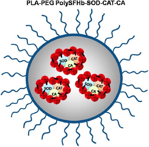
This paper combines our nanobiotechnological approaches of PolySFHb-SOD-CAT-CA (Bian et al. Citation2011) with PEG-PLA nanocapsules (Chang et al. Citation2003, Chang Citation2007, Fustier and Chang Citation2012). The details have been described in these references. We nanoencapsulated PolySFHb-SOD-CAT-CA into PEG-PLA nanocapsules to form PEG-PLA-PolySFHb-SOD-CAT-CA nanocapsules ().
Figure 1. Schematic representation of Poly(ethylene glycol)-poly(lactic acid) block-copolymer encapsulated Polystroma-free hemoglobin-superoxide dismutase-catalase-carbonic anhydrase (PEG-PLA-Poly SFHb-SOD-CAT-CA) nanocapsules.
This paper describes the preparation and characterization of PEG-PLA-PolySFHb-SOD-CAT-CA nanocapsules. To evaluate its special function of CO2 carrier, we also monitor the changes of tissue PCO2 in a lethal hemorrhagic shock model. This is the first step to be followed by much more needed research.
Materials and methods
Materials
Poly(dl-lactic acid) (PLA) (M.W. 15,000 g/mol) was purchased from Polysciences Inc. Tween 20 was obtained from Fisher. Tin 2-ethylhexanoate, methoxypolyethylene glycol (PEG) (M.W. = 5000 g/mol), Hydrogenated L-α-Phosphatidylcholine, and other analytical reagents were purchased from Sigma. Carbon film-coated TEM Grids were purchased from Science Services. The stroma-free hemoglobin (SFHb) was Hb with the original RBC enzymes extracted from bovine blood was purchased from the McGill University McDonald Campus Cattle Complex (Sainte-Anne-de-Bellevue, Canada). The ultrapure Hb with all enzymes removed was bought from Biopure to prepare PolyHb. Polyethersulfone ultrafiltration membranes with a cut-off molecular weight of 500 kDa were purchased from Millipore.
Preparation of PEG-PLA copolymer
The procedures used were referred to the methods by TMS (Chang Citation2007). Briefly, 1.5 g of DL-PLA (M.W. 15,000 g/mol) and 0.75 g of methoxypolyethylene glycol (M.W. 5,000 g/mol) were placed in a 125 mL Erlenmeyer flask and dried under vacuum overnight in a vacuum oven (Fisher isotemp) at room temperature in the presence of phosphorus pentoxide. They were dissolved in 7.5 mL of acetone. The mixture was heated in an oil bath under nitrogen. The acetone evaporated completely in 2–4 min and the flask was left in the oil bath at 180°C for two more hours. 10 μL of the catalyst stannous-2-ethylhexanoate were then added and the flask was heated for three more hours at 180°C under nitrogen. The copolymer solidified slowly at room temperature. It was collected in a vial and stored in desiccators.
Preparation of PolySFHb-SOD-CAT-CA, PolySFHb and PolyHb
The procedures used were referred to the methods by TMS (Chang Citation2007).
(1)Polyhemoglogin (PolyHb) was prepared using ultrapure Hb with all enzymes removed.
(2)PolySFHb, was prepared using 7 g/dl of stroma-free hemoglobin SFHb (extracts from red blood cells containing enzymes normally present in the red blood cell).
(3)PolySFHb-SOD-CAT-CA, was prepared using 20 mL SFHb (7 g/dL) with the addition of 5.6 mg of SOD, 110.3 mg of CAT, and 5.3 mg of CA in 50 mM potassium phosphate buffer (pH7.4). The analysis of enzyme activities have been described (Bian et al. Citation2013).
Before the initiation of the crosslinking reaction, 187 μl of 1.3 M lysine was added at a molar ration of 11.1:1 lysine/Hb, and shaken in the speed of 140 r/min for 1 h. Nitrogen gas was used to flush the reaction vessel to prevent formation of methemoglobin. Crosslinking was started with the addition of 744 μl of 0.5 M glutaraldehyde at a molar ration of 17:1 glutaraldehyde/Hb. Glutaraldehyde was added in four equal aliquots over a period of 15 minutes, and the reaction was allowed to crosslink for 24 hours at 4°C with constant stirring in the speed of 140 r/min under anaerobic conditions. Crosslinking was stopped by adding 4.47 g of lysine at a molar ratio of 1118:1 lysine/Hb and shaken in the speed of 150 r/min for 1 h. To remove free Hb and enzymes, A a Sephacryl-300 HR column was used to purify PolySFHb-SOD-CAT-CA, PolySFHb and PolyHb. The column was equilibrated with the elution buffer (0.1 M Tris-HCl and 0.15 M NaCl pH 7.4). Samples were passed through the column at a flow rate of 36 ml/hour. The molecular weights of PolySFHb-SOD-CAT-CA, PolySFHb, and PolyHb were higher than 250 kDa, so they were collected by peak identification using UV detector at 280 nm in order to exclude unlinked enzymes, such as catalase (250 kDa), superoxide dismutase (32.5 kDa), Carbonic anhydrase (30 kDa), and free tetrameric Hb (64 kDa). Then the samples were concentrated to 5 g Hb/dL using the spectral absorbent gel and stored at − 80°C.
Preparation of PLA-PEG-PolySFHb-SOD-CAT-CA nanocapsules, PLA-PEG-PolySFHb nanocapsules and PLA-PEG-PolyHb nanocapsules
The preparation was performed using the improved methods reported by TMS (Chang Citation2007). Briefly, the nanocapsules were prepared by injecting an organic phase into an aqueous phase containing PolySFHb-SOD-CAT-CA, PolySFHb, or PolyHb. The aqueous phase consisted of 10 ml of 5 g/dL PolySFHb-SOD-CAT-CA, PolySFHb, or PolyHb with 100 μl of tween 20. While the organic phase consisted of 40 mg of PEG-PLA, 20 mg of phosphatidylcholine, 3.2 ml of acetone, and 1.6 ml of ethanol. Slowly dropped the organic phase through a 26 G needle into the aqueous phase under magnetic stirring with Therm-O-Swirl Stirrer (Precision Scientific Co., Chicago) setting to 6 grade at 4°C. The nanocapsules were formed immediately, but the suspension was kept stirred for 15 min. The organic solvent was partly removed from the suspension prepared by rotary evaporator under vacuum at 0°C for about 3 h. The nanocapsules were purified by a 50 mL Millipore-stirred cells device operating at 4°C with a polyethersulfone ultrafiltration membrane under a nitrogen pressure of 69 kPa.
The FTIR of PLA-PEG and the TEM of nanocapsules
The chemical structure of PLA-PEG was determined using FTIR (Shimadzu Corporation, Japan). Solutions of PLA-PEG-PolySFHb-SOD-CAT-CA nanocapsules, PLA-PEG-PolySFHb nanocapsules, and PLA-PEG-PolyHb nanocapsules were relatively dropped on a carbon film-coated TEM grids and air dried, and then were relatively measured using transmission electron microscopy (TEM) (Philips, Holand).
Measurement of Hb concentration and enzyme activity
Based on the methods of TMS (Chang Citation2007), the Hb concentration and enzyme activity could be measured using UV-visible spectrophotometer (GE, America) as follows. The Hb concentration was determined by reaction the samples with Drabkin's reagent (Sigma-Aldrich), then measuring the concentration of the resulting cyan-methemoglobin solution by spectrophotometry at 540 nm. The hydration activity of CO2 by CA was determined by the pH of a 0.02 M Tris buffer decreasing from pH 8.3 to 6.3 per minute. The measurements in seconds were converted into W-A units according to the following formula: 1 W-A unit = [2*(T0 − T)]/T. The activity of CAT was measured by the rate of disappearance of H2O2 in UV 240 nm according to the following formula: U = 1106.301 × Log (A0/A15). The measurement for SOD activity was based on the reduction of cytochrome C.
Determination of entrapment efficiency and nanocapsule recovery
After rotary evaporation, samples (PLA-PEG-PolySFHb-SOD-CAT-CA) were centrifuged at 15000 r/min × 15 min 4°C (Beckman J2-21, USA). The volume of supernatant and its Hb concentration and enzyme activity were measured using UV-visible spectrophotometer. The sediment was lyophilized for 48 h in a Lab Con Co freeze dryer (Freeze Dry 5). The total amount of enzymes in the final PolySFHb-SOD-CAT-CA preparation consists of those normally present in the stroma-free hemoglobin (SFHb) plus the amount added. The entrapment efficiency (EE%) and nanocapsule recovery (%) of nanocapsules were calculated as follows. All procedures and measurements were repeated thrice.
The effects on a hemorrhagic shock model
McGill University ethics committee on animal research has approved our animal research. A total of 24 male Sprague–Dawley rats with a mean body weight of 250 ± 20 g were used. All rats were obtained from the comparative medicine Animal Research Center of the McGill University. The rats were maintained in the McIntyre animal center with 12 h light/dark cycles. They were provided with food pellets and water ad libitum. The rats were randomly divided into three groups (n = 6). All rats were anesthetized with intraperitoneal injection of 54.7 mg/ml × 5.5 ml/g pentobarbital solution. Then a right inguinal incision was made. Heparinized catheters were inserted into the right femoral vein and right femoral artery. A CO2 meter probe (lazer research lab, USA) was placed on the surface of the muscle at the incision site to measure tissue PCO2. Mean arterial pressure (MAP) was observed from the right femoral artery using a Biopac system (BIOPAC Systems Inc., Goleta, CA), and the result of the observation was used as a basis for the modified model of hemorrhagic shock (Chang Citation2007). When the rats recovered conjunctival reflex, blood was withdrawn from the right femoral artery and MAP was reduced to 30 mmHg and maintained for 60 min. The rats were infused from the femoral vein with the same volume of liquid that was shed from the femoral artery. The rats were divided into four groups. The first group of rats were treated with PLA-PEG encapsulated 5 g/dL of PolyHb nanocapsules (n = 6). The second group of rats were treated with PLA-PEG encapsulated 5 g/dL of PolySFHb nanocapsules (n = 6). The third group of rats was treated with 5 g/dL of PolySFHb-SOD-CAT-CA (n = 6). The fourth group of rats were treated with PLA-PEG encapsulated 5 g/dL of PolySFHb-SOD-CAT-CA nanocapsules (n = 6). After an hour of resuscitation and follow up, the rats were sacrificed using intraperitoneal nembutal.
Statistical analysis
Test data were shown as mean ± SD of three independent experiments, except when otherwise indicated. One-way ANOVA was used to analyze the statistically significant differences, followed by the Newman–Keuls post hoc test wherever appropriate. P-values less than 0.05 (P < 0.05) were considered statistically significant.
Results
The FTIR of PLA-PEG and the TEM of nanocapsules
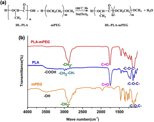
As shown in , PLA-PEG was prepared by PLA and PEG at 180°C in the presence of the catalyst stannous-2-ethylhexanoate. FTIR spectra were shown in , PEG had the peaks at 3492 cm− 1, 2889 cm− 1, and 1110 cm− 1, which were respectively assigned to at –OH, –CH2–, and –C–O–C– groups. While the peaks at 3485 cm− 1, 2995 cm− 1, 2947 cm− 1, and 1751 cm− 1 in the spectrum of PLA were due to –COOH, –CH3, –CH-, and C = O groups, respectively. Moreover, both peaks at 1125 cm− 1 and 1096 cm− 1 corresponded to –C–O–C–. PLA-PEG had all the characteristic peaks of PLA and PEG, thus proving the presence of the two component blocks in the copolymer. Meanwhile, the peaks of –OH and –COOH end groups were much smaller than those of PLA and PEG for the dehydration reaction. These results demonstrated that PLA-PEG agreed with previous literature in terms of its chemical structure (Chang Citation2007).
Figure 2. Synthesis and characterizations of PLA-PEG (a) Chemical reaction equation to synthesize PLA-PEG: the –COOH in DL-PLA reacted with –OH in mPEG, dehydrating into PLA-PEG. (b) FTIR spectra of mPEG, PLA and PLA-PEG. 0.25 mg of the dry sample was mixed with IR-grade KBr (0.1 g) and pressed (10 ton) into tablet form. The spectrum of the tablet was then recorded by an FTIR spectrometer.
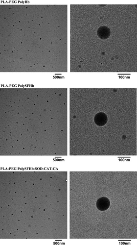
As observed through TEM in , all nanocapsules of PLA-PEG-PolyHb, PLA-PEG-PolySFHb, and PLA-PEG-PolySFHb-SOD-CAT-CA had a homogeneous particle diameter, good dispersion, and core shell structure. The mean diameters were 100 nm, and there was no difference in sizes for the different preparations.
Figure 3. TEM pictures without metal spraying of PLA-PEG-PolyHb nanoparticles, PLA-PEG-PolySFHb nanoparticles, and PLA-PEG-PolySFHb-SOD-CAT-CA nanoparticles.
EE and nanocapsule recovery
There was good EE% of Hb, SOD, CAT, and CA in PLA-PEG-PolySFHb-SOD-CAT-CA at 68.34 ± 0.85%, 68.52 ± 1.44%, 68.27 ± 1.07%, and 71.85 ± 2.51%, respectively. Nanocapsule recovery was 71.30 ± 2.97%.
Tissue PCO2 in muscle on a lethal hemorrhagic shock model ( and )
Figure 7. Muscle PCO2 of a lethal hemorrhagic shock and resuscitation for each of the four groups.
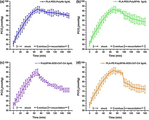
Figure 8. Combined figure of PCO2: There were no significant differences between the PolySFHb-SOD-CAT-CA group (—□—) and the PLA-PEG-PolySFHb-SOD-CAT-CA group (—■—) in terms of PCO2 at any time of resuscitation, and they had the lower PCO2 than the other two groups. PLA-PEG-PolyHb group (—•—) had higher PCO2 after 20 mins of resuscitation than PLA-PEG-PolySFHb group (—▲—). (n = 6, P < 0.05).
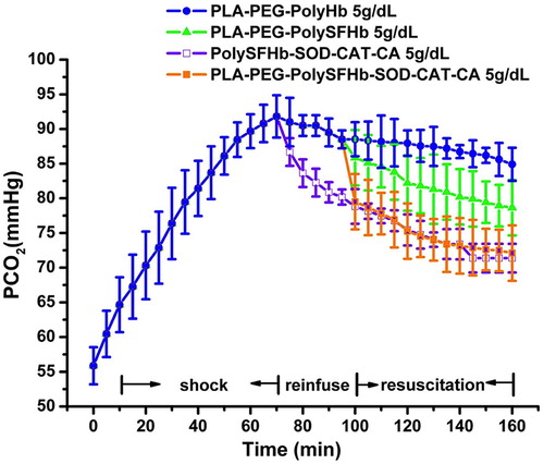
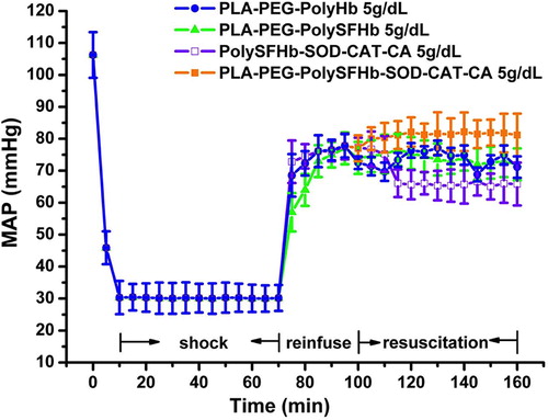
The concentrations of Hb and enzymes activities in PLA-PEG-PolyHb, PLA-PEG-PolySFHb, PolySFHb-SOD-CAT-CA, and PLA-PEG-PolySFHb-SOD-CAT-CA are shown in . There were no significant differences in MAP and PCO2 among any of the groups during the control period. The rats in all groups responded to bleeding of 67% of the total blood volume (56 mL/kg) with a decrease in MAP to 30 mmHg, which was maintained for 1 hour. After resuscitation, the MAP in all groups recovered initially. The recovery of MAP was well maintained in the PLA-PEG-PolySFHb-SOD-CAT-CA group, whereas this was not as well maintained in the PolySFHb-SOD-CAT-CA, PLA-PEG-PolySFHb, and PLA-PEG-PolyHb groups (). Meanwhile, the tissue PCO2 in all groups increased rapidly during shock. With resuscitation, the tissue PCO2 in all the groups decreased, but the PCO2 in all groups were not lowered to the same levels. Thus, PLA-PEG-PolyHb was least efficient in lowering the tissue PCO2 level. PLA-PEG-PolySFHb was lower than PLA-PEG-PolyHb. However, PolySFHb-SOD-CAT-CA and PLA-PEG-PolySFHb-SOD-CAT-CA were the most effective of the four tested. (n = 6, P < 0.05) ().
Figure 4. The concentrations of Hb and enzymes activities in PLA-PEG-PolyHb nanoparticles, PLA-PEG-PolySFHb nanoparticles, PolySFHb-SOD-CAT-CA, and PLA-PEG-PolySFHb-SOD-CAT-CA nanoparticles.
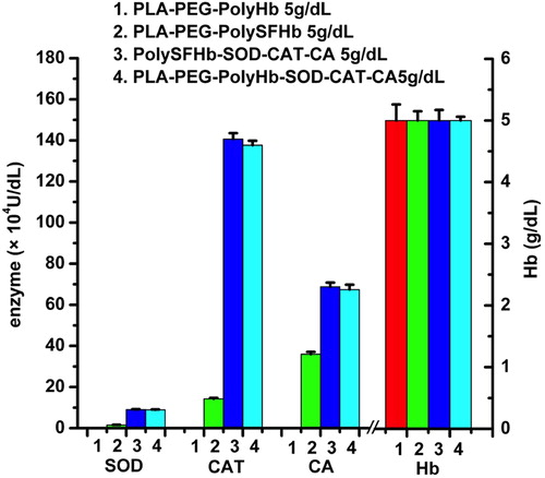
Figure 5. MAP of lethal hemorrhagic shock and resuscitation for each of the four groups.
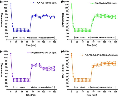
Figure 6. Combined figure of MAP: There were no significant differences between the MAP in PLA-PEG-PolySFHb group (—▲—) and PLA-PEG-PolyHb group (—•—). PLA-PEG-PolySFHb-SOD-CAT-CA group (—■—) had significantly highest MAP in all groups at any time of resuscitation, PolySFHb-SOD-CAT-CA group (—□—) had significantly lowest MAP in all groups after 15 min of resuscitation (n = 6, P < 0.05).
Discussion
Most of the current RBCs substitutes just work as O2 carriers. For example, PLA-PEG-encapsulated Hb has shown good oxygen dissociation curve (Zhang et al. Citation2009) and long circulation time (Chang et al. Citation2003). Poly SFHb-SOD-CAT has shown the dual function of an oxygen carrier and the ability to remove oxygen radicals, so it can carry oxygen to ischemic brain tissue without causing significant injury in an ischemia-reperfusion rat brain model (Powanda and Chang Citation2003). This basic research has been extended to a product which is an oxygen carrier with synthetic antioxidant properties (Buehler et al. Citation2004). However, real RBCs are not only O2 carriers, but also are CO2 carriers. As a result, a three functional O2 and CO2 carriers with enhanced antioxidant activity in the form of PolySFHb-SOD-CAT-CA were prepared (Bian et al. Citation2011) and shown to be effective in lowering PCO2 in a hemorrhagic shock rat model (Bian et al. Citation2013). We now report an extension of this to prepare nanodimension artificial RBC in the form of PLA-PEG nanocapsules containing PolySFHb-SOD-CAT-CA.
The FTIR spectrum of the PLA-PEG contained the characteristic peaks of PLA and PEG, while lowered peaks of –OH and –COOH for the dehydration reaction. These results show that PLA-PEG agrees with previous literature in terms of its chemical structure (Luo et al. Citation2002), indicating the PLA-PEG had been successfully prepared. As shown in the TEM micrographs in , all nanocapsules of PLA-PEG-PolyHb, PLA-PEG-PolySFHb, and PLA-PEG-PolySFHb-SOD-CAT-CA had a nucleus shell structure with good dispersion without aggregation. The mean diameter of the nanocapsules was 100 nm. PLA-PEG-PolySFHb-SOD-CAT-CA showed good EE% of Hb, SOD, CAT, and CA and recovery of nanocapsules. This is likely because it is easier to nanoencapsulate the larger size PolyHb-SOD-CAT-CA and the proportion of hemoglobin and enzymes remained unchanged in the crosslinked form.
Compared with arterial pressure, end-tidal CO2, cardiac index, arterial blood lactate, and base deficit, tissue PCO2 was shown to be more predictive of outcome after rapid bleeding, so it likely to serve as a useful guide for the immediate management of hemorrhagic shock (Cammarata et al. Citation2009). For rats, skeletal muscles showed better compliance than others. To optimize the compliance, PCO2 probe was placed on the surface of skeletal muscles at inguinal incision. As shown in , both the PolySFHb-SOD-CAT-CA group and the PLA-PEG-PolySFHb-SOD-CAT-CA group were as effective in lowering the elevated PCO2 at any time after resuscitation. Thus, the encapsulated procedure had not decreased CA activity. These two groups were able to lower the elevated PCO2 level much better than the PLA-PEG-PolySFHb group that had no added CA or the PLA-PEG-PolyHb group.
As stated in the introduction, the aim of the present study is to analyze the lowering of the elevated PCO2. The PLA-PEG nanocapsules groups were able to maintain the MAP better than the PolySFHb-SOD-CAT-CA group. This is likely because the circulation half time of PLA-PEG PolyHb nanocapsules is double that of the soluble PolyHb (Chang et al. Citation2003). PLA-PEG-PolySFHb-SOD-CAT-CA could maintain MAP better than the PLA-PEG-PolySFHb and PLA-PEG-PolyHb group. Further study is needed to see if this is because of the presence of CA resulting in better removal of tissue CO2 and the lowering of tissue PCO2.
Acknowledgements
Professor TMS Chang acknowledges the operating term grant from the Canadian Institute of Health Research (MOP13745) and Wei Gao's China Scholarship Council scholarship for her to come here as a PhD trainee. Jeffrey Chi Yung Tso, is a summer student who acted mostly as an observer.
Declaration of interest
The authors report no declarations of interst. The authors alone are responsible for the content and writing of the paper.
References
- Alayash AI. 2004. Oxygen therapeutics: can we tame haemoglobin?. Nat Rev Drug Discov. 3:152–159.
- Bian Y, Rong Z, Chang TMS. 2011. A novel biotechnology-based blood substitute that transports both oxygen and carbon dioxide and also acts as an antioxidant. Artif Cells Blood Substit Biotechnol. 39:127–136.
- Bian Y, Gao W, Chang TMS. 2013. Lowering of elevated tissue PCO2 in a hemorrhagic shock rat model after reinfusion of a novel nanobiotechnological polyhemoglobin-superoxide dismutase-catalase-carbonic anhydrase that is an oxygen and carbon dioxide carrier with enhanced antioxidant properties. Artif Cells Nanomedicine Biotechnol. 41:60–68.
- Biro GP, Ou C, Ryan-MacFarlane C, Anderson PJ. 1995. Oxyradicaly generation after resuscitation of hemorrhagic shock with blood or stromafree hemoglobin. Artif Cells Blood Substit Immobil Biotechnol. 23:631–645.
- Buehler PW, Haney CR, Gulati A, Ma L, Hsia CJ. 2004. Resuscitative effects of polynitroxylated alpha-alpha-cross-linked Hb following severe hemorrhage in the rat. Free Radic Biol Med. 37:124–135.
- Cammarata GA, Weil MH, Castillo CJ, Fries M, Wang H, Sun S, Tang W. 2009. Buccal capnometry for quantitating the severity of hemorrhagic shock. Shock. 31:207–211.
- Chang TM. 1964. Semipermeable microcapsules. Science. 146: 524–525.
- Chang TM. 1971. Stabilisation of enzymes by microencapsulation with a concentrated protein solution or by microencapsulation followed by cross-linking with glutaraldehyde. Biochem Biophys Res Commun. 44:1531–1536.
- Chang TMS, Powanda D, Yu WP. 2003. Analysis of polyethylene-glycol-polylactide nano-dimension artificial red blood cells in maintaining systemic hemoglobin levels and prevention of methemoglobin formation. Biotechnology. 31:231–247.
- Chang TM. 2005. Therapeutic applications of polymeric artificial cells. Nat Rev Drug Discov. 4:221–235.
- Chang TMS. 2007. Artificial Cells: Biotechnology, Nanomedicine, Regenerative Medicine, Blood Substitutes, Bioencapsulation, Cell/Stem Cell Therapy. Singapore/London: World Scientific/ Imperial College.
- Chang EJ, Lee TH, Mun KC, Kim HC, Suh SI, Bae JH, et al. 2004. Effects of polyhemoglobin-antioxidant enzyme complex on ischemia- reperfusion in kidney. Transplant Proc.;36:1952–1954.
- D’Agnillo F, Chang TM. 1998. Polyhemoglobin-superoxide dismutase-catalase as a blood substitute with antioxidant properties. Nat Biotechnol. 16:667–671.
- Geers C, Gros G. 2000. Carbon dioxide transport and carbonic anhydrase in blood and muscle. Physiol Rev. 80:681–715.
- Djordjevich L, Miller IF. 1980. Synthetic erythrocytes from lipid encapsulated hemoglobin. Exp Hematol. 8:584–592.
- Fustier C, Chang TMS. 2012. PEG-PLA nanocapsules containing a nanobiotechnological complex of polyhemoglobin-tyrosinase for the depletion of tyrosine in melanoma: preparation and in vitro characterisation. Nanomedicine Biotherapeu Discover. 2:1–6.
- Gao W, Sha B, Zou W, Liang X, Meng X, Xu H, et al. 2011. Cationic amylose-encapsulated bovine hemoglobin as a nanosized oxygen carrier. Biomaterials. 32:9425–9433.
- Jahr JS, Mackenzie C, Pearce LB, Pitman A, Greenburg AG. 2008. HBOC-201 as an alternative to blood transfusion: efficacy and safety evaluation in a multicenter phase III trial in elective orthopaedic surgery. J Traum. 64:1484–1497.
- Luo W, Li S, Bei J, Wang S. 2002. Synthesis and characterization of poly ( L-lactide ) - poly ( ethylene glycol ) multiblock copolymers. Polymer. 84:1729–1736.
- Moore EE, Moore FA, Fabian TC, Bernard AC, Fulda GJ, Hoyt DB, et al. 2009. Human polymerized hemoglobin for the treatment of hemorrhagic shock when blood is unavailable: The USA multicenter trial. Shock. 208:1–13.
- Piras AM, Dessy A, Chiellini F, Chiellini E, Farina C, Ramelli M, Della Valle E. 2008. Polymeric nanoparticles for hemoglobin-based oxygen carriers. Biochim Biophys Acta. 1784:1454–1461.
- Powanda DD, Chang TM. 2003. Cross-linked polyhemoglobin- superoxide dismutase-catalase supplies oxygen without causing blood-brain barrier disruption or brain edema in a rat model of transient global brain ischemia-reperfusion. Artif Cells Blood Substit Biotechnol. 30:23–37.
- Tsuchida E, Sou K, Nakagawa A, Sakai H, Komatsu T, Kobayashi K. 2009. Artificial oxygen carriers, hemoglobin vesicles and albumin-hemes, based on bioconjugate chemistry. Bioconjug Chem. 20:1419–1440.
- Winslow RM. Ed. 2006. Blood Substitutes. Amsterdam: Academic Press.
- Yu B, Raher MJ, Volpato GP, Bloch KD, Ichinose F, Zapol WM. 2008. Inhaled nitric oxide enables artificial blood transfusion without hypertension. Circulation. 117:1982–1990.
- Yu WP, Chang TM. 1994. Submicron biodegradable polymer membrane hemoglobin nanocapsules as potential blood substitutes: a preliminary report. Artif Cells Blood Substit Immobil Biotechnol. 22:889–893.
- Zhang X, Liu C, Yuan Y, Shan X, Sheng Y, Xu F. 2009. A noninvasive method for measuring the oxygen binding-releasing capacity of hemoglobin-loaded polymeric nanoparticles as oxygen carrier. J Mater Sci Mater Med. 20:1025–1030.
- Zhu H, Du Q, Chen C, Chang TM. 2010. The immunological properties of stroma-free polyhemolysate containing catalase and superoxide dismutase activities prepared by polymerized bovine stroma-free hemolysate. Artif Cells Blood Substit Biotechnol. 38: 57–63.