
Abstract
The objective of this study is to investigate the effects of BML-111 on acute pancreatitis-associated lung injury (APALI) induced by cerulein with subsequent an LPS administration in mice and its possible mechanisms. One hundred and twenty-eight mice were randomly allocated to four groups, namely the APALI group, the BML-111 pretreatment group, the BM-111 control group, and the control group. The ‘two-hit’ mice APALI model was established by intraperitoneal injection of cerulein 7 times at hourly intervals and Escherichia coli lipopolysaccharide (LPS) once after the last dose of cerulein immediately. The samples were taken at 3, 6, 12, and 24 h after the last injection. Serum levels of amylase, TNF-a, IL-1β and IL-10, were determined. Histological score of the pancreas and lung, the wet/dry weight ratio, and heme oxygenase-1 (HO-1) expression in the lung were also evaluated. BML-111 pretreatment significantly reduced the serum levels of amylase, TNF-α, IL-1β, the wet/dry weight ratio of lung, and the pathology injury scores of pancreas and lung, and the serum levels of IL-10 were markedly increased. The severity of pancreatic and lung histology were also significantly improved by the administration of BML-111, and the expressions of HO-1 in lung tissues also increased in the BML-111 group compared with those in the APALI group. In conclusion, BML-111 exerts protective effects on APALI induced by cerulein and LPS. In addition to its anti-inflammatory effects, the beneficial effects may also be due to the upregulation of HO-1 expression in the lung tissues.
| Abbreviations: | ||
| ALI | = | acute lung injury |
| AP | = | acute pancreatitis |
| APALI | = | acute pancreatitis-associated lung injury |
| BML-111 | = | lipoxin a4 agonist |
| ELISA | = | enzyme-linked immunosorbent assay |
| FPR-2 | = | formyl peptide receptor 2 |
| HO-1 | = | heme oxygenase-1 |
| IL-1 | = | interleukin-1 |
| IL-6 | = | interleukin-6 |
| IL-10 | = | interleukin-10 |
| LXA4 | = | lipoxin a4 |
| MODS | = | multiple organ dysfunction syndrome |
| PCR | = | polymerase chain reaction |
| SAP | = | severe acute pancreatitis |
| SIRS | = | systemic inflammatory response syndrome |
| TNF-α | = | tumor necrosis factor α |
Introduction
Severe acute pancreatitis (SAP) is an inflammatory process of the pancreas with significant morbidity and mortality rates (Shen and Lu Citation2011, Wang et al. Citation2010). Deaths associated with SAP are mainly due to its major complications, and multiple organ dysfunction syndromes (MODSs) are the major causes of SAP-related deaths (Barreto and Rodrigues Citation2008, Barreto and Rodrigues Citation2007). Acute lung injury (ALI) is a major component of this complication. About one-third of all deaths from acute pancreatitis (AP) has been reported to occur prior to admission to hospital and, in most cases, is associated with ALI. Related studies have suggested that a loss of control of inflammation and the release of systemic inflammatory cytokines, including TNF-α and IL-1β, may play a more dominant role in the pathogenesis of SAP and distant organ damage, such as APALI (Pereda et al. Citation2006). Lipoxins (LXs) are biosynthesized from arachidonic acid via lipoxygenase and are one of the most important endogenous lipid anti-inflammatory media in the body, and are regarded as “braking signal” or “stop signal” for inflammatory response (Chiang et al. Citation2005). Previous research has shown LXA4 had protective effects on experimental SAP by inhibiting the NF-κB signaling pathway and reducing the production of proinflammatory cytokines (Zhou et al. Citation2011). BML-111 is a commercially synthetic LX receptor agonist, and even more potent and stable than the original LXs molecule, and has extensive biological effects, such as anti-inflammatory, antioxidation, antitumor, and immune regulations. Those effects had been demonstrated in carbon tetrachloride-induced liver injury, zymosan-induced arthritis, and hemorrhagic shock-induced acute lung injury (Gong et al. Citation2012, Zhang et al. Citation2008, Zhang et al. Citation2007). In this study, we first examined whether BML-111 is able to attenuate APALI induced by cerulein with a subsequent LPS administration and investigated the underlying mechanisms.
Materials and methods
Animals and main reagents
BALB/c mice weighing 18–23 g from the experimental animal center of Lanzhou University were randomly allocated to four experimental groups (n = 32 for each group): the APALI group (APALI), the BML-111 pretreatment group (APALI + BML), the BM-111 control group (BML), and the control group (CON). Each group was randomly divided into four time units (3, 6, 12, and 24 h) with eight rats in each time unit. Before the experiment, the mice were housed in metabolic cages at a controlled temperature and under a 12-h light/dark cycle for at least 72 h to acclimate to the surroundings. Twelve four hours before the start of the experiments, the mice were deprived of food but allowed access to water. All experiments were performed in accordance with the experimental protocol approved by the Committee for Research and Animal Ethics of Lanzhou University, Lanzhou, China. All surgery was performed under sodium pentobarbital anesthesia, and all efforts were made to minimize suffering. Cerulein and LPS were purchased from Sigma–Aldrich, Inc. (Louis, MO, USA). BML-111 was gained from Enzo Life Sciences, Inc. (Belgium). The amylase kits were purchased from the Nanjing Jiancheng Bioengineering Institute (China). TNF-α, IL-1β, and IL-10 kits of mice were purchased from Dakewe Biotech Co., Ltd. (China). The real-time PCR kits and HO-1-primers were purchased from the TaKaRa Biotechnology (Dalian) Co., Ltd. (China). For the Western blot analysis, anti-β-actin antibody and horseradish peroxidase (HRP)-conjugated goat anti-rabbit immunoglobulin G were obtained from Beijing Biosynthesis Biotechnology Co., LTD (China). Anti -HO-1 antibody was purchased from Bioworld Technology, Inc. (USA).
Induction of the ‘two-hit’ model of APALI and pretreatments
The ‘two-hit’ mice model of APALI was established by intraperitoneal injection of cerulein (50 μg/kg body weight, 10 ml/kg of body weight) 7 times at hourly intervals and by intraperitoneal injection of Escherichia coli lipopolysaccharide (LPS, 10 mg/kg of body weight, 10 ml/kg of body weight) after the last dose of cerulein, according to the previously described method (Ding et al. Citation2003, Chen et al. Citation2009, Dixon et al. Citation2009, Yang et al. Citation2009, Elder et al. Citation2012a, Citation2012b). In the BML-111 pretreatment group, the mice were administered BML-111 (suspended in 0.4% DMSO solution, 1 mg/kg of body weight, 10 ml/kg of body weight) 1 h before the induction of APALI, and this treatment schedule and dosage were based on previous studies (Gong et al. Citation2012, Zhang et al. Citation2007, Hao et al. Citation2011) and determined with modification from preliminary experiments (unpublished data). In the BML-111 control and the control groups, mice were administered equal volume of normal saline instead of cerulein and LPS. Mice in the BML-111 control group also received BML-111 1 h before the first injection. After experimental or control treatments, all animals were returned to their cages and allowed free access to food and water. Eight mice of each group were anesthetized with sodium pentobarbital (90 mg/kg BW, i.m.) at 3, 6, 12, and 24 h after the last injection of cerulein or saline. Blood samples, lung, and pancreatic tissues were harvested for subsequent measurements.
Serum amylase estimation
Serum amylase was assayed using a commercial kit with a kinetic spectrophotometric method according to the manufacturer's instructions. The methodology is based on the enzymatic degradation of ethylidene-p-nitrophenol-G7 by amylase coupled with glucosidase, thus producing p-nitrophenol that exhibits strong absorbance at 405 nm. The concentrations of amylase were expressed as units per liter (U/L).
Serum TNF-α, IL-1β, and IL-10 estimation
Levels of serum TNF-a, IL-1β, and IL-10 were assayed with commercial kits using enzyme-linked immunosorbent assay (ELISA) method according to the manufacturers’ protocols. Levels of serum TNF-a, IL-1β, and IL-10 were expressed as picograms per milliliter (pg/ml).
Determination of the wet/dry weight ratio of lung tissues
The inferior lobe of right lung was weighed after its surface effusion, and blood was dried using filter paper, and then it was continuously dried for 72 h at 80°C and was reweighed. Lung water content was determined by calculating the wet/dry weight ratio according to the formula [water content = (wet weight-dry weight)/dry weight × 100%], where the wet weight was the original weight of the lung tissues and the weight after dried at 80°C for 72 h as the dry weight.
Pathological diagnosis of pancreas and lung injury
Tissue sections stained with hematoxylin–eosin (H&E) were evaluated blindly under light microscope by two pathologists on two separate occasions. The criteria for the histologic evaluation of pancreatic injury were based on previously described method of improved Schmidt evaluation criteria (Schmidt et al. Citation1992). The pathological changes in the lungs were based on the method of Hofbauer et al. (Citation1998).
Ultrastructural organization examination of lung tissues
The procedure was similar to that described previously (Wang et al. Citation2010). Lung sections were cut into small pieces and fixed in 2% paraformaldehyde containing 2.5% glutaraldehyde buffered by 0.1 M cacodylate (pH 7.2) overnight at 4°C, and subsequently fixed in 1% osmium tetraoxide for 2 h on ice. After dehydration, the lung sections were infiltrated with epoxy resin. Then, ultrathin sections were cut at 70 nm and stained with uranyl acetate and lead citrate. Sections were imaged on a JEM-1230 transmission electron microscope (JEOL, Tokyo, Japan).
Confirmation of HO-1 mRNA expression in lung by real-time polymerase chain reaction
The primers for HO-1 were designed from published mouse mRNA sequences. The following primers were used: for HO-1 (GenBank accession no. NM_010442.2), the sense primer sequence was 5′-TGCAGGTGATGCTGACAG AGG-3′, and the antisense primer sequence was 5′-GGGATGAGCTAGTGCTG ATCTGG-3′, with a PCR product of 144 bp; for β-actin (GenBank accession no. NM_007393.3), the sense primer sequence was 5′-CATCCGTAAAGACCTCTATGCCAAC-3′, and the antisense primer sequence was 5′-ATGGAGCCAC CGATCCACA-3′, with a PCR product of 171 bp. Total RNA was extracted from lung tissues using a commercial extract reagent RNAiso Plus. Quantitative real-time PCR was performed in a Rotor-Gene 6000 thermal cycler. Reactions were carried out in a 25-μl reaction volume. The real-time PCR was performed with the following program: 95°C for 30 s, followed by 40 cycles of denaturation at 95°C for 5 s and annealing at 60°C for 30 s. All the reactions were performed in triplicate. Data analysis was performed using the 2−ΔΔCT method, where β-actin was used as an internal standard in each experiment for quantitative evaluation of gene mRNA expression.
Confirmation of HO-1 protein expression in lung by western blotting analysis
The proteins samples (50 μg) were denatured in loading buffer and separated by 12% sodium dodecyl sulfate poly acrylamide gel electrophoresis (SDS–PAGE) at 30 mA constant current. The separated proteins were electroblotted onto a polyvinylidene difluoride (PVDF) membrane using transfer buffer at 100 V for 60 min. The membranes were blocked with 5% nonfat dry milk in Tris-buffered saline (TBS)–0.1% Tween (TBST) for 2 h at room temperature and then incubated with primary antibody/rabbit anti-β-actin (1: 1000 dilution) and rabbit anti-HO-1 (1: 500 dilution) in TBST overnight at 4°C. After five 8-min washes in TBST, the membranes were incubated with a horseradish peroxidase (HRP)-conjugated goat anti-rabbit immunoglobulin G (1:10000 dilution) for 2 h at room temperature. After five 8-min washes in TBST, the membranes were analyzed using enhanced chemiluminescence (ECL) substrate and used to expose an X-ray film according to the manufacturer's protocol. The relative expression of the protein bands of HO-1 (28 kDa) was quantified by densitometric scanning of the radiographic films with GS-700 imaging densitometer and analyzed by a computer program. The results from each experimental group were expressed as the relative integrated intensity compared with the β-actin (42 kDa) band densities in lung measured in the same batch.
Statistical analysis
All data were expressed as the mean ± standard deviation (SD). Statistics were done using Statistical Package for the Social Sciences (SPSS) software, version 16.0. Statistical significance of differences among multiple groups was evaluated using one-way ANOVA or rank-sum test. Multiple comparisons were carried out using SNK-q test or Nemenyi test. For all analyses, a p < 0.05 was considered statistically significant.
Results
Changes in serum amylase levels and histological scores of pancreatic injury
To evaluate the severity of SAP, the serum amylase levels and histological scores of pancreatic injury are summarized in . Levels of serum amylase in the control group and the BML-111 control group were low. Levels of serum amylase in the APALI group significantly elevated after 3 h, more significantly after 6 h, reached a peak after 12 h, and remained until 24 h, and the difference has statistical significance compared with that in the control group and the BML-111 control group at all corresponding time points (p < 0.05, respectively). BML-111 pretreatment significantly reduced its elevation, when compared with those in the control group at the corresponding time points (p < 0.05, respectively). In addition, the scores of pancreatic injury were reduced in the BML pretreatment group compared with those in the APALI group at the corresponding time points, which significantly increased in APALI group at each time point (p < 0.05).
Table I. Changes in the serum amylase levels (U/L) and pathological scores of pancreatic injury in four groups (n = 8).
Pathological changes in the pancreas
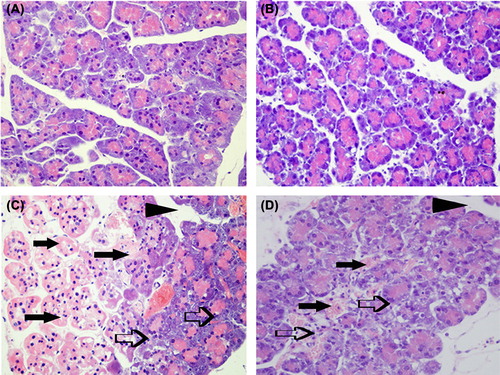
Histological examination of pancreatic tissues was stained with H&E and observed under light microscope. Representative images of histological examination of pancreas in each group at 12 h were shown in . There were no observable pathological changes in control group and BML-111 control group mice. Significant edema, mild inflammatory cell infiltration, and hemorrhage could be seen in the pancreatic tissue of APALI at 3 h. These changes were all more obvious at 6 h, and focal necrosis of pancreatic lobules could also be seen. Moderate to severe inflammatory cell infiltration, necrosis and degeneration of the large areas of the pancreas and more severe hemorrhage could be observed at 12 and 24 h. The pancreatic tissue morphological changes in mice pretreated with BML-111 included inflammatory infiltration, intralobular edema, acinar cell vacuolization and necrosis, with slight interstitial edema, but without obvious parenchyma necrosis or hemorrhage, which were significantly milder than in the APALI group at each time point.
Figure 1. Representative images of histological examination of pancreas in each group at 12 h were shown by hematoxylin and eosin staining (original magnification × 100). There were no remarkable pancreatic injury pathological changes in the control group (A) and the BML-111 control group (B). The broad necrosis of acinar (closed arrows), inflammatory cell infiltrates (open arrows), and interstitial edema (arrowheads) were observed in APALI group (C). The slight local necrosis, inflammatory cell infiltrates, and interstitial edema were only observed in the BML-111-pretreatment group (D).
Changes in the wet/dry weight ratio of lung tissues and pathological scores of the lung injury
The severity of lung injury was assessed by histological score of lung injury and the wet/dry weight ratio of lung tissues, and these changes are summarized in . The histological score of lung injury and wet/dry weight ratio of lung were significantly increased in the APALI group mice after 3 h, and reaching a climax at 12 h when compared to the control group mice. Although it declined at 24 h, the difference was not statistically significant compared with that at 12 h. BML-111 pretreatment significantly reduced histological score of lung injury and wet/dry weight ratio of lung compared to the APALI group mice at the corresponding time point, and the difference was statistically significant (p < 0.05).
Table II. Changes in the lung wet/dry weight ratio and pathological scores of lung injury in four groups (n = 8).
Pathological changes in the lung
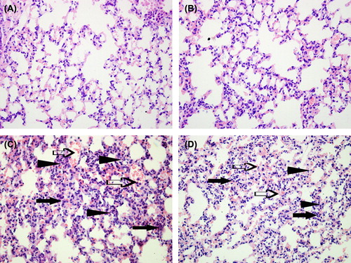
There were no remarkable pathological changes under a light microscope in two control group mice. In the APALI group mice, the lung tissue edema, congestion and inflammatory cell infiltration could be seen at 3 h. Lung tissue edema and congestion was more obviously, and little RBC exudation and severe inflammatory cell infiltration could also be seen at 6 h. The changes in lung tissue histopathology showed a widespread increase in alveolar wall thickness caused by edema, severe hemorrhage in the alveolus, alveolus collapse, and obvious inflammatory cell infiltration at 12 h and 24 h. In the BML-111 pretreatment group mice, the histopathological changes in lung tissues were minor compared with those in the APALI group mice at each time point, especially for hemorrhage in the alveolus at 12 h and 24 h. Representative images of histological examination of lung in each group at 12 h were shown in .
Figure 2. Representative images of histological examination of lung in each group at 12 h were shown by hematoxylin and eosin staining (original magnification × 100). There were no remarkable pathological changes in the control group (A) and BML-111 control group (B). In the APALI group (C), the lung tissues show widespread alveolar wall thickness (arrowheads) caused by edema, severe hemorrhage (open arrows) in the alveolus, alveolus collapse, and obvious inflammatory cell infiltration (closed arrows). In BML-111-pretreatment group (D), the changes in the lung were edema and mild hemorrhage in the alveolus.
Changes in serum TNF-α, IL-1β, and IL-10 levels
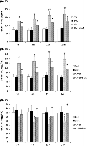
The serum levels of TNF-α and IL-1β significantly increased in the APALI group mice compared with those in the control group at each time point. The levels of TNF-α reached the highest level at 24 h. The levels of IL-1β reached the highest level at 12 h and then declined slightly at 24 h, but the difference was not statistically significant between two time points. The BML-111 pretreatment significantly reduced the serum levels of TNF-α and IL-1β compared with the APALI group at all the corresponding time points. Compared with those of the control group, the serum IL-10 levels decreased in the APALI group mice at each time point, but there was no statistical significance at 3 h. In the BML-111 pretreatment group, the IL-10 levels increased at each time point compared with those in the APALI group, but the difference has statistical significance at 6, 12, and 24 h (p < 0.05) ().
Figure 3. Changes in serum TNF-α, IL-1β, and IL-10 levels in four groups. The control group (CON), the BML-111 control group (BML), the APALI group (APALI), and the BML-111 pretreatment group (APALI + BML). A, serum TNF-α level; B, serum IL-1β level; C, serum IL-10 level; 3, 6, 12 and 24 h represent the time point at which animals were killed. Values are expressed as the means ± SD; #p < 0.05 vs. control group; ##p < 0.01 vs. control group. *p < 0.05 vs. APALI group; **p < 0.01 vs. APALI group.
Changes in lung tissue ultrastructure
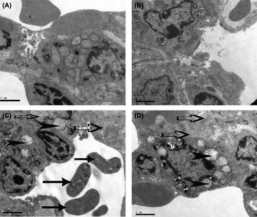
To investigate the changes in lung tissue ultrastructure, lung tissues were subjected to transmission electron microscopy. As shown in the , ultrastructure of lung tissues in the control and BML-111 control group showed complete mitochondrial outer membrane and distinct mitochondrial cristae in type Ⅱ alveolar epithelial cells. There was no evacuation phenomenon in laminated bodies. In the APALI group, alveoli were clearly dilated and merged, a large number of red blood cells and active substance leaked into the alveolar space, mitochondria in type Ⅱ alveolar epithelial cells swelled with membranes indiscernible and most of mitochondrial cristae disappeared. Vacuolar degeneration could be seen in lamellar bodies in type Ⅱ alveolar epithelial cells. In the BML-111 pretreatment group, slight swelling of mitochondria and some indistinct mitochondrial cristae can be observed, mitochondrial membranes in type Ⅱ alveolar epithelial cells were partly deliquesced, and lamellar bodies had little vacuolar degeneration.
Figure 4. Representative transmission electron microscopy images of lung tissues were shown in four groups at 12 h (original magnification of × 10000). The control group (A) and BML-111control group (B) showed complete mitochondrial outer membrane and distinct mitochondrial cristae in type Ⅱ alveolar epithelial cell. The APALI group (C) showed mitochondrial swelling and outer membrane blurred (open arrows), mitochondrial cristae disappeared, vacuolar degeneration of lamellar body (arrowheads), red blood cells (closed arrows) and the active substance were leaked into the alveolar space. The BML-111 pretreatment group (D) showed slight swelling of mitochondria, incomplete mitochondrial cristae, and little vacuolar degeneration in lamellar body.
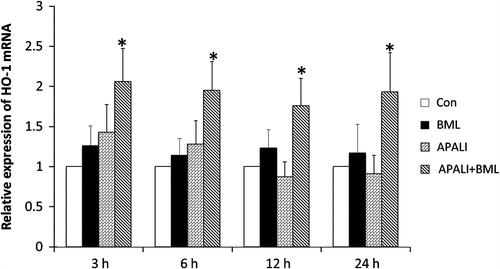
Changes in expression of HO-1 mRNA in lung tissues
As shown in , in APALI group mice, HO-1 mRNA expression in lung tissues was slightly increased at 3 and 6 h compared with that in control group at the corresponding time point, decreased somewhat at 12 h and 24 h, but there was all no statistical significance. In the BML-111 pretreatment group mice, HO-1 mRNA expression was elevated by 1.44-, 1.52-, 2.02-, and 2.12-fold at 3, 6, 12, and 24 h, respectively, compared with the APALI group at corresponding time point, and the difference has statistical significance at each time point (p < 0.05).
Figure 5. Changes in expression of HO-1 mRNA in lung tissues. Real-time PCR analysis of expression of lung HO-1 mRNA in the control group (CON), the BML-111 control group (BML), the APALI group (APALI), and the BML-111 pretreatment group (APALI + BML).The differences in the average threshold cycle (2−ΔΔCT) values were determined and normalized to the expression of β-actin; 3, 6, 12 and 24 h represent the time points at which animals were killed. Values are expressed as the means ± SD; #p < 0.05 vs. control group; ##p < 0.01 vs. control group. *p < 0.05 vs. APALI group; **p < 0.01 vs. APALI group.
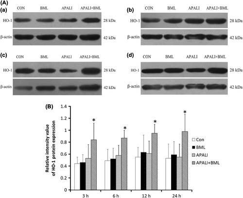
Changes in expressions of HO-1 protein in lung tissues
As shown in and , the expression of HO-1 protein in lung tissues began to increase slightly in the APALI group mice at 3 h and 6 h ( and ), when compared with the control group mice; however, its expression was decreased at 12 and 24 h compared with those of the control group mice, and there was all no statistically significant (p > 0.05) ( and ). BML-111 pretreatment significantly increased expression of HO-1, compared with the APALI group at each time point, and the difference was statistically significant (p < 0.05).
Figure 6. Changes in expressions of HO-1 protein in lung tissues. Western blotting analysis of expression of lung HO-1 in the control group (CON), the BML-111 control group (BML), the APALI group (APALI) and the BML-111 pretreatment group (APALI + BML) at four time points. β-Actin was the internal control. A a, representative image of HO-1 protein in lung tissues at 3 h in each group; A b, representative image of HO-1 protein in lung tissues at 6 h in each group; A c, representative image of HO-1 protein in lung tissues at 12 h in each group; and A d, representative image of HO-1 protein in lung tissues at 24 h in each group. B, the histogram of the average expression of HO-1 protein in lung tissue in the control group (CON), the BML-111 control group (BML), the APALI group (APALI), and the BML-111 pretreatment group (APALI + BML). Values are expressed as the means ± SD; #p < 0.05 vs. control group; ##p < 0.01 vs. control group. *p < 0.05 vs. APALI group; **p < 0.01 vs. APALI group.
Discussion
ALI is a frequent complication of AP, and is a consequence of a pronounced systemic inflammatory response with leakage of a protein-rich exudate into the alveolar space and interstitial tissues, thus compromising oxygenation and gas exchange. In SAP, the early phase is associated with systemic inflammatory response syndrome (SIRS). The magnitude of the systemic inflammatory response determines the concomitant clinical course and outcome, and one-third of the deaths occur during this early phase, and 50% of those deaths are associated with APALI (Steer Citation2001). Many investigators believe that the severity of pancreatitis and APALI results from the ratio between proinflammatory and anti-inflammatory mediators, and cytokine overproduction might participate in the pathogenesis of severe lung injury during AP. Among the anti-inflammatory mediators, IL-10 inhibits the release of proinflammatory cytokines from macrophages, such as TNF-α and IL-1β. Thus, the balance between pro-inflammatory cytokines and anti-inflammatory factors seems important in linking the pancreas and APALI (Pastor et al. Citation2003, Goodman et al. Citation2003).
Although the exact pathophysiology of clinical APALI is incompletely understood, but recent experimental work in animal models has significantly increased our understanding of the mechanistic links between AP and lung injury, and anti-inflammatory strategies may be important to the treatment of APALI, at least during the early phase of SAP. A novel treatment strategy should focus on anti-inflammatory interventions in order to improve outcome. BML-111 is a commercially stable formyl peptide receptor 2 (FPR-2) agonist, had displayed the anti-inflammatory effect, which had been demonstrated in many disease models, including carbon tetrachloride-induced liver injury (Zhang et al. Citation2007), zymosan-induced arthritis (Zhang et al. Citation2008, Conte et al. Citation2010), and haemorrhagic shock-induced acute lung injury (Gong et al. Citation2012). BML-111 could also effectively suppress the proliferation, invasion, angiogenesis, and promote the necrosis of hepatocarcinoma cells in H22 hepatocarcinoma cell bearing mice (Hao et al. Citation2011, Chen et al. Citation2010).
The present study indicated that serum amylase levels and lung wet/dry weight ratio increased significantly in the APALI group mice compared with those in the control group at all observed time points, and the pathology injury scores in the pancreas and lung were also significantly higher than those in the control group. Edema, inflammatory infiltration, hemorrhage, necrosis, and bloody ascites could be observed in APALI group mice at different time points through observation of pancreatic pathological changes. The lung tissue edema, congestion, alveolar wall thickness caused by edema, severe hemorrhage in the alveolus, alveolus collapse and obvious inflammatory cell infiltration were observed under light microscope in APALI group mice at different time points. Typical ultrastructure injuries in lung tissues were also observed using transmission electron microscopy in APALI group mice, such as mitochondrial swelling, disappearance of mitochondrial cristae, vacuolar degeneration of lamellar bodies, and so on, which were consistent with the pathological diagnosis of lung injury. Thus, those further demonstrated that the animal model of APALI was successfully established. The levels of serum amylase, wet/dry weight ratio of lung and pathology injury scores of pancreas and lung were significantly decreased after pretreatment with BML-111.When APALI mice were pretreated with BML-111, the pathological changes in lung and pancreas were all improved, observed under either transmission electron microscopy or light microscopy. These results suggested that BML-111 could ameliorate the severity of pancreatitis and APALI.
To further investigate the mechanism of effects of BML-111 on APALI, serum TNF-α, IL-1β, and IL-10 changes were also detected. The early phase of ALI is characterized by the increased permeability of the alveolar–capillary barrier. A complex network of cytokines and other pro-inflammatory compounds initiate and amplify the inflammatory response in ALI. Cytokines such as TNF-α and IL-1β are secreted by lung macrophages result in a substantial amplification of cytokine/chemokine expression by structural cells in the lung microenvironment. In the pathogenesis of APALI, the activation of proinflammatory cytokines, specifically TNF-α, IL-1β, could play major roles (Pastor and Frossard Citation2001). TNF-α has been shown to be an important initiator of local and systemic damage occurring in AP (Malmstrom et al. Citation2012), anti-TNF-α therapy in rats was able to reduce the severity and mortality resulting from AP (DiMagno and DiMagno Citation2007). Additionally, serum TNF-α level has been found to be correlated with the severity of AP in humans. IL-1β has been shown to be a significant cytokine for the development of AP, and the inhibition of its production has been shown to decrease the severity of AP. Interestingly, in the double IL-1β and TNF-α receptor knockout mice, the IV administration of sterile, cytokine-free ascitic fluid collected from rats with pancreatitis failed to induce lung injury as observed in wild-type animals, emphasizing an important role for IL-1β and TNF-α in ALI. In contrast, the anti-inflammatory cytokine IL-10 counteracts the release of pro-inflammatory cytokines from macrophages. In this study, our results demonstrated that BML-111 significantly reduced serum TNF-α and IL-1β levels that increased in the SAP group mice at each time point. Furthermore, serum IL-10 levels were increased by the BML-111 pretreatment at each time point. It was reported that BML-111-treatment decreased the concentrations of serum TNF-α and IL-6 in collagen-induced arthritis mice (Zhang et al. Citation2008) and in the haemorrhagic shock-induced ALI rats (Gong et al. Citation2012). Similarly, BML-111 decreased the TNF-α level in CCl4-treated liver injury mice (Zhang et al. Citation2007). The ratio between pro- and anti-inflammatory mediators can be of importance in predicting the severity of pancreatitis and APALI. These indicated that BML-111 has anti-inflammatory effects by inhibiting the production of pro-inflammatory mediator TNF-α and IL-1β and by promoting anti-inflammatory cytokines IL-10 in APALI mice.
In order to investigate whether other mechanisms were involved in the protective effect of BML-111 on APALI, we also observed HO-1 expression in lung tissues. Nrf2/ARE is regarded as the most important endogenous antioxidant signaling pathway in the body (Copple et al. Citation2010), and HO-1 is one of the most important antioxidant enzymes in this signal channel. HO-1 is also considered a hallmark of Nrf2/ARE activation (Chan et al. Citation2011), and pharmacologic approaches influencing HO-1 activity might have a benefical effect even in humans (Weis et al. Citation2012). HO-1 is the inducible isoform of the first and rate-limiting enzyme of heme degradation. HO-1 not only protects against oxidative stress and apoptosis, but also has received a great deal of attention in recent years because of its potent anti-inflammatory functions. Many studies also have reported that activation of Nrf2/ARE signaling pathway and its downstream genes plays a critical role in protecting a variety of organs, for example, a study showed Protandim-induced Nrf2 and HO-1 in a rat model of SU5416/hypoxia-induced pulmonary hypertension, reducing oxidative stress and cardiac fibrosis, preserving right ventricular microcirculation, maintaining right heart function, and reducing expression of osteopontin-1 (Bogaard et al. Citation2009). Our findings showed that the expression of HO-1 was increased slightly in the APALI group mice compared with the control group mice at 3 and 6 h after induction of APALI; however, they decreased at 12 and 24 h, but they were all not statistically significant. BML-111 pretreatment significantly increased expression of HO-1, compared with the APALI group at each time point, and the difference was statistically significant. In addition, the expression of HO-1 was increased slightly at 3 and 6 h after induction of APALI, which may be due to the fact that minimal stress stimuli could initiate the upregulation of HO-1. The start and self-regulation of the endogenous defense mechanisms would avoid the excessive damage to lung tissues. However, the expression of HO-1 decreased somewhat at 12 and 24 h compared with those in the control group; this may be due to the fact that certain pathogenic factors restrained self-regulation of HO-1 or resulted in deactivation of HO-1 in these stage of the disease. This may be one of the main reasons for that the lung injury was more obviously at these stage. Similarly, Kyung Hee Jung et al. (Citation2010) found that expression of HO-1 was low in AP at 12 h after cerulein-induced AP. In contrast, previous studies have observed that HO-1 was upregulated in ischemic cortex, beginning at 6 h, peaking at 48 h, and declining at 72 h after cerebral ischemia/reperfusion (Li et al. Citation2011). These differences may be related to the different types of tissues and organs, etiological factors, and time point of observation.
Our study found that expression of lung HO-1 had similar change trend over time in cerulein and LPS-induced APALI, both at the mRAN or at the protein levels; moreover, BML-111 pretreatment significantly increased the expression of HO-1 in the protein and mRNA levels compared with the APALI group at each time point. Thus, the protective effects of BML-111 on the APALI may also associate with regulating expression of HO-1. HO-1 is the inducible isoform of the first and rate-limiting enzyme of heme degradation. Studies have shown that HO-1 not only protects against oxidative stress and apoptosis, but also has received a great deal of attention in recent years because of its potent anti-inflammatory functions, and anti-inflammatory action was mediated by the reduction in leukocyte adhesion and decreasing the production of pro-inflammatory cytokines (Otterbein et al. Citation2003). Previous studies have shown that hemin, the agent for HO-1 induction, had protection against injury in experimental AP by inducing HO-1 expression (Nakamichi et al. Citation2005). In addition, Curcuma longa had protective effects on cerulein-induced APALI through the induction of HO-1 (Seo et al. Citation2011); Nardostachys jatamansi administration attenuated the severity of AP and lung injury associated with AP by inducing HO-1 (Bae et al. Citation2012); Oxymatrine protected the brain against cerebral ischemia/reperfusion injury by upregulating HO-1 expression (Li et al. Citation2011). Upregulation of HO-1 attenuated the expression of various proinflammatory genes, such as cyclooxygenase-2, TNF-α, IL-1β, and IL-6 (Immenschuh and Schroder Citation2006). Therefore, the upregulation of HO-1 by BML-111 had a protective effect on APALI, at least in part, that may be related to the anti-inflammatory effect of HO-1.
Taken together, the results suggest that the prophylactic administration of BML-111 inhibited pro-inflammatory cytokines TNF-α and IL-1β, promoted anti-inflammatory cytokine LI-10, and increased HO-1 expression in lung tissues, thus attenuating the severity of APALI induced by cerulein and LPS. Therefore, we suggest that the upregulation of HO-1 by BML-111 may provide a new idea for treatment of APALI. Of course, our study also has some limitations, and it is only a prophylactic treatment. The other routes of administration and therapeutic research will be investigated in future studies.
Acknowledgements
The authors thank Ms. Hong Yan Guo, Xiao Ni Ma, and Mr. Jie Cheng for their excellent technical assistance, and Mr. Xi Ping Shen for his help with statistical analysis during this study.
Declaration of interest
The authors report no declarations of interest. The authors alone are responsible for the content and writing of the article.
References
- Bae GS, Kim MS, Park KC, Koo BS, Jo IJ, Choi SB, et al. 2012. Effect of biologically active fraction of Nardostachys jatamansi on cerulein-induced acute pancreatitis. World J Gastroenterol. 18:3223–3234.
- Barreto SG, Rodrigues J. 2007. Comparison of APACHE II and Imrie Scoring Systems in predicting the severity of Acute Pancreatitis. World J Emerg Surg. 2:33.
- Barreto SG, Rodrigues J. 2008. Acute pancreatitis in Goa–a hospital-based study. J Indian Med Assoc. 106:575–576, 578.
- Bogaard HJ, Natarajan R, Henderson SC, Long CS, Kraskauskas D, Smithson L, et al. 2009. Chronic pulmonary artery pressure elevation is insufficient to explain right heart failure. Circulation. 120:1951–1960.
- Chan KH, Ng MK, Stocker R. 2011. Haem oxygenase-1 and cardiovascular disease: mechanisms and therapeutic potential. Clin Sci (Lond). 120:493–504.
- Chen Y, Hao H, He S, Cai L, Li Y, Hu S, et al. 2010. Lipoxin A4 and its analogue suppress the tumor growth of transplanted H22 in mice: the role of antiangiogenesis. Mol Cancer Ther. 9:2164–2174.
- Chen YP, Ning JW, Ji F. 2009. Establishment of the critical period of severe acute pancreatitis-associated lung injury. Hepatobiliary Pancreat Dis Int. 8:535–540.
- Chiang N, Arita M, Serhan CN. 2005. Anti-inflammatory circuitry: lipoxin, aspirin-triggered lipoxins and their receptor ALX. Prostaglandins Leukot Essent Fatty Acids. 73:163–177.
- Conte FP, Menezes-de-Lima O Jr, Verri WA Jr, Cunha FQ, Penido C, Henriques MG. 2010. Lipoxin A(4) attenuates zymosan-induced arthritis by modulating endothelin-1 and its effects. Br J Pharmacol. 161:911–924.
- Copple IM, Goldring CE, Kitteringham NR, Park BK. 2010. The keap1-nrf2 cellular defense pathway: mechanisms of regulation and role in protection against drug-induced toxicity. Handb Exp Pharmacol. 233–266.
- DiMagno MJ, DiMagno EP. 2007. New advances in acute pancreatitis. Curr Opin Gastroenterol. 23:494–501.
- Ding SP, Li JC, Jin C. 2003. A mouse model of severe acute pancreatitis induced with caerulein and lipopolysaccharide. World J Gastroenterol. 9:584–589.
- Dixon DL, De Smet HR, Bersten AD. 2009. Lung mechanics are both dose and tidal volume dependant in LPS-induced lung injury. Respir Physiol Neurobiol. 167:333–340.
- Elder AS, Saccone GT, Bersten AD, Dixon DL. 2012a. Evaluation of lung injury and respiratory mechanics in a rat model of acute pancreatitis complicated with endotoxin. Pancreatology. 12:240–247.
- Elder AS, Saccone GT, Dixon DL. 2012b. Lung injury in acute pancreatitis: mechanisms underlying augmented secondary injury. Pancreatology. 12:49–56.
- Gong J, Guo S, Li HB, Yuan SY, Shang Y, Yao SL. 2012. BML-111, a lipoxin receptor agonist, protects haemorrhagic shock-induced acute lung injury in rats. Resuscitation. 83:907–912.
- Gong J, Li HB, Guo S, Shang Y, Yao SL. 2012. Lipoxin receptor agonist, may be a potential treatment for hemorrhagic shock-induced acute lung injury. Med Hypotheses. 79:92–94.
- Goodman RB, Pugin J, Lee JS, Matthay MA. 2003. Cytokine-mediated inflammation in acute lung injury. Cytokine Growth Factor Rev. 14:523–535.
- Hao H, Liu M, Wu P, Cai L, Tang K, Yi P, et al. 2011. Lipoxin A4 and its analog suppress hepatocellular carcinoma via remodeling tumor microenvironment. Cancer Lett. 309:85–94.
- Hofbauer B, Saluja AK, Bhatia M, Frossard JL, Lee HS, Bhagat L, Steer ML. 1998. Effect of recombinant platelet-activating factor acetylhydrolase on two models of experimental acute pancreatitis. Gastroenterology. 115:1238–1247.
- Immenschuh S, Schroder H. 2006. Heme oxygenase-1 and cardiovascular disease. Histol Histopathol. 21:679–685.
- Jung KH, Hong SW, Zheng HM, Lee HS, Lee H, Lee DH, et al. 2010. Melatonin ameliorates cerulein-induced pancreatitis by the modulation of nuclear erythroid 2-related factor 2 and nuclear factor-kappaB in rats. J Pineal Res. 48:239–250.
- Li M, Zhang X, Cui L, Yang R, Wang L, Liu L, Du W. 2011. The neuroprotection of oxymatrine in cerebral ischemia/reperfusion is related to nuclear factor erythroid 2-related factor 2 (nrf2)-mediated antioxidant response: role of nrf2 and hemeoxygenase-1 expression. Biol Pharm Bull. 34:595–601.
- Malmstrom ML, Hansen MB, Andersen AM, Ersboll AK, Nielsen OH, Jørgensen LN, Novovic S. 2012. Cytokines and organ failure in acute pancreatitis: inflammatory response in acute pancreatitis. Pancreas. 41:271–277.
- Nakamichi I, Habtezion A, Zhong B, Contag CH, Butcher EC, Omary MB. 2005. Hemin-activated macrophages home to the pancreas and protect from acute pancreatitis via heme oxygenase-1 induction. J Clin Invest. 115:3007–3014.
- Otterbein LE, Soares MP, Yamashita K, Bach FH. 2003. Heme oxygenase-1: unleashing the protective properties of heme. Trends Immunol. 24:449–455.
- Pastor CM, Frossard JL. 2001. Are genetically modified mice useful for the understanding of acute pancreatitis. FASEB J. 15:893–897.
- Pastor CM, Matthay MA, Frossard JL. 2003. Pancreatitis-associated acute lung injury: new insights. Chest. 124:2341–2351.
- Pereda J, Sabater L, Aparisi L, Escobar J, Sandoval J, Viña J, et al. 2006. Interaction between cytokines and oxidative stress in acute pancreatitis. Curr Med Chem. 13:2775–2787.
- Schmidt J, Rattner DW, Lewandrowski K, Compton CC, Mandavilli U, Knoefel WT, Warshaw AL, et al. 1992. A better model of acute pancreatitis for evaluating therapy. Ann Surg. 215:44–56.
- Seo SW, Bae GS, Kim SG, Yun SW, Kim MS, Yun KJ, et al. 2011. Protective effects of Curcuma longa against cerulein-induced acute pancreatitis and pancreatitis-associated lung injury. Int J Mol Med. 27:53–61.
- Shen HN, Lu CL. 2011. Incidence, resource use, and outcome of acute pancreatitis with/without intensive care: a nationwide population-based study in Taiwan. Pancreas. 40:10–15.
- Steer ML. 2001. Relationship between pancreatitis and lung diseases. Respir Physiol. 128:13–16.
- Wang M, Ye R, Barron E, Baumeister P, Mao C, Luo S, et al. 2010. Essential role of the unfolded protein response regulator GRP78/BiP in protection from neuronal apoptosis. Cell Death Differ. 17:488–498.
- Wang X, Cui Z, Zhang J, Li H, Zhang D, Miao B, et al. 2010. Early predictive factors of in hospital mortality in patients with severe acute pancreatitis. Pancreas. 39:114–115.
- Weis S, Jesinghaus M, Kovacs P, Schleinitz D, Schober R, Ruffert C, et al. 2012. Genetic analyses of heme oxygenase 1 (HMOX1) in different forms of pancreatitis. PLOS ONE. 7:e37981.
- Yang R, Shaufl AL, Killeen ME, Fink MP. 2009. Ethyl pyruvate ameliorates liver injury secondary to severe acute pancreatitis. J Surg Res. 153:302–309.
- Zhang L, Wan J, Li H, Wu P, Jin S, Zhou X, et al. 2007. Protective effects of BML-111, a lipoxin A(4) receptor agonist, on carbon tetrachloride-induced liver injury in mice. Hepatol Res. 37: 948–956.
- Zhang L, Zhang X, Wu P, Li H, Jin S, Zhou X, et al. 2008. BML-111, a lipoxin receptor agonist, modulates the immune response and reduces the severity of collagen-induced arthritis. Inflamm Res. 57:157–162.
- Zhou M, Chen B, Sun H, Deng Z, Andersson R, Zhang Q. 2011. The protective effects of Lipoxin A4 during the early phase of severe acute pancreatitis in rats. Scand J Gastroenterol. 46: 211–219.