
Abstract
The present study was aimed at exploring the targeting potential of LTA-anchored chitosan nanoparticles (CH-NP) specifically to M cell following oral immunization. The lectinized CH-NP exhibited 7–29% coupling capacity depending upon the amount of glutaraldehyde added. Induction of the mucosal immunity was assessed by estimating secretory IgA level in the salivary, intestinal and vaginal secretions, and cytokine (IL-2 and IFN-γ) levels in the spleen homogenates. The results demonstrated that LTA-anchored CH-NP elicited strong humoral and cellular responses and hence could be a competent carrier-adjuvant delivery system for oral mucosal immunization against Hepatitis B.
Introduction
The most convenient way of administering a vaccine is by the oral route. It affords high patient acceptability, compliance and ease of administration when compared with traditional parental administration. Successful oral vaccination for hepatitis B certainly has a greater impact on global immunization efforts. It is generally acknowledged that oral vaccination is largely ineffective due to substantial degradation of antigens by harsh acidic condition of the stomach and enzymatic degradation in gastrointestinal tract (Gabor et al. Citation2002, Singh et al. Citation2004, Lavelle et al. Citation2004). Additionally, poor absorption of the antigens by the gut-associated lymphoid tissue (GALT) contributes to the lower efficacy and thus requires larger doses of antigen to be administered for achieving desirable level of immunity comparable with systemic administration (Gupta et al. Citation2006).
Mucosal immunization frequently results in the stimulation of both mucosal and systemic immune responses, whereas systemic immunization typically induces systemic responses without activating the mucosal immune system. Induction of mucosal response leads to production of secretory IgA (sIgA) antibodies, which are not usually produced by systemic immunization (Nugent et al. Citation1998).
Nanoparticles delivery systems are particularly useful to protect the antigen in the gastrointestinal (GI) tract and hold potential to enhance the efficacy of oral vaccine (O’Hagan Citation1998). Polymer-based carriers have gained much attention of the scientific community for safe and effective delivery of proteins since they are much more stable as compared to lipid-based delivery vehicles (Mishra et al. Citation2008). The polymer-based approaches offer several benefits such as to protect the antigen in the gut, to target the antigen to GALT and/or to increase the residence time of the antigen in the gut through bio-adhesion. M (microfold or membranous) cells are specialized epithelial cells responsible for antigen sampling at the interface of mucosal surfaces and environment. M cells-targeted oral delivery systems hold promise to improve the efficacy of the oral vaccines. It seems likely that vaccine efficacy could be enhanced by anchoring M cells-specific ligands (e.g. lectins) on to the bioactive carriers (Chen et al. Citation2008, Gupta et al. Citation2006).
Chitosan, a natural, biodegradable, biocompatible, bioadhesive polymer is gaining attention in the pharmaceutical field for a wide range of drug delivery. Chitosan is a copolymer of glucosamine and N-acetyl glucosamine linked by β 1–4 glucosidic bonds obtained by N-deacetylation of chitin (Illum Citation1998). Chitosan acts as a penetration enhancer by opening the tight epithelial junctions and hence is particularly exploited in protein and vaccine delivery (van der Lubben et al. Citation2001a). Chitosan nanoparticles (CH-NP) for delivery of polypeptides such as insulin, tetanus toxoid, and diphtheria toxoid are widely explored (van der Lubben et al. Citation2003, Xu and Du Citation2003, Calvo et al. Citation1997a, Citation1997b). Chitosan, as a cationic polysaccharide, has gained increasing attention in pharmaceutical field due to its favorable biological properties such as non-toxicity, biodegradability, mucoadhesive properties, etc. (He et al. Citation2002). Additionally, chitosan micro/nanoparticles can be easily prepared by ionic gelation method using tripolyphosphate (TPP) as precipitating agent (Gan et al. Citation2005).
Lectins, comprised of a structurally very diverse class of proteins or glycoproteins of non-immunological origin, characterized by their ability to bind sugar molecules with considerable specificity. Therefore, they are also capable of specific recognition of and reversible binding to carbohydrate determinants of complex glycoconjugates, without altering the covalent structure of the recognized glycosyl ligands (Lis and Sharon Citation1986, Kilpatric et al. Citation1985, Lehr et al. Citation1992). Lectin receptors are expressed on various cells such as endothelial cells, hepatocytes, macrophages, monocytes and lymphocytes. They are efficient in recognizing the complex oligosaccharide epitopes, which are also present on the cell surface or could be exogenous glycoconjugate ligands mimics of endogenous carbohydrate epitopes (Vyas et al. Citation2001). This property suggests that they play an important role in biological-recognition events. In recent years, lectins, described as “second generation mucoadhesives”, have been proposed as tools to offer some advantages either in specific drug delivery applications to increase the residence time of the pharmaceutical form or to resist proteolytic degradation (Clark et al. Citation2001).
There is an interest in the grafting of lectins to the surface of biodegradable polymeric particles. Their conjugation to polymeric nanoparticles has been shown to be efficient for increasing the interactions with mucus and lectins have been shown to promote particle translocation (Russell-Jones et al. Citation1999, Gao et al. Citation2006). Additionally, lectins can probably be useful for targeting specific areas of the GI tract, due to the heterogeneous distributions of sugars moieties along the normal or diseased intestine (Ponchel and Irache Citation1998). A number of polymer nanoparticles have been conjugated with the selected lectin to study their targeting potential (Gupta et al. Citation2006, Russell-Jones et al. Citation1999, Gao et al. Citation2006, Montisci et al. Citation2001a).
The Tetragonolobus purpureas or Lotus tetragonolobus (LTA) from Winged or Asparagus pea has affinity for α-L-fucosyl residues and an unusually high affinity for α-L-fucose residues on type II chain blood group oligosaccharides (Pereira and Kabat Citation1974). Therefore, it can be used for M-cell targeting. In our previous study, we have exploited M-cell targeting by successfully delivering LTA-conjugated poly(lactic-co-glyolic acid) (PLGA) orally and comparing its antibody titer and cytokine levels with plain PLGA nanoparticles against hepatitis B. It was observed that lectinized PLGA nanoparticles have the potential to elicit higher humoral as well as cellular response when compared to that of unconjugated PLGA nanoparticles (Mishra et al. Citation2010).
Thus the present study was aimed at delivering ligand anchored HBsAg-loaded CH-NP for targeted vaccine delivery. The prepared nanoparticles were prepared and characterized for evaluation of various in vitro parameters. Finally in vivo study was conducted to assess immunization potential of the developed formulation.
Materials and methods
Chitosan (Practical grade, 75–85% degree of deacetylation MW 150 kDa) was obtained from Panacea Biotech Ltd. (Lalru, Punjab, INDIA). Glutaraldehyde (25% in water) was purchased from Fluka Chemica Co. (AG CH-9470 Buchs, Switzerland). TRITC-conjugated WGA (TRITC-WGA) 3-(cholamidopropyl-o-dimethylammonio)-1-propanesulphonate (CHAPS) and bovine submaxillary mucin (BSM) were procured from Sigma Chemical Co. (St. Louis, MO, USA). Lotus tetragonolobus (LTA) and FITC-conjugated LTA (f-LTA) were procured from Hysel India, New Delhi, India. Recombinant hepatitis B surface antigen (HBsAg, MW 24 kDa) was a gift sample from Shantha Biotech Ltd. (Hydrabad, India). The protein molecular weight marker (10–250 kDa) was purchased from Genetix Biotech Asia Private Limited, New Delhi, India. RPMI-1640 was from Himedia, Mumbai. BCA protein estimation kit was purchased from Genei, Bangalore, India. Enzyme-linked immunoassay kit (AUSAB and AUSZYME) and cytokines (IL-2 and IFN-γ) estimation kit were purchased from Abbott Laboratories, USA and e-Bioscience, respectively. IgG isotyping kit was procured from Sigma-Aldrich Pvt. Ltd., USA. All other chemicals and reagents were of analytical grade, unless otherwise mentioned.
Method of preparation of chitosan nanoparticles
CH-NP were prepared by ionic gelation with TPP method Calvo et al. (Citation1997b) with slight modifications. The particles were prepared spontaneously by addition of 2 mL of TPP solution (0.5 mg/mL) through a syringe into 5 mL of chitosan solution (1 mg/mL) under magnetic string at 500 rpm at room temperature for 60 min. The chitosan solution was prepared by dissolving chitosan in dilute acetic acid (0.1% V/V) at room temperature and filtered through a 0.22 μm filter to remove insolubles.
For the association of HBsAg with chitosan nanoparticles, HBsAg at concentration 0.2 mg/mL was incorporated in the TPP solution. Nanoparticles were collected by centrifugation at 20,000 rpm for 30 min (High Speed Table Top Refrigerated Centrifuge, Hermle, Germany) and washed four times with deionized water. The supernatants were then discarded and the nanoparticles were resuspended in 100 μL of phosphate buffered saline (pH 7.4).
Preparation of LTA-anchored chitosan nanoparticles
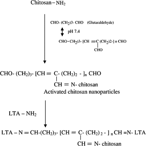
LTA were covalently conjugated to the surface of CH-NP by using glutaraldehyde as activator in two-step procedure. A two-step procedure, including the first activation step and then a coupling step, was adopted as reported previously by Montisci et al. (2001b) with slight modifications (Chang Citation1971).
Activation of chitosan nanoparticles
Very small quantity of particles was washed in milli-Q water by centrifugation (25,000 rpm for 10 min). The pellet was suspended in 500 μl of phosphate buffer saline (PBS, pH 7.4). Further microcontents of 0.25% glutaraldehyde (0–400 μl) in water were added. The mixture was then shaken gently for 3 h at 30°C to activate the hydroxyl groups, a longer time will lead to aggregation.
Conjugation of activated CH-NP to lectin
Briefly, the suspension was centrifuged to remove unreacted glutaraldehyde and further washed four times in PBS (pH 7.4) to remove any remaining traces of glutaraldehyde which might otherwise cross-link the lectin molecules. Subsequently, 500 μl of PBS containing 100 μg of lectin were added and the linkage was made by incubation overnight at room temperature. The conjugates were centrifuged to remove free lectins and incubated 1 h with 0.2 mL ethanolamine (0.1 M) to block unreacted groups on the particles. The ethanolamine was eliminated and the nanoparticles were washed thrice by centrifugation (20,000 rpm for 20 min). The lectin–Chitosan nanoparticles conjugates were finally resuspended in 1 ml of PBS and stored at 2–8°C ().
Figure 1. Schematic presentation of anchoring of LTA lectin to surface of Chitosan nanoparticles with glutaraldehyde as cross-linker.
Characterization of chitosan nanoparticles
Morphology and particle size analysis
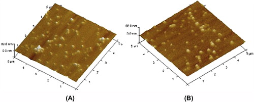
The surface morphology of HBsAg-loaded CH-NP was investigated by using atomic force microscopy (AFM) (BioscopeTM II (Veeko Asia Pvt Ltd) INDIA) in non-contact mode with a super sharp silicon cantilever (Veeco Asia Pvt Ltd) on mica substrate. The particle size and zeta potentials of nanoparticles were recorded by Zetasizer Nano ZS 90 (Malvern, UK).
Protein loading efficiency
The amount of HBsAg associated with CH-NP was estimated by method as reported van der Lubben et al. (Citation2003). The amount of antigen determined by bicinchoninic protein assay (BCA protein kit, Genie, Bangalore) method. The amount of loaded protein was calculated as the difference between the antigen content of initial solution and the antigen recovered following incubation. Similarly, protein content of HBsAg-loaded lectinized nanoparticles (a) and lectin-coupled placebo nanoparticles (without antigen) (b) was determined. Amount of HBsAg loaded in the lectinized nanoparticles was estimated by calculating difference between two values, that is (a˜b).
Determination of amount of lectin bound
The amount of lectin coupled to nanoparticles was estimated by measuring the difference between the amount of lectin added initially in coupling reaction and amount of lectin recovered in the supernatant after centrifugation of particulate dispersion. The amount of lectin was quantified by the colorimetric determination of protein in the supernatant by bicinchoninic protein assay (BCA protein kit, Genie, Bangalore) method.
In vitro release of HBsAg
The in vitro release of HBsAg from CH-NP was carried out in PBS buffer (pH 7.4). Vials containing 40 mg of nanoparticles and 5 ml of PBS (pH 7.4) were incubated at 37°C in constant shaking mixer. At appropriate intervals 1.0 ml of release medium was collected by centrifugation (22,000 × g for 20 min) and replaced by 1.0 ml of fresh PBS (pH 7.4). The amount of released HBsAg was estimated by enzyme immunoassay (EIA) kit (AUSZYME Monoclonal kit, Abbott Laboratories, Abbott Park, IL, USA). The same sample was used to measure in vitro antigenicity using an EIA kit as described by Shi et al. (Citation2002), by determining the ratio of the EIA response to protein concentration (EIA/protein).
Assessment of Structural Integrity of antigen
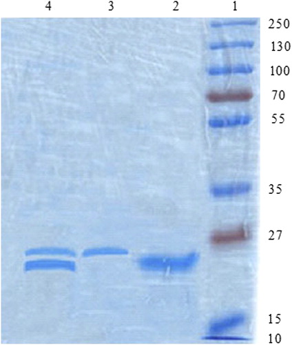
The structural integrity of HBsAg protein antigen extracted from the nanoparticles was evaluated by SDS-polyacrylamide gel electrophoresis (SDS-PAGE) and compared with native HBsAg and reference markers. HBsAg was extracted by dissolving the nanoparticles in 2 ml of 5% w/v SDS in 0.1 M sodium hydroxide solution (Singh et al. Citation1997). The extracted antigen was concentrated and loaded onto a 3.5% stacking gel and subjected to electrophoresis on a 12% separating gel at 200 V (Bio-Rad, USA) until the dye band reached the gel bottom. After migration the gel was stained with coomassie blue to detect protein band.
In vitro ligand affinity and activity studies
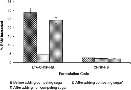
The activity of the lectinized nanoparticles towards exogenously provided bovine submaxillary gland mucin (BSM) and affinity towards competing sugar were studied to assess the targeting efficacy of ligand-anchored nanoparticles. The in vitro affinity of surface-modified nanoparticles for mucin was determined by mixing 1 ml of BSM in PBS (0.5 mg/ml) with the same volume of suspension of LTA-coupled nanoparticles in PBS. After 60 min, samples were centrifuged at 22,000 × g for 20 min. The aliquots of the supernatant were taken and the amount of BSM was quantified by HPLC, as reported by Ezpeleta et al. (Citation1996). The amount of interacted BSM was calculated as the difference between the total and the remaining BSM in the supernatant. To study the specificity for competing sugar, specific sugar (α-L-fucose; 100 mM) and non-specific sugar (D-galactose; 100 mM) was added separately to the BSM bulk solution in PBS and interaction of LTA-coupled nanoparticles and BSM was determined (Ezpeleta et al. Citation1996).
M-cell targeting study
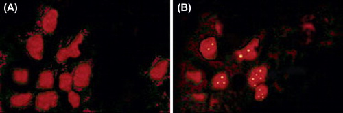
Dual staining (Clark et al. Citation1993) was performed to assess the targeting potential of the developed delivery system. Approximately 1 cm length of small intestine containing Peyer's patch were excised, opened longitudinally and pinned flat on corkboard. Tissue was rinsed thoroughly with PBS (pH 7.4) and then cut in small pieces (˜1 mm thick). The tissues were subjected to dual staining, by immersion for 60 min in TRITC-WGA, rinsing in PBS and immersion for further 60 min in FITC-LTA coupled nanoparticles. Microtomy of the tissue was performed using standard protocols and thin sections were viewed under confocal laser scanning laser microscope (Zeiss LSM 510, Jena, Germany).
Immunological studies
Female BALB/c mice was procured from DRDE, Gwalior, 8–10 weeks old, weighing 20–25 g, were used for immunization study. The study protocol was approved by Institutional Animals Ethical Committee of Dr. Hari Singh Gour University. The studies were carried as per the guidelines of Council for the Purpose of Control and Supervision of Experiments on Animals (CPCSEA), Ministry of Social Justice and Empowerment, Government of India.
Mice were housed in group of six animals for (n = 6) one week before the experiments were conducted. They were allowed free access to food and water with 12-h light/dark cycle. The mice were withdrawn of food 3 h before the immunization. The mice were immunized orally with various formulations () encapsulating HBsAg (10 μg/dose), followed by a booster dose after second week primary immunization. Single intramuscular immunization with booster dose after second weeks was also carried out with alum-HBsAg to serve as control.
Table I. HBsAg loaded vaccine formulation for immunization study.
Collection of fluid
Subsequent to immunization blood sample were collected from retro-orbital plexus of mice after 2, 4, 6 and 8 weeks. Serum was obtained by centrifugation of blood samples. Sera were stored at − 40°C until estimated for the antibody by ELISA. The salivary, intestinal and vaginal secretions were collected at Day 42 after immunization by the methods described in previous studies (Jain et al. Citation2005, Jaganathan and Vyas Citation2006). For collection of saliva, mice were injected with 0.2 ml sterile solution of pilocarpine (10 mg/ml) intraperitoneally (IP). After approximately 2 min, the saliva was collected by using capillary tube. Intestinal lavage was performed using the technique reported previously (Elson et al. Citation1984). Briefly, four doses of a 0.5 ml lavage solution (25 mM NaCl, 40 mM Na2SO4, 10 mM KCl, 10 mM NaHCO3 and 48.5 mM polyethylene glycol-MW 3350) were administered intragastrically at 15-min intervals using a blunt-tipped feeding needle. Thirty minutes after the last dose of lavage solution the mice were given 0.2 ml pilocarpine (10 mg/mL) IP. A discharge of intestinal contents occurred regularly over next 20 min, and was collected carefully.
Vaginal secretions were collected by using a pipette or to douche the mice with 0.1 ml of PBS (pH 7.4), which was then aspirated back into the pipette and used for determination of antibody levels. These fluids were stored with 100 mM phenyl methyl sulfonyl fluoride (PMSF) used as a protease inhibitor at − 40°C until estimated by ELISA for sIgA levels. Animals of a group were sacrificed after 6 weeks of booster immunization and spleens were excised and used for the determination of endogenous cytokines level (interferon-γ and interleukin-2).
Measurement of specific IgG and IgA response
The anti-HBsAg antibodies in blood samples were estimated by enzyme-linked immunoassay kit (AUSAB®, Abott laboratories, USA). Whereas IgG1 and IgG2a isotypes were estimated in samples collected at Day 42 using sigma isotyping kit (Sigma–Aldrich Pvt. Ltd., USA). Anti-HBsAg IgG present in sera was estimated at 1/100 dilution of serum. To signify actual antibody concentration (antibody titre) in mIU/ml, a standard curve was prepared using the calibrated anti-Hepatitis B panel provided by Abott Laboratories.
sIgA level in mucosal fluids was determined by ELISA. Briefly, Microtitre plates (Nunc-Immune Plate® Fb96 Maxisorb, Nunc, India) were coated with 100 μl of HBsAg solution at a concentration of 2 μg/ml in coating buffer (carbonate bicarbonate buffer) following overnight incubation at 4°C. The wells were washed thrice with PBS-T (0.05% v/v Tween 20 in PBS) and then blocked with 200 μl of blocking buffer (2% w/v BSA in PBS) via 2-hr incubation at 37°C. The wells were washed thrice with PBS-T. Serial dilutions (100 μl) of mucosal fluids in PBS-BSA (0.1% (w/v) were added and incubated for 2 h at 37°C, followed by washing thrice with PBS-T. One hundred microlitres of HRP-conjugated goat anti-mice IgA (Sigma, USA) antibodies were added to each well and again incubation was conducted for 2 h at 37°C. After three washings, 100 μl of substrate (OPD, Sigma USA) (in citrate phosphate-citrate buffer, pH 5.5, containing H2O2) was added. Colour development was stopped after 30 min by adding 50 μl of 1N H2SO4 to each well. The absorbance (OD) was measured at 490 nm. The end point titer was expressed as the reciprocal of the last dilution, which gave an optical density at 490 nm above the optical density of negative control (Khatri et al. Citation2008). Similar protocol was used for the determination of antigen-specific anti-HBsAg antibody response.
Estimation of cytokines levels
In vitro cytokoine levels in immunized animals were determined by ELISA as described previously (Sachdeva et al. Citation2004). For determination of cytokine production, 5 × 10⁶ splenocytes/mL was cultured in a final volume of 200 mL in 96-well flat-bottom plates in the presence or absence of recombinant proteins. Culture supernatants were collected after 48 h for interlukin-2 (IL-2) and IFN-γ analyses. All of these cytokines were measured by using a murine cytokine immunoassay kit (eBiosciences, USA) by following the procedure recommended by the manufacturer.
Statistical analysis
All the data were run in triplicate for each sample. All data were expressed as mean ± standard deviation (SD) for n = 3. Student's t-test analysis was done to assess the statistical significance of the data sets. A p value less than 0.05 was considered to indicate statistical significance for all comparisons.
Results and discussion
Preparation of Chitosan nanoparticles
Chitosan, mostly obtained from exoskeletons of marine arthropods, is the second abundant polysaccharide and a cationic polyelectrolyte present in nature. Its biocompatibility, biodegradability, non-toxicity and the ability to open up epithelial tight junctions have rendered it widely applicable in the pharmaceuticals as adjuvants (Illum Citation1998). The absorption-enhancing effect of chitosan is attributed to the opening of the intercellular tight junctions, thereby favoring the paracellular transport of macromolecular drugs (van der Lubben et al. Citation2001a, Citation2001b, Citation2001c). The nanoparticles of chitosan are also suitable biomaterials for amelioration of release profile of drug(s) and vaccine(s). The association of antigen(s) to the particulate systems based on chitosan has shown enhanced uptake of antigen by mucosal lymphoid tissues, thereby inducing strong systemic and mucosal immune responses against the antigens (Bramwell and Perrie Citation2006). Moreover, the specific adjuvant activity of chitosans relates and depends on the degree of deacetylation and the type of formulation (Nishimura et al. Citation1984, van der Lubben et al. Citation2001b, Citation2001c). Chitosan, being mucoadhesive in nature, provides prolonged mucoadhesion of the nanoparticles and hence longer contact time with blood-supplying capillaries, which results in a higher uptake of the protein antigen/plasmid DNA (Khatri et al. Citation2008).
Solubility of chitosan in water could be improved by the addition of an acid to aqueous medium as a result of the protonation of the amino groups. In the present study, CH-NP were prepared by using chitosan/TPP and ionic gelation method as reported by Calvo et al. (Citation1997b) with slight modifications. Nanoparticles were formed on drop-wise addition of an aqueous solution of TPP containing the antigen. The particles formation takes places due to the electrostatic attraction between the positive charged amino groups of chitosan and the negative groups of TPP. On addition of the ionic solution of TPP into the acidic solution of Chitosan, the opalescence is appeared indicating the formation of nanoparticles. These nanoparticles have shown an excellent capacity for protein entrapment and an improvement of peptide absorption by several mucosal routes, such as nasal and ocular (De Campos et al. Citation2001, Fernandez-Urrusuno et al. Citation1991, Citation1999).
The lectins can be anchored to the particulate systems through covalent linkage or using simple adsorption process. A covalent linkage method being more stable than simple adsorption is often used to bind the ligand on the particles surface. Chitosan is an excellent candidate for the preparation of conjugates owing to the presence of numerous amino groups. Hence lectin molecules can efficiently be ligated on to the surface of chitosan nanoparticles. The per cent antigen encapsulation efficiency of HBsAg-loaded CH-NP was found to be 53.2 ± 4.3% and average size and polydispersity index was measured to be 214 ± 17 nm and 0.112, respectively. Characteristics of plain and lectinized nanoparticles are compared in .
Table II. Characteristics of chitosan nanoparticles and lectinized chitosan nanoparticles.
The mean diameter, polydispersity index and zeta potential of CH-NP were determined by Zetasizer (Nano ZS90, Malvern, UK). The surface morphology of HBsAg-loaded CH-NP was investigated by using AFM. There was no change in the surface morphology of lectinized CH-NP in comparison to plain CH-NP ( and ). Protein entrapment and anchoring of lectin did not affect the spherical shape and surface visible texture of nanoparticles, however, the size of the nanoparticles varied upon lectin grafting (). This may be attributed to immobilization of the lectin on to the surface of nanoparticles. The per cent antigen encapsulation efficiency in the case of lectinized CH-NP was measure to be 42.5 ± 3.2% which is slightly lower in case of non-lectinized nanoparticles. The decrease in the per cent encapsulation efficiency could be attributed to the release of antigen from nanoparticles during incubation; employed for anchoring of lectin onto the surface of the nanoparticles. The zeta potential of lectinized (LTA-CHNP-HB) and non- lectinized (CHNP-HB) CH-NP were found to be + 23.4 ± 1.8 and + 15.6 ± 1.4 mV, respectively. The slight increase in the zeta potential of lectinized CH-NP may be because of anchoring of lectin on the surface of chitosan nanoparticles. This slight increase in size and zeta potential of lectinized CH-NP confirms the coupling of lectin on the surface of chitosan nanoparticles.
Figure 2. Atomic force Microscopy of plain Chitosan nanoparticles (A) and lectin-anchored chitosan nanoparticles (B).
Effect of glutaraldehyde content on the LTA coupling capacity of chitosan nanoparticles
Glutaraldehyde might be very efficient in cross-linking chitosan as the high content of adjacent hydroxyl groups neighboring chains. In the present study, LTA was fixed by two-stage glutaraldehyde coupling techniques. describes the reactional mechanism involved in this process. Polymerized glutaraldehyde molecules, resulted at neutral pH from an aldolic condensation, could react with the amino groups of chitosan and imine bond which is stabilized by the presence of a conjugated ethylenic bond (Monsan et al. Citation1975).
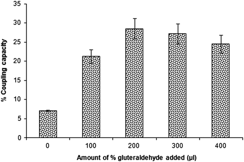
The amount of glutaraldehyde was optimized by measuring the quantity of the lectin bound to the nanoparticles. Different volumes of the glutaraldehyde (25% v/v in water) were used, i.e., 0, 100, 200, 300 and 400 μ L were used for lectin conjugation (0.25% w/v). The percentage coupling capacity was measured 7.1 ± 0.2, 21.3 ± 1.7, 28.5 ± 2.6, 27.2 ± 2.6 and 23.2 ± 2.3%, respectively (). The maximum lectin coupling capacity (28.5 ± 2.6%) was recorded with the formulation in which 200 μ L of glutaraldehyde (25%v/v in water) was used for cross-linking. When the amount of glutaraldehyde increased to above 200 μL, the coupling capacity of LTA reached to a steady state. This phenomenon was perhaps because of limitation or saturation of binding sites available on CHNP-NPs with lectin. The traces of glutaraldehyde present on the surface of lectinized CH-NP was further removed by addition of sodium bisulfite (SBS) as method reported by Jordan et al. (Citation1996) (data not shown). The amount of LTA coupled to nanoparticles was determined as the difference between the lectin added initially and the lectin recovered in the solution after incubation with the particles.(Walter et al. Citation2004) The necessary activation of CH-NP before covalent coupling of the lectin might explain this trends. Much more lectin could react with the aldehyde groups covalently, with the increasing amount of glutaraldehyde added. The higher cross-linking density of glutaraldehyde will possibly generate more reactive sites to react with lectins.
Figure 3. Effect of different amount of per cent glutaraldehyde on the coupling capacity of LTA lectin to chitosan nanoparticles (n = 3).
In vitro release and In vitro antigenicity study
The in vitro release of HBsAg from lectinized CH-NP was studied in PBS (pH 7.4). The amount of HBsAg released at different time intervals (1, 3, 5, 7 and 14 days) was estimated in supernatant by micro BCA protein assay (KT-31, Banglore Genni Pvt. Ltd, India). The cumulative antigen release after 14 days for the lectinized (LTA-CHNP-HB) and non-lectinized (CHNP-HB) nanoparticles was compared and found to be 90.6 ± 7.9% and 83.7 ± 7.5%, respectively. The lectinized nanoparticles showed initial higher release of antigen of 23.5 ± 2.2% after 24 h whereas the non-lectinized nanoparticles revealed initial antigen of 19.4 ± 1.5% (). The higher release of antigen from lectinized CH-NP as compared to non-lectinized nanoparticles may be due to the hydrophilic characteristics of lectin, which may allow easier penetration of aqueous solution into the matrix thereby dissolving the encapsulated protein.(Gupta et al. Citation2007).
Figure 4. In vitro cumulative release of HBsAg from LTA-anchored PLGA nanoparticles (with and without stabilizer) and plain nanoparticles (n = 3). A. Percentage of in vitro antigen release of HBsAg from LTA-anchored Chitosan nanoparticles and plain nanoparticles (n = 3); B. In vitro antigenicity (response of enzyme immunoassay to protein concentration) of HBsAg in lectin-coupled Chitosan nanoparticles and plain chitosan nanoparticles during in vitro release study (n = 3).
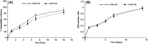
The in vitro antigenicity of the lectinized and non- lectinized nanoparticles was also determined by using an EIA kit (AUSZYME; Abbott Laboratories, Abbott Park, IL, USA) as described by Shi et al. (Citation2002). After 14 days, the EIA/protein ratio of lectinized and non-lectinized nanoparticles was found to be 0.97 ± 0.08 and 0.98 ± 0.07, respectively. There was no significant difference (P > 0.05) in the in vitro antigenicity profile of lectinized and non-lectinized nanoparticles ().
Structural integrity of antigen
In order to assess the structural integrity of antigen encapsulated in the lectin-coupled chitosan nanoparticles, SDS-PAGE measurement was employed. It can be observed from that the native HBsAg and HBsAg extracted from lectin-coupled CH-NP showed single-band marker protein corresponding to of molecular weight (MW = 24 kDa). It suggests that no chemical polymerization, non-covalent aggregation or molecular hydrolysis which could adversely affect the conformation of antigen had occurred during the preparation process ().
Figure 5. SDS-PAGE analysis of ligand-anchored nanoparticles. Lane 1: Molecular weight marker of 225 kDa, Lane 2: Naive hepatitis B antigen, Lane 3: Naive LTA lectin and Lane 4: Lectin anchored Chitosan nanoparticles.
In vitro ligand affinity and activity studies
The presence of numerous functional groups (i.e. amino and carboxylic residues) renders protein an excellent candidate for the preparation of conjugates, by coupling various ligands capable of providing specificity to the surface of nanoparticles such as lectins. In the present investigation BSM, a glycoprotein, was used as a biological model to determine the in vitro affinity and specificity of LTA-coupled nanoparticles towards sugar residue of glycoprotein. For determination of in vitro activity of LTA, experiment was conducted in the absence of specific sugar and for the determination of in vitro specificity of LTA and the experiment was carried out in presence of specific sugar (α-L-fucose) for LTA. The percentage BSM binding in the absence of specific sugar in the case of lectinized and non-lectinized nanoparticles was 28.75 ± 2.6 and 2.72 ± 0.14, respectively. In the absence of α-L-fucose, the LTA-coupled nanoparticles exhibited almost five times higher interaction with BSM than that of unmodified nanoparticles (). In the presence of α-L-fucose, interaction between LTA-coupled nanoparticles and BSM was significantly reduced. Plain nanoparticles revealed fairly comparable (P < 0.05) results in absence and in presence of specific sugar for LTA. Thus, results clearly suggest that lectinized nanoparticles retain same activity and sugar specificity as the native lectin LTA.
Figure 6 Binding of BSM to LTA-anchored Chitosan nanoparticles (LTA-CHNP-HB) and plain nanoparticles in suspension with and without competing sugar (α-L-fucose) and with non-specific or control sugar (D-galactose) (n = 3).
Targeting of nanoparticles to M cells
Confocal laser scanning microscopy (CLSM) was used to evaluate targeting potential of lectinized chitosan nanoparticles. The targeting of LTA-coupled CH-NP was confirmed by dual staining of the Peyer's patches M cells. M cells were first rinsed thoroughly with 10 mM PBS (pH 7.4) and then stained with TRITC-WGA (for localization of M cells) followed by administration of FITC-LTA-anchored nanoparticles. As shown in there was distinct binding of lectinized nanoparticles to the Peyer's patches as compared to control nanoparticles (coated with FITC-BSA) which showed little or no binding to the M cell in mice which confirms the targeting potential of lectinized nanoparticles. Thus we can assure that α-L fucose-specific, LTA-grafted CH-NP could effectively deliver the encapsulated antigen to the murine M cells. In our previous study, we have already demonstrated the M-cell targeting of α-L-fucose-specific, lectin-conjugated particles using LTA-anchored PLGA nanoparticles.(Mishra et al. Citation2010) The same strategy was also used by Roth-Walter et al. (2005) and Gupta et al. (Citation2007) to confirm the M-cell targeting of α-L-fucose-specific, lectin-conjugated particles using AAL and UEA-1 lectin, respectively.
Figure 7. Confocal laser scanning microscopy images showing targeting of the nanoparticles to the M cells of the Peyer's patches in mice by dual staining. M cells were primarily stained with TRITC-WGA (red). FITC-LTA coupled Chitosan nanoparticles stain green. Control nanoparticles (FITC-BSA coated) shows little or no binding to M cells (A). Lectinized chitosan nanoparticles (shown by arrow) were associated predominantly with M cells (B).
Immunological response
The serum anti-HBsAg antibody titer was determined by immunization with different formulations containing antigen equivalent to 10 μg of HBsAg followed by booster dose after second week with the same formulation (). The in vivo immunization study revealed that HBsAg-loaded lectinized nanoparticles (LTA-CHNP-HB) results in significantly higher (p < 0.001) anti-HBsAg titer as compared to plain nanoparticles (CHNP-HB). This may be attributed to the lectin-mediated selective targeting of HBsAg-loaded lectinized nanoparticles to the M cell of the Peyer's patches. Additionally, some of lectin molecules are highly immunostimulatory and may have potential mucosal adjuvant action.(Lavelle et al. Citation2001, Roth-Watler et al. Citation2004) The results are in accordance to our previous in vitro investigation showing that antigenicity of HBsAg in lectin-coupled nanoparticles without stabilizer which was low may be due to the denaturation of the antigen. The stabilized nanoparticles protect the antigen during stressful conditions of preparations and subsequently on oral administration.(Gupta et al. Citation2007).
Figure 8. Serum anti-HBsAg profile of mice immunized with different formulations at booster immunization which was given after 2 weeks. Values are expressed as mean ± SD (n = 6).
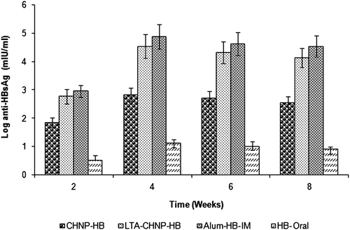
The mice were immunized on Day 0 and booster dose was given after second weeks of the primary immunization. The anti-HBsAg antibody titer was found to be comparable in animals immunized with lectinized CH-NP (LTA-CHNP-HB) (orally) and alum-HBsAg (i.m.). The antibody titer was raised substantially following booster dosing on second week in all experimental animals. The CH-NP-based formulation resulted into the induction of stronger immune response. This may be attributed to the ability of nanoparticles to enhance the uptake of antigen into the Peyer's patches. Further anchoring of the lectin may result in effective receptor-mediated uptake of the nanoparticles through the M cell.
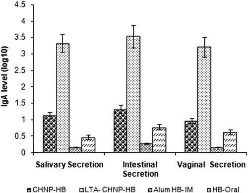
sIgA is a principle antibody isotype produced in all body secretions. It was observed that the specific antibodies in the secretions of mice immunized with alum adsorbed HBsAg (i.m.) was negligible (), whereas in the case of mucosal immunization substantially strong secretary immune response was observed. The immunization of mice with lectinized CH-NP (CHNP-LTA) induced significantly higher (p < 0.001) sIgA level as compared to plain CH-NP (CHNP-HB) in all secretions.
Figure 9. sIgA level in the intestinal, salivary and vaginal secretions of mice immunized with various formulation after 6 weeks of booster immunization. Values are expressed as mean ± SD (n = 6).
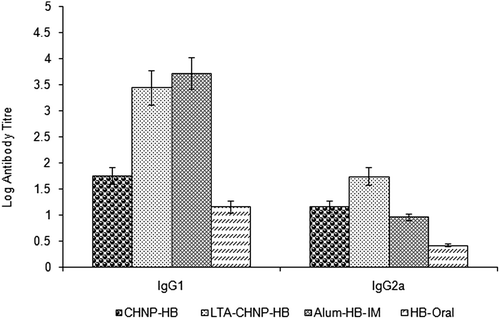
In the present study IgG1 and IgG2a isotypes were determined on Day 42 to assess the TH1/TH2 immunity pattern (). Alum-based antigen formulation elicit high level of IgG1 isotype indicating good TH2 or humoral immunity, whereas failed to generate considerable IgG2a level, that is, hallmark of TH1 immunity. Lectin-anchored nanoparticles resulted into the induction of significantly higher (p < 0.001) TH1/TH2 immune response as compared to alum-based HBsAg. A mixed TH1/TH2 response with good IgG2a/IgG1 ratio was observed with lectin-anchored nanoparticles. Therefore, this may be concluded that the developed lectin-anchored nanoparticles are relatively effective adjuvant that induces both arms of immune response following mucosal immunization.
Figure 10. IgG1 and IgG2a anti-HBsAg antibody isotypes at Day 42 in sera of mice. Values are expressed as mean ± SD (n = 6).
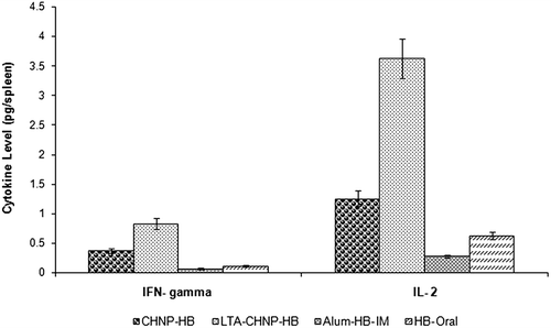
Endogenous cytokine levels (IL-2 and IFN-γ) were determined in spleen homogenate after 6 weeks of booster dose(s) of different formulations () Activation of Th1 subset is associated with production of IFN-γ and IL-2, and hence development of classical cell-mediated immune response. The results indicate that lectinized nanoparticles provoke significantly higher level (p < 0.001) of both Th1-dependent cytokines (IL-2 and IFN- γ) (). The findings are suggestive of induction of cell-mediated immune response following oral immunization with lectinized nanoparticles. This is in accordance with previous report demonstrating significant production of Th1 cytokine by lectinized microparticles.(Gupta et al. Citation2007) Further, it has been argued that lectin facilitates the accumulation of the antigen at desired mucosal sites, which may result in induction of specific Th1 antibody response as well. The generation of a dominant Th1 cytokine profile is an important biomarker suggestion of cellular immunity that eradicates HBV infection.
Figure 11. Interleukin-2 and Interferon-γ level in spleen homogenate of mice immunized with various formulations after 6 weeks of booster immunization. Values are expressed as mean ± SD (n = 6).
Similar results were also observed in the study where antigen-loaded particle was conjugated to the specific ligand such as WGA(Walter et al. Citation2004), AAL(Roth-Watler et al. Citation2005), UEA-1(Gupta et al. Citation2007) and LTA (Mishra et al. Citation2010) to target M cell. These findings revealed that M-cell targeted, antigen-loaded particles elicited strong immune response when compared to plain particles. Same type of results was also found in our study. It can be concluded from these results that M-cell targeted, LTA-anchored CH-NP can be utilized as a potent adjuvant for mucosal immunization by oral route. Moreover, developed polymeric nanocarrier systems also elicited significant mucosal and cellular immune responses, which were not elicited by conventional alum-HBsAg.
Conclusion
The study was performed to determine the targeting potential of LTA which is specific for α-L-fucose receptors that are present on the surface of M cells. The surface-engineered CH-NP efficiently carried the encapsulated antigen to desired site in intestinal Peyer's patches after oral immunization thus improving efficiency of delivered vaccine. The confocal studies confirmed targeting potential of LTA-anchored nanoparticles. Further, in vivo study revealed that lectin-appended CH-NP have proven to be efficient in stimulating mucosal, isotyping response IgG1/IgG2a and cell-mediated (IFN-γ and IL-2 level) immune responses and hold promises as carrier adjuvant for effective oral immunization against HBsAg antigen.
Acknowledgement
Authors acknowledge Prof. S. C. Lakhotia, BHU, Varanasi, India for providing confocal facility and NIPER, Mohali for providing AFM facility. Authors also acknowledge Shantha Biotech Ltd. (Hyderabad, India) for providing recombinant hepatitis B surface antigen as gift sample.
Declaration of interest
The authors report no declarations of interest. The authors alone are responsible for the content and writing of the paper.
The present work was supported by grants from Indian Council of Medical Research, New Delhi, India. One of the authors, Dr. Neeraj Mishra acknowledges ICMR- SRF (New Delhi, India) (Grant: 45/02/2007-BMS/PHA Dated 06\07\2007\H.S.Gour) for providing financial assistance.
References
- Bramwell VW, Perrie Y. 2006. Particulate delivery systems for vaccines: what can we expect. J Pharm Pharmacol. 58:717–728.
- Calvo P, Remunan-Lopez C, Vila-Jata JL, Alonso MJ. 1997a. Novel hydrophilic chitosan- polyethylene oxide nanoparticles as protein carriers. Appl Polym Sci. 63:125–132.
- Calvo P, Remunan-Lopez C, Vila-Jata JL, Alonso MJ. 1997b. Chitosan and chitosan/ethylene oxide-propylene oxide block copolymer nanoparticles as novel carriers for proteins and vaccines. Pharm Res. 14:1431–1436.
- Chang TMS. 1971. Stabilisation of enzymes by microencapsulation with a concentrated protein solution or by microencapsulation followed by cross-linking with glutaraldehyde. Biochem Biophys Res Common. 44:1531–1536.
- Chen F, Zhang ZR, Yuan F, Qin X, Wang M, Huang Y. 2008. In vitro and in vivo study of N- trimethyl chitosan nanoparticles for oral protein delivery. Int J Pharm. 349:226–233.
- Clark MA, Jepson MA, Hirst BH. 2001. Exploiting M cells for drug and vaccine delivery. Adv Drug Del Rev. 50:81–106.
- Clark MA, Jepson MA, Simmons NL, Booth TA, Hirst BH. 1993. Differential expression of lectin binding sites defines mouse intestinal M cells. J Histochem Cytochem. 41:1679–1687.
- De Campos A, Sanchez A, Alonso MJ. 2001. Chitosan nanoparticles: a new vehicle for the improvement of the delivery of drugs to the ocular surface application to cyclosporine. Int J Pharm. 224:159–168.
- Elson CO, Ealding W, Lefkowitz JJ. 1984. A lavage technique following repeated measurement of IgA antibody in mouse intestinal secretions. J Immunol Methods. 67:101–108.
- Ezpeleta I, Irache JM, Stainmesee S, Chabenat C, Gueguen J, Orecchioni AM. 1996. Peparation of lectin – vicilin nanoparticle conjugates using the carbodiimide coupling technique. Int J Pharm. 142:227–233.
- Fernandez-Urrusuno R, Calvo P, Remunan-Lopez C, Vila-Jato JL, Alonso MJ. 1991. Enhancemt of nasal absorption of insuln by using chitosan nanoparticles. Pharm Res. 16:1576–1581.
- Fernandez-Urrusuno R, Romani D, Calvo P, Vila-Jato JL, Alonso MJ. 1999. Development of freeze- dried formulation of insulin- loaded chitosan nanoparticle intended for nasal administration. Pharm Sci. 9:429–436.
- Gabor F, Scwarzbauer A, Wirth M. 2002. Lectin mediated drug delivery: binding and uptake of BSA–WGA conjugates using the caco-2 model. Int J Pharm. 237:227–239.
- Gan Q, Wang T, Cochrane C, McCarron P. 2005. Modulation of surface charge, particle size and morphological properties of chitosan-TPP nanoparticles intended for gene delivery. Colloids Surf B Biointerfaces. 44:65–73.
- Gao X, Tao W, Lu W, Zhang Q, Zhang Y, Jiang X, Fu S. 2006. Lectin – conjugated PEG- PLA nanoparticles: Preparation and brain delivery after intranasal administration. Biomaterials. 27:3482–3490.
- Gupta PN, Khatri K, Goyal AK, Mishra N, Vyas SP. 2007. M cell targeted biodegradable PLGA nanoparticles for oral immunization against hepatitis B. J Drug Target. 15:701–713.
- Gupta PN, Mahor S, Rawat A, Khatri K, Goyal A, Vyas SP. 2006. Lectin anchored stabilized biodegradable nanoparticles for oral immunizations: development and in vitro evaluation. Int J Pharm. 318:163–173.
- He Q, Mitchell A, Morcol T, Bell SJD. 2002. Calcium phosphate nanoparticles induce mucosal immunity and protection against herpes simplex virus type 2. Clin Diagn Lab Immunol. 9:1021–1024.
- Illum L. 1998. Chitosan and its use as a pharmaceutical excipients. Pharm Res. 15:1326–1331.
- Jaganathan KS, Vyas SP. 2006. Strong systemic and mucosal immune responses to surface- modified PLGA microspheres containing recombinant Hepatitis B antigen administered intranasally. Vaccine. 24:4201–4211.
- Jain S, Singh P, Mishra V, Vyas SP. 2005. Mannosylated niosomes as adjuvant carrier system for oral genetic immunization against Hepatitis B. Immunol Lett. 101:41–49.
- Jordan SL, Russo MR, Blessing RL, Theis AB. 1996. Inactivation of glutardehyde. J Toxicol Environ Health. 47:299–309.
- Khatri K, Goyal AK, Gupta PN, Mishra N, Mehta A, Vyas SP. 2008. Surface modified liposomes for nasal delivery of DNA vaccine. Vaccine. 26:2225–2233.
- Kilpatric DC, Pusztai A, Grant G, Graham C, Ewen SW. 1985. Tomato lectin resists digestion in the mammalian alimentary canal and binds to intestinal villi without deletious effects. FEBS Lett. 185: 299–305.
- Lavelle EC, Grant G, Pfuller U, O’Hagan DT. 2004. Immunological implication of the use of plant lectins for drug and vaccine targeting to the gastrointestinal tract. J Drug Target. 12:89–95.
- Lavelle EC, Grant G, Pusztai A, Fuller U, O’Hagan DT. 2001. Identification of plant lectin with mucosal adjuvant activity. Immunol. 102:77–86.
- Lehr CM, Bouwstra JA, Kok W, Noach AB, de Boer AG, Junginger HE. 1992. Bioadhesion by means of specific binding of tomato lectin. Pharm Res. 9:547–553.
- Lis B, Sharon N. 1986. Lectins as molecules and as tools. Annu Rev Biochem. 55:35–67.
- Mishra N, Goyal AK, Khatri K, Vaidya B, Paliwal R, Rai S, et al. 2008. Biodegradable polymer based particulate carrier(s) for the delivery of proteins and peptides. Antiinflammat AntiAllergy Agents Med Chem. 7:240–251.
- Mishra N, Tiwari S, Vaidya B, Agrawal GP, Vyas SP. 2010. Lectin anchored PLGA nanoparticles for oral mucosal immunization against Hepatitis B. J Drug Target. 19:67–78.
- Monsan P, Puzo G, Mazarguil H. 1975. Étude du mécanisme d’établissement des liaisons glutaraldehyde protéines. Biochimie. 57:1281–1292.
- Montisci MJ, Dembri A, Giovannuci G, Chacun H, Duchene D, Ponchel G. 2001a. Gastrointestinal transit and mucoadheasion of colloidal suspension of Lycopersicon esculentum L. and Lotus tetragonolobus Lectin- PLA microsphere conjugates in rats. Pharm Res. 18:829–837.
- Nishimura K, Nishimura S, Nishi N, Saiki I, Tokura S, Azuma I. 1984. Immunological activity of chitin and its derivatives. Vaccine. 2:93–99.
- Nugent J, Po AL, Scott EM. 1998. Design and delivery of non-parenteral vaccines. J Clin Pharm Ther. 23:257–285.
- O’Hagan DT. 1998. Microparticles and polymers for the mucosal delivery of vaccines. Adv Drug Del Rev. 34:305–320.
- Pereira MEA, Kabat EA. 1974. Specificity of purified hemagglutin in lectin from Lotus tetragonolobus. Biochemistry. 13:3184–3192.
- Ponchel G, Irache JM. 1998. Specific and non-specific bioadhesive particulate systems for oral delivery to the gastrointestinal tract. Adv Drug Del Rev. 34:191–219.
- Roth-Watler F, Scholl E, Untersmayr I, Ellinger A, Boltz-Nitulescu G, Scheiner O, et al. 2005. Mucosal targeting of allergen-loaded microspheres by Alenuria auerantia lectin. Vaccine. 23:2703–2710.
- Roth-Watler F, Scholl I, Untersmayr E, Fuchs R, Boltz-Nitulescu G, Weissenbock A, et al. 2004. M cell targeting with Alenuria auerantia lectin as a novel approach for oral allergen immunotherapy. J Allergy Clin Immunol. 114:1362–1368.
- Russell-Jones GJ, Veitch H, Arthur L. 1999. Lectin-mediated transport of nanoparticles across Caco-2 and OK cells. Int J Pharm. 190:165–174.
- Sachdeva S, Ahmad G, Malhotra P, Mukherjee P, Chauhan VS. 2004. Comparison of immunogenicities of recombinant plasmodium vivax merozoite surface protein 119 and 42 kilodalton fragments expressed in Escherichia coli. Infect Immun. 70:5775–5785.
- Shi L, Caulfield MJ, Chern RT, Wilson RA, Sanyal G, Volkin DB. 2002. Pharmaceutical and immunological evaluation of single-shot hepatitis B vaccine formulated with PLGA microspheres. J Pharm Sci. 91:1019–1035.
- Singh M, Li X, McGhee JP, Zamb T, Koff W, Wang CY, O’Hagan DT. 1997. Controlled release microparticles as a single dose hepatitis B vaccine: evaluation of immunogenicity in mice. Vaccine. 15: 475–481.
- Singh P, Prabakaran D, Jain S, Mishra V, Jaganathan KS, Vyas SP. 2004. Cholera toxin B sub-unit conjugated bile salt stabilized vesicles (bilosomes) for oral immunization. Int J Pharm. 278:379–390.
- van der Lubben IM, Kersten G, Fretz MM, Beuvery C, Coos Verhoef J, Junginger HE. 2003. Chitosan microparticles for mucosal vaccination against diphtheria: oral and nasal efficacy studies in mice. Vaccine. 21:1400–1408.
- van der Lubben IM, Konings FA, Borchard G, Verhoef JC, Junginger HE. 2001a. In vivo uptake of chitosan microparticles by murine Peyer's patches: visualization studies using confocal laser scanning microscopy and immunohistochemistry. J Drug Target. 9:39–47.
- van der Lubben IM, Verhoef JC, Borchard G, Junginger HE. 2001b. Chitosan for mucosal vaccination. Adv Drug Del Rev. 52:139–144.
- van der Lubben IM, Verhoef JC, van Aelst AC, Borchard G, Junginger HE. 2001c. Chitosan microparticles for oral vaccination: preparation, characterization and preliminary in vivo uptake studies in murine Peyer's patches. Biomaterials. 22:687–694.
- Vyas SP, Sihorkar V, Dubey PK. 2001. Preparation, characterization and in-vitro antimicrobial activity of metronidazole bearing lectinized liposomes for intra peridontal pocket delivery. Pharmazie. 56:554–560.
- Walter F, Scholl I, Untersmayr E, Ellinger A, Boltz-nitulescu G, Scheiner Q, et al. 2004. Functionalization of allergen loaded microspheres with wheat germ agglutinin for targeting enterocytes. Biochem Biophys Res Comm. 315:281–287.
- Xu Y, Du Y. 2003. Effect of molecular structure of chitosan on protein delivery properties of chitosan nanoparticles. Int J Pharm. 250: 215–226.