
Abstract
In this study, a novel amperometric glucose biosensor with immobilization of glucose oxidase on electrochemically polymerized polyaniline–polyvinylsulphonate–potassium ferricyanide (Pani–Pvs–Fc) films has been accomplished via the entrapment technique. Potassium ferricyanide was used as the mediator. Determination of glucose was carried out by the oxidation of potassium ferrocyanide at 0.3 V vs. Ag/AgCl. The effects of pH and temperature were investigated, and the optimum pH value was found to be 7.5. The storage stability and the operational stability of the enzyme electrode were also studied.
Introduction
Glucose is a major component of animal and plant carbohydrates. Quantitative determination of glucose is of paramount importance in biochemistry, clinical chemistry and food analysis (Wu et al. Citation2004).
Numerous efforts have been devoted to develop glucose biosensor with fast and accurate response. The conducting polymers are being widely used in biosensor applications because they provide stable and porous matrix for the immobilization of biocomponent, and they also facilitate the electron transfer process. The widely used conducting polymers for immobilization of enzyme are polyaniline, polypyrrole, polythiophene, etc. (Bidan Citation1992, Gaikwad et al. Citation2005, Barlett and Cooper Citation1993, Shirale et al. Citation2005, Cosiner Citation1999, Colak et al. Citation2012, Arslan et al. Citation2012).
Mediators are artificial electron transferring agents that can readily participate in the redox reaction with the biological component, and thus help in the rapid electron transfer. Mediated enzyme electrodes are known to be less susceptible to interfering substances due to lower electrode potentials. Because of this reason, lots of mediated enzyme electrodes are prepared (Ricci and Palleschi Citation2005, Ghica and Brett Citation2005, Pate et al. Citation2003, Chaubey and Malhotra Citation2002).
One of the most important sensing materials widely used in the glucose biosensor is glucose oxidase (GOx). Most of the electrochemical glucose biosensors are based on the glucose oxidase (GOx) enzyme, which catalyzes the oxidation of glucose to gluconolactone that was hydrolyzed to gluconic acid in water (Norouzi et al. Citation2010).
In this study, a novel amperometric glucose biosensor with immobilization of glucose oxidase on electrochemically polymerized polyaniline–polyvinylsulphonate–potassium ferricyanide (Pani–Pvs–Fc) films has been accomplished via the entrapment technique. Potassium ferricyanide was used as the mediator. Effects of the immobilization process on kinetic parameters, storage and reuse capability of the enzyme were investigated. The optimum working conditions of biosensor with respect to the substrate concentrations, the pH and temperature were investigated.
Experimental section
Equipment and reagents
The electrochemical studies were carried out using Epsilon EC electrochemical analyzer with a three-electrode cell. The working electrode was a Pt plate (0.5 cm2). The auxiliary and the reference electrodes were a Pt wire and a Ag/AgCl electrode (3 M KCl), respectively. The pH values of the buffer solutions are measured with ORION Model 720A pH/ionmeter. Temperature control was achieved with Grant W14 thermostat.
Glucose oxidase (EC 1.1.3.22, purified from the Aspergillus Niger and with an activity of 10 unit ml− 1) and glucose were purchased from Sigma. Aniline from Merck and sodium polyvinylsulphonate (Pvs) from Aldrich were supplied. All other chemicals were obtained from Sigma. All the solutions were prepared using double-distilled water. Glucose stock solution was allowed to mutarotate for at least 24 h at room temperature prior to use and stored at 4°C.
Preparation of Pt/Pani–Pvs electrode
The surface of the Pt was mechanically, chemically and electrochemically pretreated prior to the coating process. The surface was first polished with silicon carbide 1200/P2500 grinding paper and subjected to a bunsen flame. It was then dipped in acetonitrile, ethanol, concentrated HCl and concentrated nitric acid for 5 min each. The surface was then washed thoroughly with double-distilled water, before being scanned in 5 M H2SO4 between − 2.0 and 2.0 V for electrochemical treatment. It was again washed with double-distilled water and then with acetone to remove any traces of water which may have remained on the surface. This process was repeated prior to each coverage (Gros et al. Citation2000). The surface of the pretreated electrode was covered with a conducting polyaniline in polyvinylsulphonate medium. A 10 mL solution containing 0.2 M aniline and 2.5 mL (25%) Pvs was prepared. The electrode was immersed into this solution and the system was purged with purified argon for 10 min in order to remove any traces of oxygen. The polymerization was carried out using constant potential of 0.75 V for 60 min [vs. Ag/AgCl electrode (3 M KCl)] (Arslan et al. Citation2011). The electrode was washed with buffer solution after the coating process.
Preparation of Pt/Pani–Pvs–Fc–GOx electrode
Immobilization of glucose oxidase was carried out by the physical entrapment technique. The Pt electrode was immersed in 0.2 M aniline, 0.2 M potassium ferricyanide (K3Fe(CN)6 and 2.5 mL aqueous solution of the sodium salt of polyvinylsulphonate (25%). Then 1 mL of GOx enzyme (10 U/mL) was added into the solution, which was purged with argon for the removal of oxygen before electropolymerization. The polymerization was carried out at a constant potential of 0.75 V for 60 min [vs. Ag/AgCl electrode (3 M KCl)]. After entrapping the glucose oxidase onto the Pani–Pvs–Fc film, the electrode was rinsed with deionized water to remove the unreacted aniline monomer and free glucose oxidase. Immobilized enzyme electrodes were kept in a refrigerator at 4°C in phosphate buffer when not in use.
Amperometric biosensor measurements
Amperometric response studies were carried out in phosphate buffer (0.1 M, pH 7.5). The Pt/Pani–Pvs–Fc–GOx electrode was immersed into the phosphate buffer (0.1 M) of pH 7.5. The solution contained 0.1 M sodiumperchlorate as a supporting electrolyte. The electrode was brought to equilibrium by keeping it at 0.3 V [vs. Ag/AgCl electrode (3 M KCl)]. Steady-state current (ia) was recorded. Glucose was added to the cell from stock solution and the system was stirred. The currents (ib) obtained at 0.3 V were recorded. The current values (Δi = ib− ia) were plotted against the glucose concentration to determine the working range of the electrode. Research into the operational and storage stability, as well as the effects of pH and temperature was carried out using 5.0 × 10− 5 M glucose.
Results and discussion
In this study, a new glucose-sensitive amperometric enzyme electrode was prepared using Pani–Pvs–Fc film. Potassium ferricyanide was used as the mediator. When there is no mediator in the medium, molecular oxygen takes the electrons coming from the substrate and then hydrogen peroxide is formed. However, in this study potassium ferricyanide also exists in oxygenated medium. Both oxygen and potassium ferricyanide are electron acceptors. Because reduction potential of potassium ferricyanide [+ 0.075 V vs. Ag/AgCl electrode (3 M KCl)] is more positive than reduction potential of oxygen [− 0.1 V vs. Ag/AgCl electrode (3 M KCl)], ferricyanide takes the electrons coming from substrate and is reducted to ferrocyanide. As a result, glucose determination is done by oxidation of ferrocyanide on electrode surface. Reactions that occur are shown below:
Fe(CN)63− → Fe(CN)64− (before applying the potential)
Fe(CN)64−→ Fe(CN)63− (after applying potential, on the electrode surface, 0.3 V)

The parameters effecting the performance of the biosensor were investigated.
The effect of mediator
In this study the effect of two electron transfer mediators [potassium ferricyanide (Fc) and methylene blue (Mb)] was investigated. Amperometric responses of the Pt/Pani–Pvs, Pt/Pani–Pvs–Fc and Pt/Pani–Pvs–Mb electrodes to glucose in the presence of glucose oxidase (50 μL) were determined at different glucose concentrations. In both cases as shown in , when Pt/Pani–Pvs–Fc electrode was used, current differences were obtained highest. It is seen that the mediators which were used increased the electron transfer. When potassium ferricyanide was used, electron transfer occurred faster compared with methylene blue. Therefore, ferricyanide was used as the mediator in the following studies.
Figure 1. Amperometric response of the Pt/Pani–Pvs, Pt/Pani–Pvs–Fc and Pt/Pani–Pvs–Mb electrodes to glucose [in the phosphate buffer (pH 7.5), 50 μL glucose oxidase (10 U/mL), 0.3 V].
![Figure 1. Amperometric response of the Pt/Pani–Pvs, Pt/Pani–Pvs–Fc and Pt/Pani–Pvs–Mb electrodes to glucose [in the phosphate buffer (pH 7.5), 50 μL glucose oxidase (10 U/mL), 0.3 V].](/cms/asset/60649e03-a01a-4d7a-beac-7a6183278c16/ianb_a_812650_f0001_b.jpg)
The effect of working potential
After preparing Pt/Pani–Pvs–Fc electrode, the glucose determination was carried out at different potentials (0.2, 0.3 and 0.4 V) ().
Figure 2. The effect of potential on the response of the Pt/Pani–Pvs–Fc electrode to glucose [in the Phosphate buffer (pH 7.5), 50 μL glucose oxidase (10 U/mL)].
![Figure 2. The effect of potential on the response of the Pt/Pani–Pvs–Fc electrode to glucose [in the Phosphate buffer (pH 7.5), 50 μL glucose oxidase (10 U/mL)].](/cms/asset/826c2f4c-dd6a-423c-92b8-36e82f8b19b9/ianb_a_812650_f0002_b.jpg)
Potentials higher than 0.4 V were not studied, because that interference effect could be more in higher potentials (Zhang et al. Citation2007). Highest currents were seen at 0.4 V. Currents were too low at 0.2 V. A low potential of 0.3 V was chosen as working potential, at which currents higher than 0.2 V were observed.
The effect of potassium ferricyanide concentration on the response of the Pt/Pani–Pvs–Fc electrode to glucose
Mediators are added to the polymerization medium of the biosensors prepared by the use of conducting polymer-covered electrodes in order to facilitate the electron transfer (Chaubey and Malhotra Citation2002, Garcia et al. Citation1998, Zhao et al. Citation2002). In this study, potassium ferricyanide was used as the mediator. The responses of electrodes to glucose were determined in order to elucidate the role of potassium ferricyanide concentration. Five different potassium ferricyanide concentrations (0.05, 0.1, 0.15, 0.2 and 0.3 M) were used (). In , it can be seen that response currents are increased with ferricyanide concentration.
Figure 3. The effect of potassium ferricyanide concentration on the response of the Pt/Pani–Pvs–Fc electrode to glucose [in the phosphate buffer (pH 7.5), 50 μL glucose oxidase (10 U/mL), 0.3 V].
![Figure 3. The effect of potassium ferricyanide concentration on the response of the Pt/Pani–Pvs–Fc electrode to glucose [in the phosphate buffer (pH 7.5), 50 μL glucose oxidase (10 U/mL), 0.3 V].](/cms/asset/7f5c2b2e-a7fd-48c9-af6f-35c67f7f9803/ianb_a_812650_f0003_b.jpg)
indicates that the current response of the electrode covered in 0.3 M potassium ferricyanide is the lowest, and the surface of this electrode is not homogeneous. The polymer coverage easily peeled from the surface. The polymer on the electrode surface was thicker than the others. Increase in the thickness of the polymer decreased the conductivity; therefore, currents were also decreased (Arslan et al. Citation2006). It was concluded that a high potassium ferricyanide concentration was not suitable for polymer coverage. The electrode, which gave the best linear response, was the one prepared with the use of 0.2 M potassium ferricyanide. That is why all the experiments were carried out with Pt/PPy–Fc electrodes prepared using the potassium ferricyanide concentration at 0.2 M.
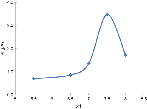
The effect of pH on amperometric response of biosensors
Buffers solutions (phosphate buffer) at various pH values were tested to investigate the effect of pH. The pH values of the buffer solutions were varied: 5.5, 6.5, 7.0, 7.5 and 8.0. The measurements were performed at a constant glucose concentration of 5.0 × 10− 5 M. shows that the maximum response was obtained at pH 7.5. For glucose biosensor, pH values different from 7.5 were employed in the literature (pH 6.5, 7.4) (Shan et al. Citation2008, Gaikwad et al. Citation2007). This was attributed to the fact that the used polymer and the type of immobilization were different.
Figure 4. The effect of pH on the response of the biosensor at (25°C 5.0 × 10− 5 M glucose in operating potential 0.3 V).
The effect of temperature on amperometric response of biosensors
Temperature has a great effect on enzyme activity, and it is important to investigate temperature's dependence to the response of the enzyme electrode. Temperature's influence on the response of glucose enzyme electrode was tested between 20 and 60°C at pH 7.5, using constant glucose concentration of 5.0 × 10− 5 M ().
Figure 5. The effect of temperature on the response of the biosensor at (pH 7.5, 5.0 × 10− 5 M glucose in operating potential 0.3 V).
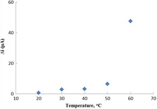
In , it can be seen that response currents are increased with temperature. Because of the increase in temperature, an optimum value could not be seen. For glucose biosensor, temperature values were employed in the literature (33°C, 40°C) (Shan et al. Citation2008, Xue et al. Citation2005).
When literatures were examined, it was seen that the polyaniline film which was obtained by the electropolymerization of aniline became a good micro environment around the enzyme. It was observed that the enzyme was stronger even in high temperatures because of this micro environment (Arslan et al. Citation2011, Shi et al. Citation2009, Chen et al. Citation2006). Therefore, the temperature of 25°C was chosen as working temperature for all further experiments.
Substrate concentration and calibration curves
There is a linear part ranging between 5.0 × 10− 6 and 1.0 × 10− 3 M (R2 = 0.962). The graph for the calibration curve is given in . It is shown that the linearity of graph is highly satisfactory, and it could be used for the quantitative determination of glucose. The detection limit of the biosensor was 5.0 × 10− 7 M and the response time of the biosensor was 200 s. Kinetic parameters Km(app) and Imax for the enzyme biosensor were detected at constant temperature (25°C) and pH (pH 7.5) in varying substrate concentration. Km(app) and Imax were calculated as 2.42 mM and 0.0042 μA/min, respectively using 1/[glucose]− 1/Δi graph (Lineweaver–Burk plot) (). Km values for immobilized glucose oxidase presented in the literature are 18.0, 11.9 and 9.34 mM (Shan et al. Citation2008, Xue etal. Citation2001, Zhou et al. Citation2005). This was attributed to the fact that the used polymer and the type of immobilization were different.
Figure 6. The calibration curve of glucose biosensor (pH 7.5 phosphate buffer 25°C operating potential 0.3 V).
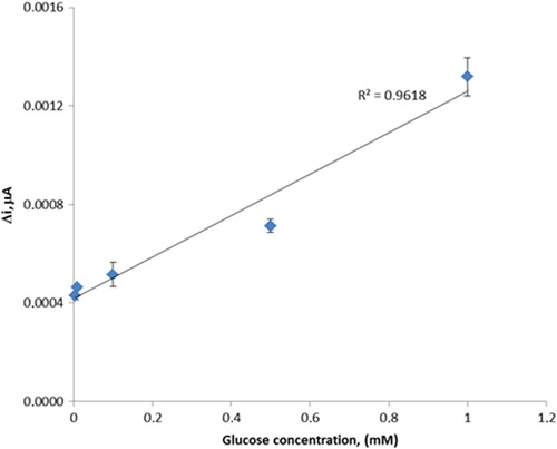
Figure 7. The effect of glucose concentration upon the amperometric response of the biosensor. [Lineweaver–Burk plot, in the phosphate buffer (pH 7.5) and operating potential 0.3 V, 25°C].
![Figure 7. The effect of glucose concentration upon the amperometric response of the biosensor. [Lineweaver–Burk plot, in the phosphate buffer (pH 7.5) and operating potential 0.3 V, 25°C].](/cms/asset/5aab7a45-ea1c-48bc-9f10-cfcdf9304058/ianb_a_812650_f0007_b.jpg)
Operational stability and storage stability
The biosensor was used at optimum activity conditions for 10 activity assays in 1 day to determine the operational stability. The operational stability was studied by applying activity assay (under optimum conditions) 10 times on the same day at constant temperature, pH and substrate concentration (5.0 × 10− 5 M). At the end of the 10 measurements, the biosensor lost 8% of its initial activity (). The relative standard deviation obtained after 10 measurements at a constant glucose concentration of 5.0 × 10− 5 M was found to be 4.9%. Storage stability of the biosensor was determined by performing activity assays within 35 days (). The prepared glucose biosensor retained 47% of initial activity after 35 days, when stored in 0.1 M phosphate buffer solution at 4°C.
Figure 8. Measure number of the biosensor [in the phosphate buffer (pH 7.5), in operating potential 0.3 V, 25°C].
![Figure 8. Measure number of the biosensor [in the phosphate buffer (pH 7.5), in operating potential 0.3 V, 25°C].](/cms/asset/86aacfc5-d19b-48f7-8734-540812356eab/ianb_a_812650_f0008_b.jpg)
Figure 9. Storage stabilization of the biosensor [in the phosphate buffer (pH 7.5), in operating potential 0.3 V, 25°C and substrate concentration 5.0 × 10− 5 M].
![Figure 9. Storage stabilization of the biosensor [in the phosphate buffer (pH 7.5), in operating potential 0.3 V, 25°C and substrate concentration 5.0 × 10− 5 M].](/cms/asset/8b7bd32e-14f9-41df-8b20-d28357a8723d/ianb_a_812650_f0009_b.jpg)
Interference effect
A few common substances found in serum or urine were studied for any interfering effect on the analysis of glucose. Known concentrations of ascorbic acid, uric acid and paracetamol (acetaminophen) were added. It was observed that paracetamol, concentration 1.0 × 10− 5 M, had no interfering effects on the analysis of glucose. But interference effects of ascorbic acid (in 1.0 × 10− 5 M) and uric acid (in 1.0 × 10− 5 M) on the analysis of glucose were found to be 40% and 30%, respectively. These interferences were almost removed by dilution of solution in cell.
Conclusions
In this study, glucose oxidase was successfully immobilized on a Pani–Pvs–Fc film. Potassium ferricyanide was used as the mediator. The experimental results clearly showed that the biosensor exhibited good performance in the determination of glucose. It was seen that glucose biosensor was sensitive, and operational stability and long-term storage stability were found to be good.
Glucose biosensor prepared in this study is useable in a large concentration range 5.0 × 10− 6–1.0 × 10− 3 M (R2 = 0.962). It has a very low detection limit (5.0 × 10− 7 M) and an acceptable response time for a biosensor (200 s). It gives perfect reproducible results (the relative standard deviation is 4.9% after 10 measurements, the standard deviation obtained). Also, it has a good storage stabilization (gives 47% of the initial amperometric response at the end of the 35th day). The Km(app) and Imax(app) values of glucose oxidase enzyme immobilized in Pani–Pvs–Fc were 2.42 mM and 0.0042 μA/min, respectively. Glucose biosensor prepared in this study is easy to prepare and highly cost effective.
Declaration of interest
The authors report no declarations of interest. The authors alone are responsible for the content and writing of the paper.
References
- Arslan F, Ustabas S, Arslan H. 2011. An amperometric biosensor for glucose determination prepared from glucose oxidase immobilized in polyaniline-polyvinylsulfonate film. Sensors. 11:8152–8153.
- Arslan F, Yasar A, Kiliç E. 2006. Preparation of Pt/Polypyrrole– Ferrocene hydrogen peroxide sensitive electrode for the use as a biosensor. Russ J Electrochem. 42:137–140.
- Arslan H, Özdemir M, Zengin H, Zengin G. 2012. Glucose biosensing at carbon paste electrodes containing polyaniline-silicon dioxide composite. Int J Electrochem Sci. 7:10205–10214.
- Barlett PN, Cooper JM. 1993. A review of the immobilization of enzymes in electropolymerized films. J Electroanal Chem. 362:1–12.
- Bidan G. 1992. Electroconducting conjugated polymers: new sensitive matrices to build up chemical or electrochemical sensors. Sens Actuators B. 6:45–56.
- Chaubey A, Malhotra BD. 2002. Review: mediated biosensors. Biosens Bioelectron. 17:441–456.
- Chen C, Jiang Y, Kan J. 2006. A noninterference polypyrrole glucose biosensor. Biosens Bioelectron. 22:639–643.
- Colak O, Arslan H, Zengin H, Zengin G. 2012. Amperometric detection of glucose by polyaniline-activated carbon composite carbon paste electrode. Int J Electrochem Sci. 7:6988–6997.
- Cosiner S. 1999. Biomolecule immobilization on electrode surfaces by entrapment or attachment to electrochemically polymerized films. A review. Biosens Bioelectron. 14:443–456.
- Gaikwad PD, Savale PA, Shirale DJ, Kharat HJ, Kakde KP, Gade VK, Shirsat MD. 2005. Effect of electrolyte on optical properties of potentiostatic electro-deposited conducting polymer films for biosensor applications. Microwaves and Optoelectronics. UK: Anshan Ltd, Tunbridge Wells, pp. 450–454.
- Gaikwad PD, Shirale DJ, Savale PA, Datta K, Ghosh P, Shirsat MD. 2007. Development of PANI-PVS-GOD electrode by potentiometric method for determination of glucose. Int J Electrochem Sci. 2: 488–497.
- Garcia CAB, Neto GO, Kubota LT. 1998. New fructose biosensors utilizing a polypyrrole film and D-fructose 5-dehydrogenase immobilized by different processes. Anal Chim Acta. 374:201–208.
- Ghica ME, Brett CM. 2005. A glucose biosensor using methyl viologen redox mediator on carbon film electrodes. Anal Chim Acta. 532: 145–151.
- Gros P, Durliat H, Comtat M. 2000. Use of polypyrrole film containing Fe(CN)63− as pseudo-reference electrode: application for amperometric biosensors. Electrochim Acta. 46:643–650.
- Norouzi P, Faridbod F, Larijani B, Ganjali MR. 2010. Glucose biosensor based on MWCNTs-Gold nanoparticles in a nafion film on the glassy carbon electrode using flow injection FFT continuous cyclic voltammetry. Int J Electrochem Sci. 5:1213–1224.
- Pate H, Li X, Karana HI. 2003. Amperometric glucose sensors based on ferrocene containing polymeric electron transfer systems. Biosens Bioelectron. 18:1073–1076.
- Ricci F, Palleschi G. 2005. Review: sensor and biosensor preparation, optimisation and applications of Prussian Blue modified electrodes. Biosens Bioelectron. 21:389–407.
- Shan D, Wang S, He Y, Xue H. 2008. Amperometric glucose biosensor based on in situ electropolymerized polyaniline/poly (acrylonitrile-co-acrylic acid) composite film. Mater Sci Eng C. 28:213–217.
- Shi Q, Wang P, Jiang Y, Kan J. 2009. Glucose biosensor based on polyaniline synthesized in ionic liquid. Biocatal Biotransformation. 27:54–59.
- Shirale DJ, Bhalerao AS, Kharat HJ, Gaikwad PD, Kakde KP, Savale PA, Gade VK, Shirsat MD. 2005. Influence of pH on optical properties of conducting polyaniline film for biosensor applications. In: Shirsat MD, Ed. Microwaves and Optoelectronics. UK: Anshan Ltd, Tunbridge Wells, pp. 455–458.
- Wu B, Zhang G, Shuang S, Choi MM. 2004. Biosensors for determination of glucose with glucose oxidase immobilized on an eggshell membrane. Talanta. 64:546–553.
- Xue H, Shen Z, Li Y. 2001. Polyaniline-polyisoprene composite film based glucose biosensor with high permselectivity. Synthetic Metals. 124:345–349.
- Xue H, Shen Z, Li C. 2005. Improved selectivity and stability of glucose biosensor based on in situ electropolymerized polyaniline-polyacrylonitrile composite film. Biosens Bioelectron. 20:2330–2334.
- Zhang Y, Wen G, Zhou Y, Shuang S, Dong C, Choi MM. 2007. Development and analytical application of an uric acid biosensor using an uricase-immobilized eggshell membrane. Biosens Bioelectron. 22:1791–1797.
- Zhao H, Yuan Y, Adelojue S, Wallace GG. 2002. Study on the formation of the Prussian blue films on the polypyrrole surface as a potential mediator system for biosensing applications. Anal Chim Acta. 472:113–121.
- Zhou H, Chen H, Luo S, Chen J, Wei W. 2005. Glucose biosensor based on platinum microparticles dispersed in nano-fibrous polyaniline. Biosens Bioelectron. 20:1305–1311.