
Abstract
We present a rare case of benign tumor of the hand.
Keywords:
Introduction
A variety of benign tumors arise from the synovial membrane of the tendon. Lipomas are one of the uncommon benign tumors that arise from the synovial membrane of the tendon sheath, mostly occurring in the wrist and hand when they do Citation[1]. The treatment of synovial lipoma consists of resection of the mass Citation[1].
As a subtype of lipomas, angiofibrolipomas consist of mature adipocytes, vascular tissue, and collagenous connective tissue Citation[2,3]. These tumors are very rare and have been reported in different parts of the body Citation[2–10]. To the best of our knowledge, we present the first case in the literature of an angiofibrolipoma of the tendon sheath.
Case report
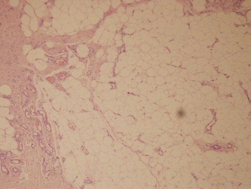
In March 2012, a 24-year-old woman presented with a 2-year slow-growing lesion on the dorsum of her left hand. The patient did not complain of pain or functional deficiency. Examination revealed a nodular subcutaneous mass, which moved with the tendon sheath. Magnetic resonance (MR) imaging showed a 1.5 × 2 × 1.5 cm sized heterogeneous hyperintense mass in contact with the extensor digitorum tendon, with intensity similar to subcutaneous fatty tissue on T1- and T2-weighted sequences (). Intravenous regional anesthesia was administered. After the skin incision, a mass of lipoma-like small vascular structures was encountered under the subdermal tissue (). The mass in the tendon sheath was dissected uneventfully by total excision. The procedure was performed under a tourniquet. Histopathology examination revealed fibrolipomatous areas and a dense vascular structure consistent with the diagnosis of angiofibrolipoma (). At the 6-month follow-up, no recurrence was present.
Figure 1. (a) Preoperative axial T1-weighted (b) and T2-weighted MR images showing heterogeneous hyperintense well-defined mass (arrows), which has similar intensity with subcutaneous fat tissue, adjacent to extensor digitorum tendon.
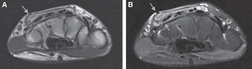
Figure 2. Intraoperative view of the angiofibrolipoma.
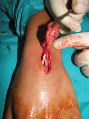
Figure 3. Histological view of the angiofibrolipoma.
Discussion
Tumors of the tendon sheath are uncommon. Local symptoms, which appear as the mass grows, are one of the most important characteristics of tendon sheath tumors. Although common in other parts of the body, lipomas are rarely seen in tendon sheaths Citation[1]. When they occur, they cause local, nonspecific symptoms (trigger finger, carpal tunnel syndrome, and tendon rupture) Citation[1]. They may also cause neurovascular compression symptoms like the other tumors Citation[11]. Preoperative differentiation of the mass should be done with an X-ray, ultrasonography, or an MR scan. In our case, the patient presented to our clinic with a mass on her hand. We performed an X-ray to differentiate the lesion from bone and cartilaginous masses and an MR scan to reveal the dimensions and for exact localization of the mass.
Histological variants of lipomas include fibrolipomas, angiolipomas, angiofibrolipomas, angiomyolipomas, and infiltrating angiolipomas Citation[3]. An angiofibrolipoma is one of the rarest histopathological variants of a lipoma Citation[3]. It is composed of mature adipocytes, vascular tissue, and collagenous connective tissue Citation[3].
As for all lipomas, angiofibrolipomas are treated with surgery, and recurrence rates are very low Citation[2,10]. However, infiltrating angiolipomas have a high recurrence rate Citation[10]. Meticulous dissection should be done to prevent bleeding and recurrence.
In conclusion, it should be kept in mind that angiofibrolipomas, a rare type of lipoma, can cause tendon sheath masses. Radiological and histological examination is important for differential diagnosis. The treatment of all types of lipomas is surgical excision.
Declaration of interest: The authors report no conflicts of interest. The authors alone are responsible for the content and writing of the paper.
Notes
References
- Garner HW, Bestic JM. Benign synovial tumors and proliferative processes. Semin Musculoskelet Radiol 2013;17:177–8
- Brkić A, Ozçamur C, Gürkan-Köseoğlu B, Olgac V. Angiofibrolipoma of the buccal mucosa: a case report. J Oral Sci 2010;52:173–6
- Jacob A, Kneile J, Welling DB. Angiofibrolipoma of the ear canal. Laryngoscope 2005;115:1461–2
- Liu QL, Tian B, Zhang H, Qiao DS. Angiofibrolipoma of the spermatic cord. Asian J Androl 2009;11:746–7
- Pérez-Navarro JV, Flores-Cardoza A, Anaya-Prado R, et al. Angiofibrolipoma of the greater omentum: case report and literature review. Cir Cir 2009;77:229–32
- Novozhilov VN, Dolidze UR, Degterev DB, et al. Angiofibrolipoma of the transverse colon. Vestn Khir Im I I Grek 2006;165:102–3
- Krausen C, Becker K, Hamann KF. Angiofibrolipoma of the tonsil. Laryngol Rhinol Otol (Stuttg) 1986;65:355–6
- Tóth C. Kidney angiofibrolipoma. Z Urol Nephrol 1975;68:279–82
- Hojman D. Mixed tumor of the pericardium; angiofibrolipoma. Prensa Med Argent 1949;36:2478–81
- Uwale Eyesan S, Christopher Ayeni S, Adesope Adesina S, et al. Angiofibrolipoma of the calf. Rare Tumors 2013;5(3):e48
- Santanelli F, Paolini G, Longo B, et al. Compression of the digital nerves by a giant periosteal chondroma. J Plast Surg Hand Surg 2013;47(2):155–7