
Abstract
Bilobed flaps were first introduced to close small nasal defects. We reconstructed a defect of the popliteal fossa using a random-pattern bilobed flap. We recommend the use of random-pattern bilobed flaps as a reliable technique for covering defects of the popliteal fossa.
Introduction
Although the bilobed flap was first introduced to repair small (<1 cm) nasal defects, it has since been shown to be a reliable and versatile tool for repairing variously sized defects in different anatomical regions [Citation1]. Karacalar [Citation2] reported the first use of the bilobed flap for popliteal reconstruction in a patient with a burn contracture. In his report, the bilobed flap was used in an axial manner based on the median superficial sural arteries [Citation2].
We herein present a case involving a young male with a popliteal defect that was reconstructed with a random-pattern bilobed flap.
Case report
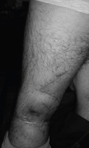
A 26-year-old otherwise healthy male presented with a traumatic wound to his left popliteal region secondary to a traffic accident. He had also sustained severe popliteal artery injury and a patellar fracture. After arterial repair and establishment of limb perfusion, he was referred for repair of the tissue defect in the left popliteal region. Physical examination revealed a 5 × 5 cm defect on the midline of the left popliteal fossa. The wound was necrotic, but no arteries or nerves were exposed. Daily dressing changes were performed. Following wound bed preparation, reconstruction was planned. The operation was performed under spinal anesthesia. The defect was covered with a 45°-angled random-pattern bilobed flap (primary lobe measured 9 × 6 cm, and secondary lobe measured 9 × 3 cm) () and two Penrose drains were placed (). The patient was followed up for 3 months. No complications occurred, including infection, dehiscence, or flap loss. Healing progressed uneventfully and he had no difficulties sitting, standing, or walking ().
Figure 1. Preoperative view of the defect and flap design.
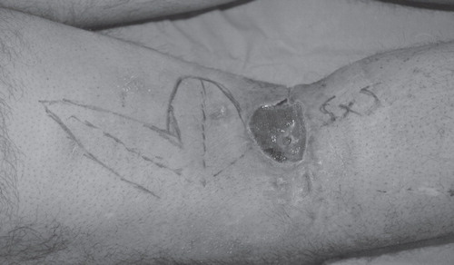
Figure 2. Immediate postoperative view.
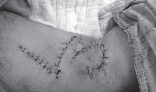
Figure 3. Three-month postoperative view.
Discussion
Popliteal skin defects are not suitable for primary closure. Secondary healing of wounds in this area usually results in severe contracture. Similarly, skin grafting may result in contracture, delayed healing, and aberrant scarring. Prolonged splinting and meticulous dressing changes should be performed to avoid contracture after skin grafting [Citation2]. Because of these disadvantages, flap surgery is a reasonable option for reconstruction in this region. Such flaps may be prepared as axial musculocutaneous, fasciocutaneous, or perforator-based local flaps or free flaps.
Esser in 1918 first described and used the bilobed flap to repair nasal tip cutaneous defects [Citation3]. It comprises of two transposition flaps with a single pedicle. The first flap is used for reconstructing the defect area and the second is used for closing the donor area of the first flap.
In 1953, Zimany [Citation4] made the first modification to this technique by creating a smaller second flap. The second major modification was performed by Zitelli in 1989 [Citation5,6]. Zitelli changed the rotational angle to 50°. A number of authors have recommended different interlobar angles for different anatomical regions [Citation5,6]. In the present case, we planned a 45° angle to prevent distortion and increase flap movement.
Repair using the bilobed flap has proven to be a versatile, reliable, and easy technique. Karacalar and Güner as well as Smith and Lee introduced the axial bilobed flap technique to increase the flap dimensions and viability [Citation7,8]. Bilobed flaps may be used to reconstruct major defects in different anatomical regions [Citation1,7–13]. Due to the high flexibility and freedom of movement of the lobes, the bilobed design has been demonstrated to be versatile and reliable not only for random-pattern flaps but also in microsurgery; thus, free bilobed flaps are commonly used [Citation14–16]. The sizes of the lobes may vary depending on the tissue elasticity of the relevant anatomical region. In general, the size of the first lobe is planned to be equal to the defect size, whereas the second is narrower, enabling primary closure. In the present case, however, the first lobe was planned to be wider than the original defect to permit further knee extension. The second lobe was planned to be smaller than the first.
In this case, the popliteal wound dimension was 5 × 5 cm, which can be classified as a moderate-sized defect. We harvested a random-pattern bilobed flap from the upper region of the defect as an alternative to an axial flap. Axial flaps have disadvantages, including donor area problems, longer operation times, and technical challenges. The use of a bilobed flap in this region enabled us to obtain a good outcome by using an inferior method with respect to the reconstructive ladder of plastic surgery.
Conclusion
We conclude that the random-pattern bilobed flap can be used as an easy, reliable, and versatile alternative to axial-pattern local or distant flaps to cover small defects in the popliteal region.
Declaration of interest: The authors report no conflicts of interest. The authors alone are responsible for the content and writing of the paper.
References
- Tissiani LAL, Alonso N, Carneiro MH, Bazzi K, Rocco M. Versatility of the bilobed flap. Rev Bras Cir Plást 2011;26:411–17.
- Karacalar A. Axial bilobed flap based on the median and medial superficial sural arteries: a case report. Scand J Plast Reconstr Surg Hand Surg 2001;35:207–10.
- Esser JFS. Gestielte lokale nasenplastik mit zweizipfligen lappen: de- ckung des sekunderen defectes vom ersten zipfel durch den zweiten. Dtsch Z Chir 1918;143:385–90.
- Zimany A. The bilobed flap. Plast Reconstr Surg 1953;11:424–34.
- Zitelli JA. The bilobed flap for nasal reconstruction. Arch Dermatol 1989;125:957–9.
- Cho M, Kim DW. Modification of the Zitelli bilobed flap: a comparison of flap dynamics in human cadavers. Arch Facial Plast Surg 2006;8:404–9; discussion: 410.
- Karacalar A, Güner H. The axial bilobed flap for burn contractures of the axilla. Burns 2000;26:628–33.
- Smith ML, Lee JC. Bilobed flap for axillary reconstruction. Plast Reconstr Surg 2009;124:179e–80e.
- Sampaio FM, Miller MD, Gualberto GV, Galhardo MC, do Valle AC, de Souza PR. Use of the bilobed flap in the pubic region after tumoral lesion excision. An Bras Dermatol 2013;88:224–6.
- Cordeiro CN, McCarthy CM, Mastorakos DP, Cordeiro PG. Repair of postauricular defects using cervical donor skin: a novel use of the bilobed flap. Ann Plast Surg 2007;59:451–2.
- Yenidunya MO. Axial pattern bilobed flap for the reconstruction of the midline forehead defects. Plast Reconstr Surg 1999;103:737.
- Yenidunya MO, Demirseren ME, Ceran C. Bilobed flap reconstruction in infraorbital skin defects. Plast Reconstr Surg 2007;119:145–50.
- El-Khatib HA. Bilobed fasciocutaneous flap for reconstruction of the posterior neck after necrotizing fasciitis. Plast Reconstr Surg 2004;114:885–9.
- Urken ML, Biller HF. A new bilobed design for the sensate radial forearm flap to preserve tongue mobility following significant glossectomy. Arch Otolaryngol Head Neck Surg 1994;120:26–31.
- Longo B, Ferri G, Fiorillo A, Rubino C, Santanelli F. Bilobed perforator free flaps for combined hemitongue and floor of the mouth defects. J Plast Reconstr Aesthet Surg 2013;66:1464–9.
- Longo B, Belli E, Pugliese P, Ferri G, Santanelli F. Bilobed skin paddle fibula flap for large oromandibular defects. J Craniofac Surg 2013;24:e327–30.