
Abstract
Bortezomib (BTZ) is the first proteasome inhibitor entered in clinical practice. Peripheral neuropathy is likely to be a class side effect of these drugs, although its severity is largely variable, and it deserves to be further investigated, since the mechanisms of BTZ-induced peripheral neurotoxicity (BiPN) are still unknown.
In our study, we investigated in vivo and in vitro possible pathogenic events relevant to BiPN using a well-established rat model, with particular reference to the extent of proteasome inhibition and the effects on α-tubulin polymerization in sciatic nerves and dorsal root ganglia specimens obtained from animals treated with chronic regimens at a dose of 0.2 mg/kg intravenously. The same assessments were also performed after a single injection. Moreover, these studies were replicated in vitro using embryonic DRG neurons exposed to 100 nM BTZ and adult DRG neurons exposed to 10–50 nM BTZ for 24 h and 48 h.
A significant increase in the polymerized fraction of α-tubulin and prolonged proteasome inhibition were observed after the chronic BTZ treatment in vivo. Recovery to physiological levels was observed after a 4-week follow-up post-treatment period. Proteasome inhibition and increased α-tubulin polymerization were also observed following BTZ treatment of both embryonic and adult DRG neurons in vitro.
Our in vivo results suggest that proteasome inhibition and alteration of tubulin dynamics contribute to BiPN. The in vitro systems here described reliably replicate the in vivo results, and might therefore be used for further mechanistic studies on the effects of proteasome inhibitors on neurons.
Background
The proteasome is an intracellular complex based on multicatalytic protease activities, including chymotryptic-like, tryptic-like, and post-glutamyl peptide hydrolyzing activities.Citation1 It is essential for the rapid elimination of abnormal proteins caused by mutation or post-translational damage. Interruption of proteasome activity can induce the activation of an apoptotic cascade and the accumulation of misfolded regulatory proteins.Citation1 In in vitro and in in vivo xenograft murine models, several proteasome inhibitors induce apoptosis of tumor cells, resulting in tumor regression.Citation2 Bortezomib (BTZ), a dipeptide boronic acid analog, is an agent selectively targeting the β5-subunit of 20S proteasome, largely used for the treatment of multiple myeloma. The most clearly demonstrated anticancer effect of BTZ, described in human multiple myeloma cells, is inactivation of the nuclear factor-kB (NFkB) pathway.Citation3 NFkB is a heterodimeric transcription factor localized in the cytoplasm bound to its inhibitory regulatory protein IkB in inactive state. Cell stimulation with pro-inflammatory signals and other forms of cellular stress induce the ubiquitination and degradation of IkB, leading to release and translocation of NFkB to the nucleus, where it induces the expression of specific proinflammatory and antiapoptotic genes. BTZ treatment prevents the degradation of IkB and the consequent activation of NFkB, resulting in cell cycle arrest and the induction of apoptotic cascade.Citation4 However, other antitumor mechanisms of cytotoxicity have been investigated in several cell lines and xenograft models, including activation of essential cellular pathways (e.g., AKT, MAPK, p53, and c-Jun), upregulation of proapoptotic proteins (e.g., Noxa), and dysregulation of the cell cycle increasing p21- and p27-mediated induction of cell cycle arrest. Moreover, downregulation of proteins involved in DNA repair pathways and induction of endoplasmic reticulum (ER) stress were described.Citation5 BTZ-induced peripheral neurotoxicity (BiPN) is a frequent side effect, usually manifesting as a painful, sensory axonal neuropathy. BiPN is generally dose-dependent,Citation6-Citation8 but it can occur even after a few cycles of treatment,Citation9 while, in contrast, some exposed patients are clinically unaffected.Citation10 Patients experiencing neuropathic pain generally require pharmacologic intervention (e.g., using anticonvulsant, tricyclic antidepressants and other analgesic drugs).Citation11 In the most severe cases of BiPN dose reduction or even treatment discontinuation is required in order to prevent irreversible damage, and specific dose modification guidelines have been released.Citation12 Recently implemented subcutaneous delivery of BTZ seems to be associated with a reduced severity of BiPN.Citation13
The pathogenesis of BiPN is still unclear, and it is possible that peripheral neurotoxicity is a class effect for proteasome inhibitors, since it has been reported also with other similar and effective drugs (although to a more limited extent).Citation14,Citation15 Among the proposed possibilities, interference with tubulin dynamics has been proposed both in cancer cells and in neurons.Citation16,Citation17 Pathogenic hypotheses can be reliably investigated in vivo using well-characterized animal models reproducing chronic BiPN as well as neuronal systems in vitro.Citation18-Citation21 In this context, we have studied for the first time, using parallel in vivo and in vitro experiments, the possible correlation existing between proteasome inhibition and α-tubulin polymerization in order to understand the pathogenic basis of BiPN onset.
Results
In vivo studies
Assessment of general toxicity
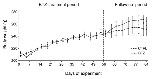
During all experimental studies, none of the animals treated with BTZ showed any evidence of distress. BTZ-treated animals showed a slight decrease in body weight after the second injection when compared with control animals () and recovered soon after, as already observed in previous experiments.Citation18 Overall, no statistically significant difference in body weight between treated and untreated animals was observed at any time of observation.
Figure 1. Body weight. Effect of BTZ (0.20 mg/kg) treatment on the body weight of rats during the chronic in vivo study. No significant difference of body weight as observed at any time point (mean ± SEM).
Electrophysiological studies on caudal nerve
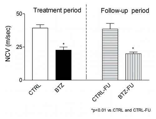
Evaluation of NCV was performed on the caudal nerve as a measure of neurophysiological function. No difference between groups was observed at baseline (data not shown). By contrast, at the end of the treatment, BTZ induced a significant reduction of the NCV. As expected, statistically significant impairment of the NCV was still observed at the end of the 4-wk follow-up period (, P < 0.01 vs. CTRL and CTRL-FU).Citation18
Figure 2. Neurophysiological studies. BTZ-induced decrease of the NCV was observed after 8 wk of chronic administration as well as at the end of the 4-wk follow up period (P < 0.01 vs. CTRL and CTRL-FU) (mean ± SEM).
Histopathology
Caudal and sciatic nerves
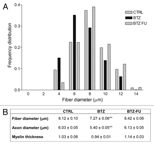
After 8 wk of BTZ-treatment, the caudal and sciatic nerve histopathological examinations showed different degrees of axonal degeneration. The most relevant pathological changes were evident in the caudal nerves with respect to control animals (). After the follow-up period, initial recovery in myelinated fibers density was evident also in this more severely affected nerve (), although this improvement was not yet relevant enough to allow recovery in NCV. The histogram of myelinated fibers distribution showed a marked decrease in the percentage of large fibers in BTZ-treated rats with respect to control animals (). Accordingly, the morphometric analysis of the sciatic nerves showed a significant decrease in both fiber and axonal size in treated animals compared with the untreated rats at the end of BTZ-treatment (, P < 0.001 vs. CTRL). After the follow-up period, the decrease of the axon and fiber diameter was no longer present in the sciatic nerve, and an increase in the percentage of largest myelinated fibers was also observed ().
Figure 3. Light microscopy analysis. BTZ-induced morphological alteration in sciatic and caudal nerves. Axonal degeneration with loss of fiber density was evident in myelinated fibers of peripheral nerves, particularly in the caudal nerve at the end of BTZ treatment. CTRL (A and B), 8 wk-BTZ-treated (C and D) and follow-up rats (E and F). The arrows show axonal degeneration of myelinated fibers (scale bar = 10 μm).
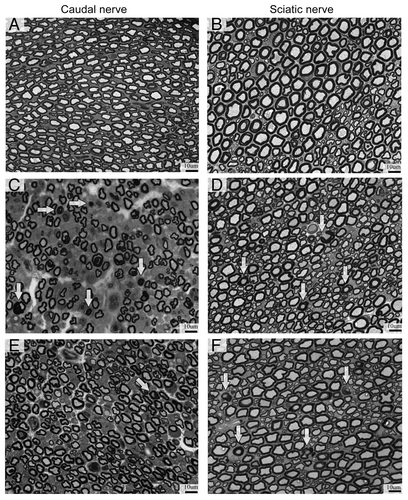
Figure 4. Effect of BTZ on frequency distribution (A) and morphometrical measures (B) of myelinated fibers in the sciatic nerve in control and BTZ-treated groups. CTRL, control; BTZ, animals treated for 8 wk; BTZ-FU, animals after 4 wk of washout period.*P < 0.001 vs. CTRL; °P < 0.001 vs. BTZ-FU.
DRG
As previously reported,Citation18 the observations at the light microscope of DRG collected from treated rats at the end of BTZ-treatment showed a cytoplasmic vacuolization in satellite cells, while only occasional alterations were observed in neurons (data not shown). However, the DRG formal evaluation performed by morphometric analysis showed a statistically significant decrease in somatic (P < 0.001), nuclear (P < 0.001) and nucleolar (P < 0.01) area in DRG neurons of BTZ-treated animals compared with the controls. After the follow-up period, the somatic and nuclear area of DRG neurons, but not nucleolar area, recovered in BTZ-treated animals ().
Table 1. Morphometrical analysis of DRG neurons
Pharmacodynamics profile of BTZ-induced proteasome inhibition
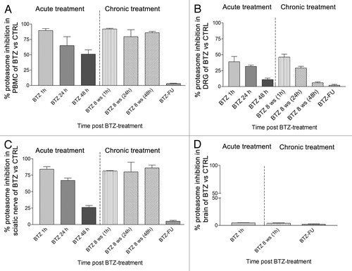
To determine the pharmacodynamics of BTZ administration, PBMC, brain, DRG, and sciatic nerve specimens were collected at different time points after BTZ treatment, and the residual chymotrypsin-like proteasome activity was determined in the acute schedule or after 8 wk of treatment and follow-up period in the chronic schedule (). The results showed that in the acute schedule, BTZ elicited a time-dependent decrease in 20S proteasome inhibition in PBMC, DRG, and sciatic nerve that tended to return to baseline values within 48 h. Different effects were observed in the chronic schedule, where BTZ administration determined a persistent inhibition of the proteasome activity until 48 h after the last BTZ administration in PBMC and sciatic nerve (). Overall the effect in DRG was less marked and persistent (). In agreement with the incapacity of BTZ to cross the blood–brain barrier, no proteasome inhibition was detected in the brain at 1 h (when we would expect peak effects), and so following time points were not investigated ()
Figure 5. Characterization of the time course of proteasome inhibition after single and repeated BTZ administrationsin different tissues. The graph shows the % of proteasome inhibition after single BTZ administration (acute treatment), 1 h after the 24th administration (8th week) (chronic treatment) and at the end of the 4-wk follow-up period (BTZ-FU). Proteasome inhibition rate (PI%) in sample tissue lysates from treated vs. control rats was calculated as % A = FBTZ-FSUBSTRATE/FCTRL-FSUBSTRATE, and the inhibition was obtained as 100*(1-A). (A) Peripheral blood mononuclear cell (PBMC), (B) DRG, (C) sciatic nerve, (D) brain tissues (mean ± SEM).
In vivo immunoblotting for α-tubulin
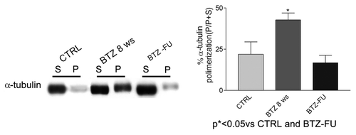
Western blot analysis was performed to investigate α-tubulin polymerization in the sciatic nerve. In the acute schedule of treatment, a single dose of BTZ was not able to induce any alteration in the polymerization 48 h and 72 h after the administration (data not shown). However, after the chronic treatment a significant increase of polymerized α-tubulin expression was observed after 8 wk of BTZ treatment (P < 0.05 vs. CTRL and BTZ-FU) (). After the follow up period the tubulin polymerization extent was similar in BTZ-treated and control rats.
Figure 6. In vivo tubulin polymerization in sciatic nerve after chronic BTZ treatment. The immunoblotting analysis performed on sciatic nerve showed a significant increase of polymerized tubulin after 8 wk of BTZ treatment compared with the controls and the follow-up rats (*P < 0.05 vs. CTRL and BTZ-FU) (P, polymerized tubulin fraction; S, soluble free tubulin fraction). Representative blots are shown on the left. On the right, western blot values of α-tubulin polymerization are expressed as mean ± SEM.
In vitro studies
BTZ-induced proteasome inhibition on DRG neuron cultures
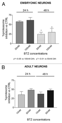
To analyze the effect of BTZ on DRG cultures, embryonic DRG neurons were treated with BTZ 10–100 nM, and adult DRG neurons were exposed to BTZ 10–50 nM for 24 h or 48 h.
In embryonic neurons 24 h after BTZ treatment, proteasome activity markedly decreased with respect to animal control (), while after 48 h of BTZ prolonged exposure, inhibition of the proteasome was significantly reduced with respect to the 24-h BTZ-treatment. The lowest dose of BTZ (10 nM) did not induce any appreciable proteasome inhibition (data not shown). Adult DRG neurons exposed to BTZ for 24 h show a remarkable proteasome inhibition, persistent up to 48 h of drug treatment ().
Figure 7. Effect of BTZ treatment on DRG neurons protesom activity. Embryonic (A) and adult (B) DRG neurons exposed to the indicated concentration of BTZ for 24 h and 48 h show a remarkable proteasome inhibition, persistent up to 48 h in adult neurons. Data are expressed as mean ± SEM.
In vitro immunoblotting for α-tubulin
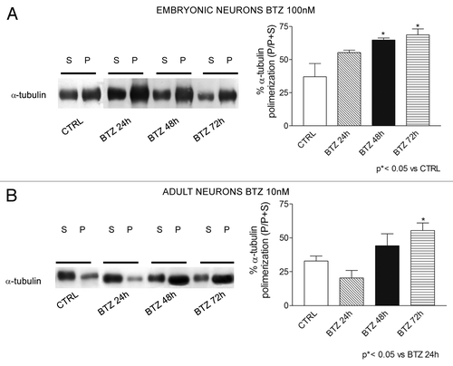
α-tubulin polymerization was evaluated by immunoblotting analysis also in embryonic and adult neuron cultures exposed to 100 nM or 10 nM BTZ, respectively. In embryonic neuron cultures, we observed an increase of polymerized α-tubulin expression beginning at 24 h of treatment with BTZ () that became significant after 48 h. In adult neurons, a less marked effect was present, although the trend was similar to that observed in embryonic neurons ().
Figure 8. Effect of BTZ treatment on tubulin polymerization in DRG neuron cultures. Immunoblotting revealed induction of polymerized tubulin in neurons obtained from E15 embryonic neurons (A) and neurons from adult rats (B) after BTZ treatment. Embryonic and adult cells were exposed to 100 nM or 10 nM BTZ, respectively, for 24 h, 48 h, or 72 h, and polymerization of α-tubulin was assessed using western blot analysis. (P = polymerized tubulin fraction and S = soluble free tubulin fraction). Representative blots are shown on the left. On the right, western blot data were quantified and expressed as % P/(P+S) in BTZ treated cultures compared with control ones (mean ± SEM).
Discussion
BiPN is a dose-limiting toxicity that impact on patients’ quality of life,Citation8,Citation22 although the use of subcutaneous schedules of administration has been reported to be less neurotoxic.Citation1,Citation13 Several preclinical models reproducing the features of BiPN have been reported, and they can be reliably used to investigate the molecular mechanisms underlying BiPN.Citation18,Citation20,Citation23 In this study we explored the effects of acute and protracted proteasome inhibition on the modulation of tubulin dynamics. Altered dynamics of α-tubulin polymerization has an important effect on microtubule function and may be an important pathogenic mechanism underlying peripheral neurotoxicity in addition to the proposed effects of proteasome inhibition on apoptosis.Citation24 However, despite preliminary evidence in different cellular systems, the possible correlation existing in vivo between the time course of proteasome inhibition, α-tubulin dynamics, and neurotoxicity has not yet been assessed.Citation17,Citation25 The rat model used in this study has already been thoroughly assessed, and it reproduces the hallmarks of BiPN in humans, i.e., painful sensory neuropathy with nerve axonopathy, a distal-to-proximal severity gradient, and tendency to recovery in the follow-up period after BTZ withdrawal.Citation18,Citation26 Most of these results, included the involvement of DRG, were independently confirmed in similar studies in mice.Citation19,Citation20
In this model we assessed for the first time the temporal profile of proteasome inhibition within PBMC and nervous tissue following BTZ administration. As expected, after the acute delivery, maximal proteasome inhibition (i.e., more than 80%) was present in PBMC, with a progressive reduction of the inhibition (however still present) during the subsequent 48 h.Citation27,Citation28 Remarkably, a similar extent of inhibition and time course was observed in the sciatic nerve. By contrast, brain proteasomal activity was unaffected, and an intermediate inhibition was observed in the DRG. The pharmacodynamics of proteasome inhibition after chronic BTZ treatment was completely different, with a sustained and prolonged effect in PBMC and sciatic nerve until the final time point of planned observation, while in the DRG this trend was also present but less marked. In a study performed in DRG harvested from Sprague–Dawley rats the authors demonstrated that after 24 h of BTZ treatment using a dose higher than the dose used in our study and not suitable for a long-term animal experiment (i.e., 0.50 mg/kg), the proteasome activity was reduced approximately to 50% after acute exposure.Citation29 In a subsequent set of experiments, where the animals were subcutaneously treated with BTZ for 4 wk, they evidenced an increase of expression of proteasome subunit genes as a compensatory response against “proteotoxic” stress in DRG.Citation30 These results might explain the course of recovery from proteasomal inhibition 48 h after treatment in chronically treated DRG observed in our in vivo study, while PBMC and sciatic nerve proteasomal activity is still inhibited. It is remarkable that the extent of proteasome inhibition achieved in our animal study reflects the human condition. In fact, in patients affected by relapsed myeloma and treated with different BTZ regimens levels of proteasome inhibition ranging between 70 and 84% were measured after 1 cycle of treatment, and proteasomal activity recovered within 48 h from BTZ administration.Citation31 The prolonged proteasome inhibition observed in our study after 8 wk of treatment is substantially in agreement with the results of a preliminary study in multiple myeloma patients, where it was demonstrated that BTZ pharmacokinetic changes with repeated dose administration following reduction in plasma clearance and associated increase in systemic exposure.Citation32 This cumulative effect has not been completely explained so far, but it might be due to misfolded protein accumulation leading to “proteotoxic” event.Citation32 This hypothesis was supported by previous studies in human cell lines, in which it was indicated that proteasome inhibitors caused a proteotoxic stress with accumulation of oxidant-damaged proteins.Citation33
Based on the reported evidence of changes in tubulin dynamics obtained in different cellular systems and on the relevance of tubulin interference in the pathogenesis of toxic peripheral neuropathies,Citation21,Citation34,Citation35 we focused on α-tubulin polymerization as a possible mechanism of neurotoxicity in BiPN. Despite the fact that the severity of nerve damage was more marked in the caudal nerve than the sciatic nerve, we selected the latter tissue to assay proteasome inhibition. Our rationale was that in this tissue the course of recovery after drug withdrawal was more evident, allowing a more proper comparison with possible changes in α-tubulin dynamics occurring in the follow-up period of observation. Immunoblotting analysis performed on sciatic nerve specimens after 8 wk of BTZ administration showed a marked increase in the amount of polymerized α-tubulin with respect to untreated controls, which recovered within the follow-up period. Therefore, in our in vivo study BTZ significantly enhances α-tubulin polymerization at a dose that can induce a clinically relevant proteasome inhibition and peripheral neuropathy. We propose that enhanced α-tubulin polymerization is a specific neurotoxicity mechanism of BTZ administration, since this effect has not been observed with other neurotoxic agents such as cisplatin both in vitroCitation36 and in peripheral nerves obtained from chronically treated rats with a demonstrated peripheral neurotoxicity (Meregalli C, personal observation).
Since animal models, despite their unsurpassed relevance to clinical conditions, are not easily available for detailed mechanistic studies due to the presence of several confounding factors and possible uncontrollable bias, our additional aim was to set up a reliable cellular model able to reproduce the events already demonstrated in vivo. For the in vitro studies, we used concentrations comparable to peak levels observed in multiple myeloma patients.Citation37,Citation38 We aimed at achieving proteasomal inhibition in a range close to that observed in our rat model. Based on the available results regarding BTZ neurotoxicity and cellular models of chemotherapy-induced peripheral neurotoxicity,Citation18,Citation19,Citation39 we selected DRG neurons for the in vitro studies and to rule out possible effects due to incomplete maturation of the proteasomal activity; we tested in parallel the most commonly used E15 and adult neuronal cultures.
During the screening studies designed to select the optimal BTZ dose range it appeared that adult neurons are more vulnerable than E15 neurons to cytotoxic BTZ activity, and they did not survive at the highest doses tested in E15 neurons. After BTZ concentration adjustment, the use of non-lethal doses of BTZ was able to induce a clear proteasome activity inhibition in both cellular systems, although the time course and extent of inhibition were not identical, with an earliest and more severe effect in embryonic vs. adult neurons. Accordingly, sustained and prolonged α-tubulin polymeration could also be demonstrated in the selected cellular systems, and also in this case the time course and severity of the effect was different in E15 and adult neurons. Overall, the sequence of events observed in our cellular systems is consistent with that observed in neuroblastoma and neuronal origin cell lines, where progressive increase of α-tubulin polymerization following BTZ administration occurs.Citation25 Moreover, our results obtained in E15 neuronal cultures are in substantial agreement with those reported in a similar model after only 24 h of BTZ exposure,Citation17 while no comparison is still available for longer observations or for adult neurons. Since at the same BTZ concentration used in our experiments functional impairment of axonal transport has been demonstrated in E15 neuronal cultures,Citation17 our results support the hypothesis that the toxicity of proteasome inhibition might result, at least in part, from the accumulation of microtubule-associated proteins, as suggested by Poruchynsky and colleagues.Citation25
In conclusion, our experimental data provide evidence for a remarkable effect of BTZ on α-tubulin dynamics in a well-characterized in vivo model of BiPN and further suggest that this mechanism might be a major determinant in its pathogenesis. The in vitro cellular systems established in this study may be reliably used to further investigate this effect and to deepen the current knowledge on BiPN pathogenesis, as well as to compare the effects of other proteasome inhibitors.
Materials and Methods
Experimental design
To evaluate the molecular mechanisms of BiPN we used in vivo and in vitro models previously characterized in our laboratory.Citation18,Citation40 Using these models we studied BTZ-induced α-tubulin polymerization and proteasome inhibition in rat peripheral nervous tissues and in in vitro-cultured dissociated dorsal root ganglia (DRG) sensory neurons obtained from embryonic and adult rats.
Drug
BTZ was from LC Laboratories. For the in vivo study, BTZ was reconstituted in a vehicle solution composed by absolute ethanol/tween80/saline (5%/5%/90%) and administered intravenously via the caudal vein as published.Citation18 This solvent has already been tested for the absence of general toxicity and of any effect of the peripheral nervous system.Citation41 For in vitro experiments, the drug was dissolved in DMSO and diluted in culture medium before use.
Experimental models
In vivo studies
Animals
All experimental procedures were approved by the Ethics Committee for Animal Studies of the University of Milan Bicocca (protocol approved, number: 0015349/12). In this study, female Wistar rats (175–200 g, Harlan) were used, and we provided them with food and water ad libitum. Rats were housed in a limited-access animal facility, and they were maintained under an artificial 12-h light/dark cycle, in agreement with national (Italian Legislative Decree 116/92, Gazzetta Ufficiale della Repubblica Italiana, no 40, February 18,1992 and subsequent modifications) and international laws (EEC Council Directive 86/609; Italian, 1992; Guide for the Use of Laboratory Animals, US National Research Council, 8th ed. 2011). All procedures were performed in accordance with the ethical guidelines of the International Association for the Study of Pain.Citation42 Rats were deeply anesthetized for electrophysiology procedures, with 3% isoflurane carried in medical air followed by 1–1.5% isoflurane for maintenance during the procedures. At sacrifice, the animals were euthanized under deep xylazine/ketamine anesthesia and used for biological sampling.
The experimental in vivo plan was performed using an acute schedule of treatment, in which BTZ was administered as a single dose (0.20 mg/kg), and a chronic schedule, which consisted of repeated administrations of BTZ (0.20 mg/kg 3 times a week, for 8 wk) as published.Citation18 After the end of BTZ-treatment, one group of animals was left untreated for 4 wk (follow-up period).
Assessment of general toxicity
All rats were examined daily for clinical distress signs. Body weight changes were measured twice weekly for the assessment of the general health and for drug dose adjustment in BTZ-treated rats.
Electrophysiological studies on caudal nerve
At baseline, at the end of the chronic treatment, and after the follow-up period, caudal nerve conduction velocity (NCV) was determined employing an electromyography apparatus (Myto2 ABN Neuro) as previously described.Citation18 Briefly, caudal NCV was measured by placing 2 recording needle electrodes distally on the tail and 2 stimulating needle electrodes 5 and 10 cm proximal to the recording points. The caudal NCV was calculated as a ratio of the distance between stimulating and recording electrodes, and the latency obtained from the measure between the 2 peaks of recorded potentials. The intensity, duration and frequency of stimulation were set up in order to obtain optimal results.
Histopathological analysis on DRG, sciatic and caudal nerves
In both treatment schedules, after the sacrifice DRG, sciatic, and caudal nerves were harvested and processed accordingly to previously reported protocols.Citation18,Citation19,Citation43 Briefly, tissues were removed and fixed for 3 h at room temperature in 3% glutaraldehyde, post-fixed in OsO4, and resin embedded. Morphological analysis was performed on 1-µm semi-thin sections stained with toluidine blue. At least 2 tissue blocks for each animal were sectioned and then examined with a Nikon Eclipse E200 light microscope (Leica Microsystems GmbH). In the morphometric evaluation of DRG, performed according to the method previously described,Citation44 the somatic, nuclear, and nucleolar sizes of DRG sensory neurons were measured in randomly selected sections on at least 200 DRG neurons/animal. Entire fiber, axonal, and myelin area of sciatic nerve myelinated fibers were also calculated using a QWin automatic image analyzer (Leica Microsystems GmbH).Citation41 Histograms of fiber population distribution, separated into class intervals increasing by 1.0 μm, were obtained.
In vitro studies
DRG dissociated neurons from embryonic rats
DRG from 15-d-old (E15) embryonic Wistar rats (Harlan) were obtained as previously reported.Citation40,Citation45 Briefly, DRG neurons were aseptically removed, pelleted, and dissociated with trypsin. Neuronal cells were cultured in AN2 medium prepared with MEM (Life Technologies) plus 15% calf bovine serum (Hyclone), 50 µg/ml ascorbic acid (Sigma), 1.4 mM L-glutamine (Life Technologies), 0.6% glucose (Sigma) supplemented with 5 ng/ml NGF (Life Technologies), and plated onto collagen-coated 35-mm dishes. After 24 h, neurons were treated for 5 d with AN2 medium, to which was added 5 ng/ml NGF and 10−5 M 2′-Deoxy-5-fluorouridine (Fudr) (Sigma) to remove satellite cells, which were less than 5% at the end of treatment. Neurons were then kept in AN2 medium with 5 ng/ml NGF for 24 h, then neurons were exposed to BTZ. DRG neurons cultured in AN2 medium with 5 ng/ml NGF were used as controls.
DRG dissociated neurons from adult rats
DRG obtained from adult Wistar rats (8-wk-old) were aseptically removed and collected in a dish containing cold F12 medium (Euroclone). Working under a dissecting microscope and using fine forceps, the surrounding membrane was gently teased away from each DRG; nerves and sheath were cut. All desheathed DRG were then transferred into a sterile dish containing collagenase 0.125% (Sigma) and DNase (Sigma) in F12 medium and incubated at 37 °C for 2 h. After incubation, DRG were triturated using a tip p1000. Myelin and nerve debris were eliminated by centrifugation trough a bovine serum albumin (BSA) cushion. Cell pellet were re-suspended in Bottenstein and Sato medium (BS) prepared with F12 supplemented with N2 supplement (Life Technologies), 3% BSA, penicillin/streptomycin and plated in 24 multi-well plate pre-coated with laminin (Sigma). The day after cells were treated for 3 d with BS medium containing 100 μM Fudr to eliminate satellite cells. Neurons were kept in BS medium for 24 h, then neurons were exposed to BTZ. DRG neurons treated with BS medium were used as controls.
In vivo and in vitro proteasome activity measurement
To evaluate the proteasome inhibition profile in in vivo models, several tissues from acute and chronic schedule were collected at specific time points. Blood sample, sciatic nerve, DRG and brain specimens were harvested 1–24–48 h after single and chronic BTZ treatment and after the 4 wk of follow up. Peripheral blood mononuclear cells (PBMC) were isolated using a Ficoll–Hypaque density separation. Later, the PBMCs were added with lysis solution (50 mM Hepes, 5 mM EDTA, 150 mM NaCl, Triton-X100 1% in H2O) and extracted. The lysates obtained were then spinned at 13 500 rpm for 15 min at 4 °C. Protein extracts from tissue samples were solubilized in the lysis buffer (10% glycerol, 25 mM TRIS-HCl pH 7.5, 1% triton X-100, 5 mM EDTA pH 8, 1 mM EGTA pH 8) without added protease and phosphatase inhibitors, and centrifuged at 14 000 rpm for 10 min at 4 °C. Protein concentration was determined by the Bradford assay using a Coomassie® Protein Assay Reagent Kit (Pierce). In in vitro models, sensory neuron cultures from both adult and embryonic DRG were processed, similar to the method of protein extraction used during tissue extract, without adding protease and phosphatase inhibitors to lysis buffer. For proteasome inhibition assay, BTZ concentrations were differently defined based on neurotoxicity preliminary data (data not reported). Due to different sensibility of E15 and adult neurons evidenced by these preliminary studies, the concentrations were reduced accordingly in adult DRG cultures. Protein extracts obtained from embryonic and adult neurons were performed after 24 h and 48 h of treatment, with 10–100 nM BTZ and with 10–50 nM BTZ, respectively. Finally, to evaluate the proteasomal activity a fluorometric assay was used, and the protein extract from tissue was incubated with the N-succinyl-Leu-Leu-Val-Tyr-7-Amido-4-Methylcoumarin substrate (Sigma) for 2 h, as published.Citation46 The proteasome activity was detected as the relative light unit generated from the cleaved substrate in the reagent. Fluorescence generated from each reaction was detected with a fluorometer (Wallac 1420 multilabel counter, PerkinElmer). The proteasome activity (A) was calculated as following: % A = FBTZ-FSUBSTRATE/FCTRL-FSUBSTRATE and the inhibition was obtained as 100*(1-A).
In vivo and in vitro assessment of tubulin polymerization
To evaluate α-tubulin polymerization in vivo, the sciatic nerves were collected 48 and 72 h after a single dose of BTZ (acute schedule) or after the administration at the end of treatment and of the 4-wk follow-up period (chronic schedule). In in vitro models, sensory neuron cultures from both adult and embryonic DRG were processed. Preliminary neurotoxicity experiments gave the optimal BTZ concentrations to obtain suitable cultures. To study the effect of BTZ on α-tubulin polymerization, one dose of the drug (defined on best condition of growth of the cultures) was used. In particular, adult cultures were treated with 10 nM BTZ, while embryonic neurons were treated with 100 nM BTZ. DRG cultures extracts were obtained at the same time points after single BTZ-treatment (i.e., 24, 48, and 72 h). Protein extracts from both nervous tissue samples and neuron cell cultures were obtained as previously described for proteasome assay, with the exception that the lysis buffer contained freshly added protease and phosphatase inhibitors (10 mM sodium orthovanadate, 4 mM phenylmethylsulfonyl fluoride, 1% aprotinin and 20 mM sodium pyrophosphate). The protein extracts were centrifuged at 14 000 rpm for 10 min at 4 °C to separate the soluble free tubulin fraction (S) from the polymerized ones (P). The supernatants, containing free tubulin, were collected, and the pellets of polymerized tubulin were resuspended by sonication for 20 s in a volume of lysis buffer, supplemented with 0.5% sodium deoxycholate, equal to the S fraction. Protein aliquots (10 μg/ml) were loaded onto 13% SDS-PAGE, and, after electrophoresis, they were transferred to nitrocellulose filters, and immunoblotting analysis was performed using rabbit anti-α-tubulin antibody from Cell Signaling Technologies. After incubation with primary antibody, membrane was washed and then incubated with appropriate horseradish peroxidase conjugated to goat anti-rabbit IgG, PerkinElmer, and ECL chemiluminescence system (Amersham) was used. Bands intensity were quantified using Gel Logic 100 Image System (Eastman Kodak Co). Final mean values were obtained from triplicate experiments. Data were expressed as a percentage of (P/[P+S]) in BTZ-treated rats compared with untreated controls.
Statistical analysis
The differences between all experimental groups in the in vivo studies were analyzed by Student t test for the body weight and NCV analysis, while analysis of variance (1-way ANOVA, Turkey post hoc-test) was used for DRG and sciatic nerve morphometry data assessments using the GraphPad 3.0 software (GraphPad Software; significance level set at P < 0.05). Tubulin polymerization in the in vivo and in vitro studies were evaluated by Student t test or ANOVA as opportune.
| Abbreviations: | ||
| BTZ | = | bortezomib |
| BTZ-FU | = | bortezomib follow up |
| BiPN | = | bortezomib-induced peripheral neuropathy |
| DRG | = | dorsal root ganglion |
| DMSO | = | dymethil sulfoxide |
| NCV | = | nerve conduction velocity |
| BS | = | medium Bottenstein and Sato medium |
| PBMC | = | peripheral blood mononuclear cell |
| P | = | polymerized tubulin fraction |
| S | = | soluble free tubulin fraction |
| E15 | = | neurons embryonic neurons |
Disclosure of Potential Conflicts of Interest
No potential conflicts of interest were disclosed.
Aknowledgements
The authors wish to thank Dr Roberta Rigolio and Dr Virginia Rodriguez-Menendez for their assistance with the interpretation of data and histological evaluation.
This work was supported in part by a research grant from Fondazione Banca del Monte di Lombardia to PM.
References
- Moreau P, Karamanesht II, Domnikova N, Kyselyova MY, Vilchevska KV, Doronin VA, Schmidt A, Hulin C, Leleu X, Esseltine DL, et al. Pharmacokinetic, pharmacodynamic and covariate analysis of subcutaneous versus intravenous administration of bortezomib in patients with relapsed multiple myeloma. Clin Pharmacokinet 2012; 51:823 - 9; http://dx.doi.org/10.1007/s40262-012-0010-0; PMID: 23018466
- Bortezomib EH. Recent Results Cancer Res 2010; 184:173 - 87; PMID: 20072838
- Driscoll JJ, Woodle ES. Targeting the ubiquitin+proteasome system in solid tumors. Semin Hematol 2012; 49:277 - 83; http://dx.doi.org/10.1053/j.seminhematol.2012.04.002; PMID: 22726552
- Hayden MS, Ghosh S. Shared principles in NFkappaB signaling. Cell 2008; 132:344 - 62; http://dx.doi.org/10.1016/j.cell.2008.01.020; PMID: 18267068
- Schwartz R, Davidson T. Pharmacology, pharmacokinetics, and practical applications of bortezomib. Oncology (Williston Park) 2004; 18:Suppl 11 14 - 21; PMID: 15688598
- Cavaletti G, Jakubowiak AJ. Peripheral neuropathy during bortezomib treatment of multiple myeloma: a review of recent studies. Leuk Lymphoma 2010; 51:1178 - 87; http://dx.doi.org/10.3109/10428194.2010.483303; PMID: 20497001
- Berkowitz A, Walker S. Bortezomib-induced peripheral neuropathy in patients with multiple myeloma. Clin J Oncol Nurs 2012; 16:86 - 9; http://dx.doi.org/10.1188/12.CJON.86-89; PMID: 22297012
- Argyriou AA, Bruna J, Marmiroli P, Cavaletti G. Chemotherapy-induced peripheral neurotoxicity (CIPN): an update. Crit Rev Oncol Hematol 2012; 82:51 - 77; http://dx.doi.org/10.1016/j.critrevonc.2011.04.012; PMID: 21908200
- Lanzani F, Mattavelli L, Frigeni B, Rossini F, Cammarota S, Petrò D, Jann S, Cavaletti G. Role of a pre-existing neuropathy on the course of bortezomib-induced peripheral neurotoxicity. J Peripher Nerv Syst 2008; 13:267 - 74; http://dx.doi.org/10.1111/j.1529-8027.2008.00192.x; PMID: 19192066
- Dimopoulos MA, San-Miguel JF, Anderson KC. Emerging therapies for the treatment of relapsed or refractory multiple myeloma. Eur J Haematol 2011; 86:1 - 15; http://dx.doi.org/10.1111/j.1600-0609.2010.01542.x; PMID: 20942854
- Brix Finnerup N, Hein Sindrup S, Staehelin Jensen T. Management of painful neuropathies. Handb Clin Neurol 2013; 115:279 - 90; http://dx.doi.org/10.1016/B978-0-444-52902-2.00017-5; PMID: 23931787
- Richardson PG, Xie W, Mitsiades C, Chanan-Khan AA, Lonial S, Hassoun H, Avigan DE, Oaklander AL, Kuter DJ, Wen PY, et al. Single-agent bortezomib in previously untreated multiple myeloma: efficacy, characterization of peripheral neuropathy, and molecular correlations with response and neuropathy. J Clin Oncol 2009; 27:3518 - 25; http://dx.doi.org/10.1200/JCO.2008.18.3087; PMID: 19528374
- Mateos MV, San Miguel JF. Safety and efficacy of subcutaneous formulation of bortezomib versus the conventional intravenous formulation in multiple myeloma. Ther Adv Hematol 2012; 3:117 - 24; http://dx.doi.org/10.1177/2040620711432020; PMID: 23556118
- Thompson JL. Carfilzomib: a second-generation proteasome inhibitor for the treatment of relapsed and refractory multiple myeloma. Ann Pharmacother 2013; 47:56 - 62; http://dx.doi.org/10.1345/aph.1R561; PMID: 23300152
- Pautasso C, Bringhen S, Cerrato C, Magarotto V, Palumbo A. The mechanism of action, pharmacokinetics, and clinical efficacy of carfilzomib for the treatment of multiple myeloma. Expert Opin Drug Metab Toxicol 2013; 9:1371 - 9; http://dx.doi.org/10.1517/17425255.2013.817556; PMID: 23834482
- Bazzaro M, Lin Z, Santillan A, Lee MK, Wang MC, Chan KC, Bristow RE, Mazitschek R, Bradner J, Roden RB. Ubiquitin proteasome system stress underlies synergistic killing of ovarian cancer cells by bortezomib and a novel HDAC6 inhibitor. Clin Cancer Res 2008; 14:7340 - 7; http://dx.doi.org/10.1158/1078-0432.CCR-08-0642; PMID: 19010849
- Staff NP, Podratz JL, Grassner L, Bader M, Paz J, Knight AM, Loprinzi CL, Trushina E, Windebank AJ. Bortezomib alters microtubule polymerization and axonal transport in rat dorsal root ganglion neurons. Neurotoxicology 2013; 39:124 - 31; http://dx.doi.org/10.1016/j.neuro.2013.09.001; PMID: 24035926
- Meregalli C, Canta A, Carozzi VA, Chiorazzi A, Oggioni N, Gilardini A, Ceresa C, Avezza F, Crippa L, Marmiroli P, et al. Bortezomib-induced painful neuropathy in rats: a behavioral, neurophysiological and pathological study in rats. Eur J Pain 2010; 14:343 - 50; http://dx.doi.org/10.1016/j.ejpain.2009.07.001; PMID: 19695912
- Carozzi VA, Canta A, Oggioni N, Sala B, Chiorazzi A, Meregalli C, Bossi M, Marmiroli P, Cavaletti G. Neurophysiological and neuropathological characterization of new murine models of chemotherapy-induced chronic peripheral neuropathies. Exp Neurol 2010; 226:301 - 9; http://dx.doi.org/10.1016/j.expneurol.2010.09.004; PMID: 20832406
- Bruna J, Udina E, Alé A, Vilches JJ, Vynckier A, Monbaliu J, Silverman L, Navarro X. Neurophysiological, histological and immunohistochemical characterization of bortezomib-induced neuropathy in mice. Exp Neurol 2010; 223:599 - 608; http://dx.doi.org/10.1016/j.expneurol.2010.02.006; PMID: 20188093
- Huff LM, Sackett DL, Poruchynsky MS, Fojo T. Microtubule-disrupting chemotherapeutics result in enhanced proteasome-mediated degradation and disappearance of tubulin in neural cells. Cancer Res 2010; 70:5870 - 9; http://dx.doi.org/10.1158/0008-5472.CAN-09-4281; PMID: 20587529
- Cavaletti G, Frigeni B, Lanzani F, Mattavelli L, Susani E, Alberti P, Cortinovis D, Bidoli P. Chemotherapy-Induced Peripheral Neurotoxicity assessment: a critical revision of the currently available tools. Eur J Cancer 2010; 46:479 - 94; http://dx.doi.org/10.1016/j.ejca.2009.12.008; PMID: 20045310
- Shin YK, Jang SY, Lee HK, Jung J, Suh DJ, Seo SY, Park HT. Pathological adaptive responses of Schwann cells to endoplasmic reticulum stress in bortezomib-induced peripheral neuropathy. Glia 2010; 58:1961 - 76; http://dx.doi.org/10.1002/glia.21065; PMID: 20830808
- Windebank AJ, Grisold W. Chemotherapy-induced neuropathy. J Peripher Nerv Syst 2008; 13:27 - 46; http://dx.doi.org/10.1111/j.1529-8027.2008.00156.x; PMID: 18346229
- Poruchynsky MS, Sackett DL, Robey RW, Ward Y, Annunziata C, Fojo T. Proteasome inhibitors increase tubulin polymerization and stabilization in tissue culture cells: a possible mechanism contributing to peripheral neuropathy and cellular toxicity following proteasome inhibition. Cell Cycle 2008; 7:940 - 9; http://dx.doi.org/10.4161/cc.7.7.5625; PMID: 18414063
- Cavaletti G, Gilardini A, Canta A, Rigamonti L, Rodriguez-Menendez V, Ceresa C, Marmiroli P, Bossi M, Oggioni N, D’Incalci M, et al. Bortezomib-induced peripheral neurotoxicity: a neurophysiological and pathological study in the rat. Exp Neurol 2007; 204:317 - 25; http://dx.doi.org/10.1016/j.expneurol.2006.11.010; PMID: 17214983
- Chauhan D, Catley L, Li G, Podar K, Hideshima T, Velankar M, Mitsiades C, Mitsiades N, Yasui H, Letai A, et al. A novel orally active proteasome inhibitor induces apoptosis in multiple myeloma cells with mechanisms distinct from Bortezomib. Cancer Cell 2005; 8:407 - 19; http://dx.doi.org/10.1016/j.ccr.2005.10.013; PMID: 16286248
- Adams J, Kauffman M. Development of the proteasome inhibitor Velcade (Bortezomib). Cancer Invest 2004; 22:304 - 11; http://dx.doi.org/10.1081/CNV-120030218; PMID: 15199612
- Casafont I, Berciano MT, Lafarga M. Bortezomib induces the formation of nuclear poly(A) RNA granules enriched in Sam68 and PABPN1 in sensory ganglia neurons. Neurotox Res 2010; 17:167 - 78; http://dx.doi.org/10.1007/s12640-009-9086-1; PMID: 19609631
- Palanca A, Casafont I, Berciano MT, Lafarga M. Proteasome inhibition induces DNA damage and reorganizes nuclear architecture and protein synthesis machinery in sensory ganglion neurons. Cell Mol Life Sci 2013; http://dx.doi.org/10.1007/s00018-013-1474-2; PMID: 24061536
- Reece DE, Sullivan D, Lonial S, Mohrbacher AF, Chatta G, Shustik C, Burris H 3rd, Venkatakrishnan K, Neuwirth R, Riordan WJ, et al. Pharmacokinetic and pharmacodynamic study of two doses of bortezomib in patients with relapsed multiple myeloma. Cancer Chemother Pharmacol 2011; 67:57 - 67; http://dx.doi.org/10.1007/s00280-010-1283-3; PMID: 20306195
- McConkey DJ, White M, Yan W. HDAC inhibitor modulation of proteotoxicity as a therapeutic approach in cancer. Adv Cancer Res 2012; 116:131 - 63; PMID: 23088870
- Steffen J, Seeger M, Koch A, Krüger E. Proteasomal degradation is transcriptionally controlled by TCF11 via an ERAD-dependent feedback loop. Mol Cell 2010; 40:147 - 58; http://dx.doi.org/10.1016/j.molcel.2010.09.012; PMID: 20932482
- Contini A, Cappelletti G, Cartelli D, Fontana G, Gelmi ML. Molecular dynamics and tubulin polymerization kinetics study on 1,14-heterofused taxanes: evidence of stabilization of the tubulin head-to-tail dimer-dimer interaction. Mol Biosyst 2012; 8:3254 - 61; http://dx.doi.org/10.1016/j.molcel.2010.09.012; PMID: 20932482
- LaPointe NE, Morfini G, Brady ST, Feinstein SC, Wilson L, Jordan MA. Effects of eribulin, vincristine, paclitaxel and ixabepilone on fast axonal transport and kinesin-1 driven microtubule gliding: implications for chemotherapy-induced peripheral neuropathy. Neurotoxicology 2013; 37:231 - 9; http://dx.doi.org/10.1016/j.neuro.2013.05.008; PMID: 23711742
- Tulub AA, Stefanov VE. Cisplatin stops tubulin assembly into microtubules. A new insight into the mechanism of antitumor activity of platinum complexes. Int J Biol Macromol 2001; 28:191 - 8; http://dx.doi.org/10.1016/S0141-8130(00)00159-8; PMID: 11251225
- Ceresa C, Giovannetti E, Voortman J, Laan AC, Honeywell R, Giaccone G, Peters GJ. Bortezomib induces schedule-dependent modulation of gemcitabine pharmacokinetics and pharmacodynamics in non-small cell lung cancer and blood mononuclear cells. Mol Cancer Ther 2009; 8:1026 - 36; http://dx.doi.org/10.1158/1535-7163.MCT-08-0700; PMID: 19383850
- Papandreou CN, Daliani DD, Nix D, Yang H, Madden T, Wang X, Pien CS, Millikan RE, Tu SM, Pagliaro L, et al. Phase I trial of the proteasome inhibitor bortezomib in patients with advanced solid tumors with observations in androgen-independent prostate cancer. J Clin Oncol 2004; 22:2108 - 21; http://dx.doi.org/10.1200/JCO.2004.02.106; PMID: 15169797
- Maggioni D, Nicolini G, Chiorazzi A, Meregalli C, Cavaletti G, Tredici G. Different effects of erythropoietin in cisplatin- and docetaxel-induced neurotoxicity: an in vitro study. J Neurosci Res 2010; 88:3171 - 9; http://dx.doi.org/10.1002/jnr.22465; PMID: 20722073
- Scuteri A, Donzelli E, Ravasi M, Tredici G. Adult mesenchymal stem cells support cisplatin-treated dorsal root ganglion survival. Neurosci Lett 2008; 445:68 - 72; http://dx.doi.org/10.1016/j.neulet.2008.08.056; PMID: 18771708
- Persohn E, Canta A, Schoepfer S, Traebert M, Mueller L, Gilardini A, Galbiati S, Nicolini G, Scuteri A, Lanzani F, et al. Morphological and morphometric analysis of paclitaxel and docetaxel-induced peripheral neuropathy in rats. Eur J Cancer 2005; 41:1460 - 6; http://dx.doi.org/10.1016/j.ejca.2005.04.006; PMID: 15913989
- Zimmermann M. Ethical considerations in relation to pain in animal experimentation. Acta Physiol Scand Suppl 1986; 554:221 - 33; PMID: 3469880
- Marmiroli P, Rodriguez-Menendez V, Rigamonti L, Tonoli E, Rigolio R, Cavaletti G, Tredici G, Vercelli A. Neuropathological changes in the peripheral nervous system and spinal cord in a transgenic mouse model of Niemann-Pick disease type A. Clin Neuropathol 2009; 28:263 - 74; PMID: 19642505
- Cavaletti G, Tredici G, Marmiroli P, Petruccioli MG, Barajon I, Fabbrica D. Morphometric study of the sensory neuron and peripheral nerve changes induced by chronic cisplatin (DDP) administration in rats. Acta Neuropathol 1992; 84:364 - 71; http://dx.doi.org/10.1007/BF00227662; PMID: 1441917
- Scuteri A, Nicolini G, Miloso M, Bossi M, Cavaletti G, Windebank AJ, Tredici G. Paclitaxel toxicity in post-mitotic dorsal root ganglion (DRG) cells. Anticancer Res 2006; 26:2A 1065 - 70; PMID: 16619507
- Berkers CR, Verdoes M, Lichtman E, Fiebiger E, Kessler BM, Anderson KC, Ploegh HL, Ovaa H, Galardy PJ. Activity probe for in vivo profiling of the specificity of proteasome inhibitor bortezomib. Nat Methods 2005; 2:357 - 62; http://dx.doi.org/10.1038/nmeth759; PMID: 15846363