
Abstract
Background
Urolithiasis is one of the most common urinary tract diseases worldwide, with a wide range of affected age groups. Non-contrast CT examination of the urinary tract is the gold-standard examination for detection and characterization of urinary tract stones, with great impact upon the choice of method of management. Aside from detection of stones, non-contrast CT examination of the abdomen and pelvis also offers a valuable overlook upon the other abdominal organs, pathologies of which may simulate a stone disease, or accompany stone disease and can be detected incidentally, which may shift management plan dramatically.
Aim of work
To demonstrate the use of non-contrast CT examinations (stone protocol) in the detection of abdominal pathologies other than stones, whether or not simulating the clinical picture of urolithiasis, and its impact upon patient management.
Patients and methods
Assessment of the non-contrast examinations of the urinary tract of patients referred for suspected stone urolithiasis recording any incidental finding and follow-up of the impact of these incidental findings upon the management delivered to the patient.
Results
A total of 719 examinations were performed, of which 334 had urinary tract stones only, 211 had incidental finding beside urinary tract stones, 170 had an incidental finding with no urinary tract stones, and four patients had neither stones nor incidental findings. A total number of 381 patients had incidental findings, 198 (47%) of which had an impact upon the management.
Conclusion
Non-contrast CT examination of the urinary tract (stone protocol) is a valuable tool in the detection of incidental findings which may simulate, or coincide with urolithiasis and it has a significant impact upon the management of the patients.
1 Introduction
Urolithiasis is a common urinary tract pathology that affects a wide range of age group.Citation1 Multiple treatment options are available for management of urinary tract stones, including medical treatment, Shock Wave lithotripsy, percutaneous nephrolithotomy, as well as open surgery.Citation2 The major determinants of treatment options are the stone number, site, size, attenuation, as well as the presence or absence of obstruction.Citation2 Multiple radiological techniques can be used to detect and characterize urinary tract stones, including plain X-ray, intravenous urography, ultrasonography, and computed tomography.Citation3
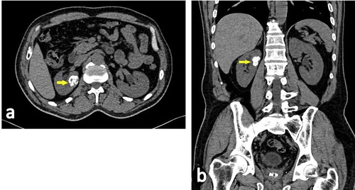
Figure 8 Sixty-one year old female patient with right flank pain. CT stone protocol through the right kidney in axial (a) and coronal (b) planes shows dense parenchymal calcifications (arrows). No underlying masses or stones were found and stationary size was found on follow-up, and so considered as granuloma.
Pain and hematuria are the most common presentations of urinary tract stones. Site and character of pain as well as the amount of hematuria, being gross or microscopic, depend upon the site and shape of the stone among other factors.Citation4,Citation5 However, pain and hematuria are also common presentation of other urinary tract diseases, and of diseases other than the urinary tract. Gynecological disorders may present with pelvic pain, dysuria and even hematuria,Citation6 and colonic diseases may give abdominal pain, confusable with renal colic.Citation7,Citation8 Appendicitis is a common differential diagnosis of an acute abdominal pain, together with right ureteric stone.Citation8
Ever since its introduction, computed tomography examination of the urinary system with no contrast, known as CT stone protocol, has become the gold-standard examination for detection and characterization of urinary tract stones, with a sensitivity and specificity approaching 100%, which lead to a breakthrough in the management.Citation9–Citation12
Another advantage in CT stone protocol is that it gives an overview of the other abdominal organs and of the peritoneal cavity with possible detection of other incidental pathological processes that may gain a priority in its management over the urinary tract stones, with early detection and hence early management, resulting in better prognosis. CT stone protocol also enables detection of other pathologies that mimic urinary tract stone in its symptoms and signs, and so redirecting the management plan to its correct path.Citation13–Citation15
2 Methods and materials
This is a prospective study that included a total of 719 patients who had CT stone protocol examinations performed by a 16-detector CT Siemens Somatom Sensation, with no oral or intravenous contrast medium administration, during the period between May 2012 and December 2014, with clinically suspected urinary tract stone disease.
No patient preparation was required, apart from assuring a full urinary bladder.
Patients lied on CT table in supine position, with elevated arms behind the head. Initially a topogram in antero-posterior view was extended from the lower chest down to the upper thighs. Then, scans were obtained from the dome of the liver to below the ischial tuberosities using 1.5 mm slice collimation and images were reconstructed at 1 mm slice thickness and 0.75 intervals. Setting of the exposure factors had been 130 KVp and 200 mAS.
Exclusion criteria included any patient with a known disease (either urinary or extra-urinary) other than the suspected urinary tract stone.
Follow-up of the cases with incidental findings was done with documentation of the impact of detection of the incidental pathology upon management.
Informed consent was obtained from all individual participants included in the study.
3 Results
A total of 719 CT stone protocol examinations were obtained. 467 of the patients were males (65%) and 252 were females (35%), age ranged from 15 years and 68 years.
334 patients (46%) had urinary tract stones only with no other associated pathologies detected by non-contrast CT, and 211 patients (29%) had incidental finding beside urinary tract stones, 170 patients (24%) had an incidental finding with no urinary tract stones, and four patients (1%) had neither stones nor incidental findings seen in non-contrast CT study .
Table 1 Distribution of stones.
Table 2 Distribution of all cases.
The most common symptom encountered in the study was flank pain (right in n = 280, left in n = 226, bilateral in n = 129), followed by hematuria (n = 110) and finally dysuria (n = 21).
A total number of 381 patients (53% of total patients) had incidental findings, and these incidental findings were divided into two groups: group 1 with incidental findings related to the urinary system (66%), and group 2 related to organs other than the urinary system (34%); .
Table 3 Distribution of incidental findings between Group 1 and Group 2.
Considering the patients with extra-urinary incidental findings (group 2), the most of the incidental findings were related to the hepato-biliary system; 106 patients (62%) (), followed by the bowel ( and ); 28 patients (16%), adrenal masses; 23 patients (14%) (), bone deposits; seven patients (4%), followed by gynecological disorders; five patients () and one patient with situs inversus.
Figure 1 Fifty-two year old female patient with bilateral flank pain. CT stone protocol in axial plane at the level of gall bladder (a) showing gall bladder stones (arrow). (b) Axial scan at the left of left renal hilum shows a left renal pelvic stone (arrow).
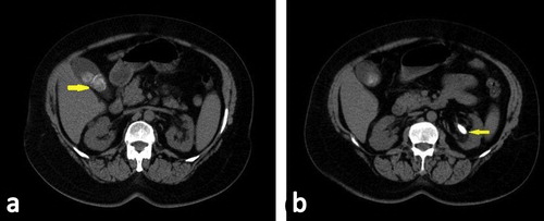
Figure 2 Fifty-six year old male patient with left flank pain. CT stone protocol shows small diverticula in the sigmoid colon (arrows). No stones were found.
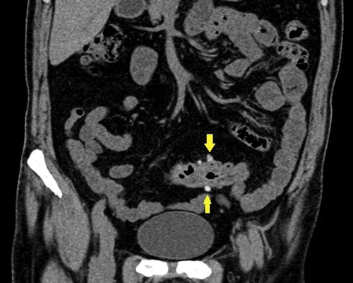
Figure 3 Thirty-four year old female patient with right sided abdominal pain and dysuria. CT stone protocol revealed an oblong tubular structure related to the ileal loops, with stranded adjacent fascial planes, proven at surgery to be an inflamed Meckel’s diverticulum.
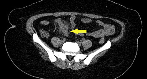
Figure 4 Thirty-four year old male patient with bilateral flank pain. In addition to bilateral renal stones, CT stone protocol shows a left adrenal lipid-rich adenoma (arrow).
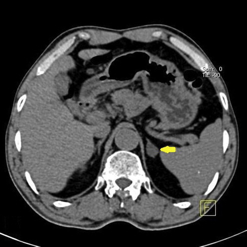
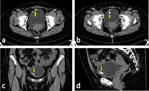
Figure 5 Forty-one year old female patient with dysuria and hematuria. CT stone protocol revealed a contraceptive device that penetrated into the urinary bladder lumen. (a) Axial cut at the urinary bladder showing the two limbs of the device (arrow). (b) Axial cut at a lower level showing the stem of the device (arrow). Coronal (c) and sagittal (d) showing the contraceptive device (arrows).
On the other hand, renal cysts were the urinary tract related incidental finding most commonly encountered (only one cyst of about 6 cm diameter caused renal pain and hence required intervention) (); 161 patients (62%), followed by renal infections; 29 patients (11%) (–), urinary tract tumors; 24 patients (9%) ( and ), and double-moiety; 17 patients (7%).
Figure 6 Thirty-four year old female patient with left flank pain. CT stone protocol in sagittal oblique plane through the left kidney shows a parapelvic cyst causing mild dilatation of the upper calyx with no stones. Cyst aspiration and sclerotherapy relieved the pain.
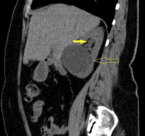
Figure 7 Fifty-three year old diabetic female patient with left flank pain. CT stone protocol in axial (a) and coronal (b) planes through the kidneys shows left renal abscess (arrows). No stones were found.
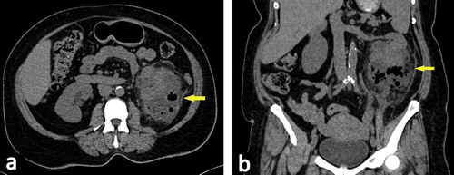
Figure 9 Sixty-eight year old female patient with left flank pain. CT stone protocol through the left kidney in (a) axial, (b) coronal, and (c) sagittal planes revealed a small calcified left kidney, consistent with putty kidney.
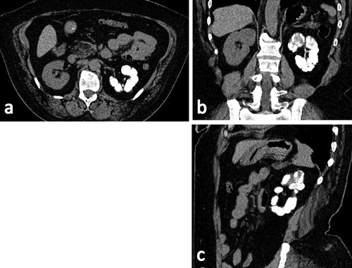
Figure 10 Sixty-one year old male patient with right flank pain and hematuria. CT stone protocol shows urinary bladder mass at the right vesico-ureteric junction (large yellow arrow) with obstruction of the right ureter, which is filled by mass tissues (small yellow arrow). Small lower calyceal stone was found in the left kidney (white arrow).
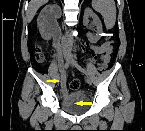
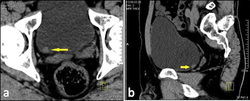
Figure 11 Forty-three year old male patient with left flank pain. CT stone protocol though the urinary bladder in axial (a) and sagittal (b) planes shows small mass lesion in right postero-lateral wall (arrows), turned out by cystoscopy to be a small urothelial tumor.
From the total 381 patients with incidental findings, the discovered incidental findings had an impact on the management in 198 patients (47%), either in the form of diet adjustment, medical treatment or even surgical intervention (open surgery or endoscopy). Two patients had medullary sponge kidneys ().
Figure 12 Fifteen year old female patient with bilateral flank pain. CT stone protocol through right kidney in oblique coronal (a) and oblique sagittal (b) and through the left kidney in oblique coronal (c) and oblique sagittal (d) planes, shows bilateral renal medullary calcifications with no stones, diagnosed as medullary cystic kidneys.

shows the patients with incidental findings, and the modification of management they received
Table 4 The patients with incidental findings, and the modification of management they received.
4 Discussion
Flank pain and hematuria are common presentations of urinary tract calculi. However, a number of other pathologies in different abdominal organs and in the urinary tract itself can give a similar presentation. We aimed in our study to assess the utility of non-contract CT examination of the urinary tract (stone protocol) in the detection of the pathologies other than urinary tract stones which mimic their symptoms and signs, and comparing our results with other similar studies.
Ather et al.Citation13 studied 4000 patients suspected to have urinary tract stone, and found an alternate diagnosis in 398 patients (9.9%), which is different than our finding of 24% stone-free patients, and it should be noted that in this study – in addition to the different sample size - the search was for a cause for the complaint other than stone; however, in our study the search was for concomitant as well as for alternate diagnosis. Ather et al. also noted a wide spectrum of significant alternate diagnoses including urogenital (76.6%) and non-urogenital (23.4%) conditions that could be reliably established or suggested on spiral CTs performed for suspected renal colic cases. However, Ather et al. included ovarian lesions in the genitor-urinary group.Citation13
In a study conducted by Katz et al.,Citation14 1000 stone protocol examinations were reviewed, ureteric stones were found on 557 examinations, findings consistent with a recently passed stone were found on 67 examinations, and 275 CT examinations were free. An alternative or additional diagnosis was found or suggested on 101 examinations (10%), including 26 patients having both urinary tract stone and an incidental pathology. Again, different sample size than that in our study may cause the different percentage of patients with incidental findings. In Katz et al. study, there were 62 incidental findings related to genitourinary system and 39 findings not related to the genitourinary tract. Katz et al. included pathologies related to the female genital system to the urinary tract group, which was separate in our study.
Studying incidental diseases on 233 unenhanced helical computed tomography examinations performed for ureteric colic, Ahmad et al.Citation16 found stones-only in 148 examinations (64%), findings of recent passage of calculi in 10 examinations (4%) and no calculus in 75 examinations (32%). Overall the incidental findings (additional or alternative diagnosis) were found in 28 (12%) CT scans. They grouped the different incidental diagnoses according to the pathology into inflammatory conditions (n = 12), tumors and masses (n = 12), and other urological diseases (n = 4). However, by analyzing the different incidental pathologies, those related to the urinary tract were 9 (32%), while those not related to the urinary tract were 19 (68%), and the most common extra-urinary pathologies were in adnexal masses and cysts (n = 6), followed by gall bladder diseases (n = 4) and bowel diseases (n = 4).
In a study conducted by Hoppe et al.,Citation17 1500 patients underwent unenhanced CT due to acute flank pain. 1035 (69%) had urinary tract calculi. Stones alone were found in 331 of these patients (32%) and additional pathological conditions were noted in 704 (68%). Of all patients 1064 (71%) had other or additional CT findings. Of all patients 207 (14%) had non-stone related CT findings requiring immediate or deferred treatment, 464 (31%) had pathological conditions of little clinical importance and 393 (26%) had pathological conditions of no clinical relevance. CT was normal in 105 of all patients (7%).
The different sample sizes in our study and in the mentioned studies contribute to the different percentages of patients with stone-only, stone with incidental findings and patients with alternate diseases. Also the fact that different studies (including our study), different categorization of the individual incidental/alternate findings also contributed to the apparently different results.
Still all studies have agreed that the stone protocol examination adds to the detection of pathologies other than urinary tract stones, which may mimic their presentations, or be incidentally found with urolithiasis.
5 Conclusion
Non-enhanced CT examination of the urinary tract offers the highest sensitivity and specificity in the detection and characterization of urinary tract stones, and is also valuable in the detection of both incidental and alternate pathologies that may be incidentally found, with great impact on patient diagnosis and management.
Conflict of interest
The authors declared that there is no conflict of interest.
Notes
Peer review under responsibility of Alexandria University Faculty of Medicine.
References
- B.W.TurneyJ.M.ReynardJ.G.NobleS.R.KeoghaneTrends in urological stone diseaseBJU Int109201210821087
- G.M.PremingerH.G.TiseliusD.G.AssimosP.AlkenC.BuckM.Gallucci2007 guideline for the management of ureteral calculiJ Urol178200724182434
- P.BhargavaM.K.DigheJ.H.LeeC.WangMultimodality imaging of ureteric diseaseRadiol Clin North Am5022012271299
- S.N.KaziR.L.BenzS.N.KaziR.L.BenzWork-up of hematuriaPrim Care4142014737748
- R.FlanniganW.H.ChoyB.ChewD.LangeRenal struvite stones–pathogenesis, microbiology, and management strategiesNat Rev Urol1162014333341
- P.Cordeiro GonzálezA.Puñal PereiraB.Blanco GómezJ.Lema GrilleBladder endometriosis: report of 7 new cases and review of the literatureArch Esp Urol6772014646649
- G.NigriN.PetruccianiG.GianniniP.AurelloP.MagistriM.GasparriniGiant colonic diverticulum: clinical presentation, diagnosis and treatment: systematic review of 166 casesWorld J Gastroenterol2112015360368
- J.BrownDiagnostic and treatment patterns for renal colic in US emergency departmentsInt Urol Nephrol3820068792
- G.E.TasianL.CopelovitchEvaluation and medical management of kidney stones in childrenJ Urol1925201413291336
- N.A.AhmadM.H.AtherJ.ReesUnenhanced helical computed tomography in the evaluation of acute flank painInt J Urol102003287292
- A.S.LarsenR.PedersenG.SandbaekComputed tomography of the urinary tract: optimalization of low-dose stone protocol in a clinical settingActa Radiol4672005764768
- A.KirpalaniK.KhaliliS.LeeM.A.HaiderRenal colic: comparison of use and outcomes of unenhanced helical CT for emergency investigation in 1998 and 2002Radiology2362005554558
- M.H.AtherK.FaizullahE.AchakzaiAlternate and incidental diagnoses on non contrast enhanced spiral computed tomography for acute flank painUrol J620091418
- D.S.KatzM.ScheerJ.H.LumermanB.C.MellingerC.A.StillmanM.J.LaneAlternative or additional diagnoses on unenhanced helical computed tomography for suspected renal colic: experience with 1000 consecutive examinationsUrology5620005357
- M.H.AtherW.MemonJ.ReesClinical impact of incidental diagnosis of disease on non-contrast enhanced helical CT for acute ureteral colicSemin Ultrasound CT MR2620052023
- N.A.AhmadM.H.AtherJ.ReesIncidental diagnosis of diseases on un-enhanced helical computed tomography performed for ureteric colicBMC Urol320032
- H.HoppeR.StuderT.M.KesslerP.VockU.E.StuderH.C.ThoenyAlternate or additional findings to stone disease on unenhanced computerized tomography for acute flank pain can impact managementJ Urol175200617251730