Abstract
Objectives
Assessment of the value of using mannitol for the reduction of intracranial pressure and optimizing surgical condition during awake craniotomy.
Methods
Forty patients; 21 males and 19 females; 21 ASA I and 19 ASA II patients. Twenty patients had left hemispheric tumors and 14 patients had right hemispheric tumors, while six patients suffered from epilepsy. Patients were randomly allocated into two equal groups. Group A, was given mannitol, while to group B no mannitol was given (but kept as a rescue drug). Intracranial pressure (ICP) and blood gases were recorded every 15 min till the end of surgery. Surgeon satisfaction regarding brain status, tense or slack was recorded. Postoperative nausea and vomiting (PONV), fits and electrolyte disturbances were noted.
Results
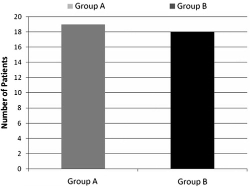
Intracranial pressure (ICP) readings were comparable between the two groups at baseline, skin incision and 15 min after. Mannitol effect on ICP appeared as a lower reading of ICP in group A from 30 min after skin incision till dural exposure and incision. Impact of hyperventilation on ICP measures was evident in both groups since prior to dural incision till after dural closure. However, there was no difference regarding brain status judged by the surgeon between the two groups as brain was found to be slack in 19 patients versus18 patients in groups A and B, respectively. Blood CO2 levels in blood gases showed progressive declination in both groups from the start of hyperventilation till the end of surgery. Potassium (K+) correction was needed in four patients in the mannitol group. Three patients in group A suffered from nausea versus one patient in group B. A single patient in each group suffered from fits.
Conclusion
Usage of mannitol did not add much benefit over ICP perception and brain status in elective awake craniotomy and mannitol should be kept as a rescue drug if needed.
1 Introduction
Mannitol has long been considered as a golden tool for the reduction of ICP, and for minimizing brain edema in patients undergoing surgeries for brain tumor resection. However, the clinical use of mannitol is faced by its side effects including its nephrotoxic effects, diuretic effect, nausea, vomiting, pain or swelling at the injection site and the fast increase of the osmotic gradient followed by its reversal due to disruption of the Blood–Brain Barrier (BBB) [Citation1,Citation2]. Recently, it was found that the hyperosmotic stress itself can activate the process of apoptotic cell death [Citation3]. These side effects would add much stress to the awake patients and the whole team of awake craniotomy that might lead to failure of the whole procedure and end up in the conversion into general anesthesia. Optimization of the operative conditions during awake craniotomy is the main challenge faced by the anesthesiologist who should provide comfortable stable patients to remain immobile throughout the whole procedure, yet sufficiently alert to comply with the neurological testing during surgery [Citation4].
Awake craniotomy started in the 2nd half of the 19th century and it was mainly used for epilepsy surgery [Citation5]. Nowadays, awake craniotomy is also used for the resection of tumors located in the eloquent cortex to allow optimal tumor resection with minimal postoperative neurologic dysfunction [Citation6]. Elevation of intracranial pressure caused by the presence of intracranial space occupying lesion is the main problem faced by the surgeon during tumor resection. This problem is the main target of the anesthesiologist to solve. Hyperventilation of some degree is still provided to facilitate intracranial surgery [Citation7]. It is generally believed that deliberate hyperventilation facilitates surgical access to the tumor and reduces brain edema due to reduction of ICP and cerebral blood volume [Citation8].
Due to the aforementioned mannitol side effects we conducted the present study to assess the value of using mannitol in awake patients and to assess its privilege over ICP, brain status and surgeon satisfaction.
2 Materials and methods
The study was performed in El Kasr El Aini Hospital, Cairo University from 2006 to 2009. After obtaining local institutional approval of the study protocol and fully informed written patients consent. Forty patients (ASA I and II), physically able to tolerate awake surgery, assigned for craniotomies for either epilepsy surgery or excision of small sized tumors with minimal brain edema located in eloquent brain areas were enrolled in this study. Exclusion criteria included obese patients (body mass index >30), patients with communication difficulties and those with difficult airway (Malampatti III and IV), patients having respiratory problems, surgeries done in positions other than supine position and those with massive increase in ICP proved clinically (nausea, vomiting and headache) and/or radiologically (presence of hydrocephalus or severe mid-line shift >1 cm).
Preoperative visit was done for all patients the day before surgery to gain their confidence, reassurance and explain the steps of the procedure. All medications taken by the patients for concurrent diseases were continued to the day of surgery as usual. In the operating room, patients were randomly allocated into two groups, group A (mannitol group) where patients received mannitol 20% in a dose of 0.5 g/kg after urinary catheterization whereas, patients in group B did not receive mannitol unless the surgeon asked for the treatment of tense brain. Intravenous access was inserted under local anesthesia for administration of intravenous dexamethasone 8 mg that was already started to be given to the patients 2 days before surgery, ondansetron 8 mg, ranitidine 50 mg and slow intravenous injection of phenytoin 250 mg. Injection of diclofenac sodium 75 mg and atropine 0.5 mg intramuscularly was done. Basic monitoring that included non-invasive blood pressure monitoring, 5-leads electrocardiography; pulse oximetry and respiratory rate were attached to the patients. Bispectral index electrodes (by Aspect Medical System, Natick, MA, USA) were placed on the skin of the forehead after cleansing with alcohol at the contra lateral side of the site of surgery to assess the degree of sedation throughout the whole procedure. Supplemental O2 was delivered at a rate of 3–4 l/min via an oxygen mask. Initially propofol 1 mg/kg was given, then infusion was started at a rate of 1–3 mg/kg/h and fentanyl bolus (0.5–1 μg/kg/h) was then given. Thereafter, two wide bore cannulae and left radial arterial cannula 20 gauge were inserted under local anesthesia for invasive blood pressure monitoring and withdrawal of samples for blood gas analysis. All patients in mannitol group were subjected to insertion of Foley’s urinary catheter using lidocaine gel. This was not the rule in the other group where catheterization only occurred if the patient had a sensation of full bladder or if diuretic therapy was needed.
Skull block involved bilateral infiltration of the nerves supplying the scalp. This block allowed the application of head clamp pins, raising scalp flaps and the application of Codmann® Microsensor™ ICP monitor probe into the contra lateral frontal lobe. The nerves involved were the supraorbital and supratrochlear nerves (2–3 ml) each, zygomatico-temporal nerves (2–3 ml), as well as, auriculo-temporal nerves 1.5 cm anterior to the ear at the level of tragus (4–5 ml) and post-auricular branches of great auricular nerves (3–4 ml) posterior to the ear at the level of tragus. The third, lesser and greater occipital nerves were blocked in a band extending laterally from the inion in the superior nuchal line to just behind the ear. The subcutaneous tissue of the anterior temporal region was then injected. Skull block was performed using a solution made of a mixture of 0.25% bupivacaine with adrenaline 1:200,000. All patients were positioned 20–30° head elevation in supine position. After positioning, the proposed site for skin incision was infiltrated with a solution of 2% lidocaine with adrenaline 1:200,000. All patients were kept sedated during skin incision and craniotomy and allowed to regain their conscious level slowly and gradually to be fully awake upon reaching the dura where all drugs were stopped. The dura was anesthetized by placing gauze soaked with lidocaine 1% with no adrenaline for 15 min. All patients in both groups were encouraged to hyperventilate by counting from 1 to 10 and after each count the patients took a deep breath. This procedure was repeated every 15 min throughout the duration of surgery.
2.1 Data collected
| • | ICP measurements and blood CO2 levels were monitored every 15 min from prior to skin incision till end of surgery. | ||||
| • | Surgeon satisfaction regarding brain status, tense or slack, since dural exposure till closure every 15 min, and his overall satisfaction were expressed as yes or no. | ||||
| • | Number of patients suffered from complications (nausea, vomiting, fits) in both groups and their management. | ||||
| • | Number of patients needed urinary catheterization in the second group and those suffered from its presence in the 1st group. | ||||
| • | The average duration of surgery in each group was also calculated to know whether mannitol accelerated the rate of tumor resection or not. | ||||
| • | Number of patients needed correction of serum Na+ and K+ levels in both groups. Serum Na+ and K+ levels at the beginning and end of surgery. | ||||
2.2 Statistical analysis
Obtained data were presented as mean ± SD, ranges, numbers and ratios as appropriate. Categorical data were analyzed using χ2 test or fisher exact test as appropriate. Continuous data were analyzed using unpaired t-test or univariate two-group repeated measures analysis of variance (ANOVA) with post hoc Dunnett as appropriate. Statistical calculations were performed using SPSS (Version 10, 2002) for windows statistical package, p-value <0.05 was considered statistically significant.
3 Results
The study included 40 patients; 21males and 19 females. All the 40 patients completed the study. The demographic data of patients in the two groups were comparable ().
Table 1 Patients’ characteristics.
Table 2 Presenting symptoms.
The patients presented most frequently with seizures followed by symptoms of headache and hemiparesis ().
There was no significant difference between both study groups as regards patients’ demographic data, lesion side or presenting symptoms.
All procedures were completed under local anesthesia with conscious sedation within a comparable time. Regarding complication, only three patients suffered from nausea in group A and one patient in group B and they were managed by supplemental dose of ondansetron 4 mg. However, vomiting did not occur in any patient. Two patients, one in each group, developed a fit and it was controlled successfully by irrigating the cortex with ice cold saline and the supplemental dose of phenytoin 5 mg/kg ().
Table 3 Intraoperative data.
Regarding the number of patients that needed urinary catheterization in group B, there were only two patients whom the surgeon asked for more brain relaxation and mannitol 0.5 g/kg was given as a rescue drug. Four patients in group A suffered from the presence of urinary catheter that presented in the form of severe desire to void urine in one patient who was persuaded to void as there is a catheter in place. Meanwhile, the other three patients suffered from severe burning pain, so the catheter was removed to solve the problem. Fortunately, this occurred by the end of surgery and none of our patients required conversion to general anesthesia.
Relative to baseline, intracranial pressure measurement showed a statistically significant decrease in group A, 30 min after the administration of mannitol and continued till the end of surgery, while in group B intracranial pressure measurements showed a significant decrease starting from 45 min of the start till the end of surgery. Relative to the other group, group A showed a statistically significant difference at 30 min after the administration of mannitol which was lost at postdural incision till the end of surgery, as shown in .
Table 4 ICP reading in the two groups. Data are presented as mean (SD).
We relied upon surgeon satisfaction after dural incision (as ICP reading dropped to zero) that showed no statistically significant difference between both groups as 19 patients in group A found to be slack versus 18 patients in group B. Additional dose of mannitol 0.25 and 0.5 g/kg was given to the patients with tense brain in group A and group B, respectively ().
Relative to baseline, arterial CO2 levels in blood gases readings decreased significantly in both groups with the onset of burr hole with the greatest drop noticed with the start of hyperventilation at dural exposure ().
Table 5 CO2 measurements (mmHg) throughout the observation period.
Regarding electrolyte imbalance, four patients in the mannitol group suffered from hypokalemia (serum level <3.5 Mmol/l) that was corrected by I.V. KCl supplement. Serum Na level was not affected.
4 Discussion
Although mannitol has long been used for many years as a golden tool for the reduction of ICP in craniotomy surgeries, its value of usage in cases of awake craniotomy is not the same. No study had discussed the value of using mannitol in the cases of awake craniotomy and whether it really improves ICP and surgeon perception of the brain status or not.
The main finding of our study was that the use of mannitol in patients undergoing awake craniotomy does not improve surgeon-assessed brain bulk.
Although mannitol significantly reduced ICP readings, its impact on brain status perception by the surgeon was not that evident in awake craniotomy, where 95% versus 90% of patients were found to have slack brains in groups A and B, respectively. This could be attributed to the unique selection criteria of patient candidate for awake craniotomy and the adoption of the voluntary hyperventilation technique that lowered ICP readings and improved brain status perception by the surgeon.
Both ICP measurement and surgeon assessed brain bulk are important in intracranial surgery. At first we depend on ICP measurement that became effectively zero when dural incision was made after which we depended on the surgeon perception. There was an assumption of a direct correlation between these two factors and found to be closest when ICP was ranging from 6 to 17 mmHg [Citation9]. This highlights the subjective nature of brain bulk assessment because there are other factors that contribute to this assessment as firmness of the tumor and the amount of bulging relative to the craniotomy size. Nevertheless, we must respond to the surgeon assessment of operating condition. Therefore, the result of the current study relied on the evaluation of the effect of mannitol on these two factors as measurement end-points.
In our study, mannitol effect on ICP started to be evident in group A in the form of a statistically significant decrease in ICP readings starting from 30 min after administration till the end of surgery. However, in comparison to the other group there were no statistically significant differences between groups from postdural incision. This could be attributed to the effect of hyperventilation on ICP [Citation10].
Our result goes in hand with the result of Blanshard et al. [Citation11], who reviewed 241 patients who underwent awake craniotomy on ambulatory basis and stated that exclusion of obese patients and those with respiratory problems helped in controlling hypercarbia and hence better control of ICP.
This also goes in hand with our results which showed that arterial CO2 levels decreased significantly in both groups with the start of hyperventilation upon dural exposure. This result was consistent with the results of Robertson [Citation12] who stated that hyperventilation could rapidly lower ICP, but it induced a consistent decrease in cerebral blood volume and its effects on ICP were transient. Hence, we adopted the technique of repeated hyperventilation every 15 min till end of surgery to control ICP. This also goes in hand with the result of Gelb et al. [Citation13], who found that hyperventilation decreased the risk of increased brain bulk by 45% with significantly lower levels of mean ICP during hyperventilation compared to normo-ventilation. Moreover, the anesthetic regimen did not affect brain bulk assessment or ICP.
Also in our study we chose the conscious sedation technique, not the asleep awake asleep technique, in which the patients remained fully conscious starting from dural exposure till the end of surgery and were encouraged to hyperventilate to allow better control upon ICP. This was guided by the result of Fukaya et al. [Citation14], who found that the asleep awake asleep technique was occasionally associated with difficulty in controlling brain volume especially in brain tumors with large mass effects as sedation with propofol tended to cause hypercapnia.
Moreover, in our study we excluded all positions except supine position with head elevation of 20–30° guided by the result of Hung et al. [Citation15], who found that changes in ICP were proportional to head elevation and rotation. As head elevation above 20° reduced ICP and maximal reduction at 40° elevation and head elevation to 30° reduced the intracranial hypertension associated with brain rotation.
The result of this study was also consistent with the results obtained by the Palazon et al. [Citation16], who found mean ICP values were lower in semi-sitting positions than in supine position and attributed this to the reduction of mean arterial blood pressure. Also in our present study, our patients were either suffering from epilepsy (having normal ICP) or small low grade gliomas located near eloquent brain areas with minimal brain edema that is often controlled clinically by corticosteroids alone. All these factors made mannitol much less needed in the cases of awake craniotomy.
In our study, one patient in group A suffered from tense brain (meaning that 95% of patients’ brains were slack) versus two patients in group B (meaning that 90% of patients’ brains were slack).
Zorzi et al. [Citation17] reported in their review article that brain swelling was never a problem and according to their routine practice invasive monitoring including urinary catheter was not used unless huge tumors and prolonged surgery with aggressive diuresis was anticipated. Skucas et al. [Citation18] reported that the incidence of tight brain was 0.6%. They studied the complication in over 300 patients who underwent awake craniotomy for epilepsy surgery. The very low incidence of 0.6% could be explained by the type of patients who were suffering from epilepsy and these patients had already normal to near normal ICP.
Archer et al. [Citation19] reported an incidence of 1.4% after reviewing the perioperative records of 354 patients, retrospectively. In consistent also with the result of our study was that of Blanshard et al. [Citation11], who needed mannitol only in four patient with an incidence of 1.6% and also reported that most patient did not receive mannitol even with huge tumors with large mass effect and mid-line shift especially if a good surgical decompression was anticipated.
In line with our result, Manninen et al. [Citation20], who studied awake craniotomy for tumors and reported that four patients only in the study needed mannitol, three of them were according to surgeon request while the 4th was due to tight brain. It also goes in hand with the result of See et al. [Citation21], who studied awake craniotomy for tumor resection and found that the brain was slightly swollen in only one patient who needed mannitol and the resection was not affected.
In contrast to our study was that of Sinha et al. [Citation22], who found that 14.2% of the awake patients had tight brain and this high result was explained by the subjective nature of assessment and also the vague definition of tight brain.
Operating surgeons found the brain slack in 19 of 20 patients in group A and in 18 of 20 patients in group B, thus surgeon satisfaction rate was 95% in group A and 90% in group B. Meanwhile, mannitol did not shorten the duration of surgery as the operative time was comparable between the two groups.
One patient in group B suffered from PONV with an incidence of (5%) versus three patients in groupA (15%). This incidence was lessened by the prophylactic use of ondansetron as well as the antiemetic effects of propofol. Actually, the difference between groups could not be attributed to the usage of mannitol due to the smaller sample size used in the current study.
Also, our results showed an important drawback of mannitol which was the necessity of insertion of urinary catheter that added stress and discomfort to the patient inspite of the usage of lidocaine jelly for insertion as four patients suffered from its presence with an incidence of 20%. Another drawback was the need for correction of hypokalemia that occurred in four patients with an incidence of 20% due to the diuretic effect of mannitol.
Successful awake craniotomy needs a lot of factors; the most important is the presence of stable, comfortable, alert and co-operative patients. Mannitol side effects, starting from massive diuresis necessitating urinary catheterization, increased sensation of nausea and vomiting, pain at the site of injection and also electrolyte disturbances, may add much stress to the awake patient and the whole team of awake craniotomy. Therefore, many anesthesiologists limited the use of mannitol in awake craniotomy, in addition to the fact that awake craniotomy has different patients’ selection criteria that lessen the need for the usage of mannitol.
In conclusion, this study clearly showed that proper selection of patient, proper positioning, and adoption of the conscious sedation technique with voluntary hyperventilation made mannitol much less needed and better to be reserved as a rescue drug in the cases of awake craniotomy.
Notes
Available online 16 February 2011
References
- R. Garcia-Sola P. Pulido P. Capilla The immediate and long-term effects of mannitol and glycerol. A comparative experimental study Acta Neurochir 109 1991 114 121
- Y. Node K. Yajima S. Nakazawa Rebound phenomenon of mannitol and glycerol: clinical studies No To Shinkei 35 1983 1241 1246
- G. Famularo The puzzle of neuronal death and life: is mannitol the right drug for the treatment of brain oedema associated with ischaemic stroke? Eur J Emerg Med 6 1999 363 368
- K.R. Bulsara J. Johnson A.T. Villavicencio Improvement in brain tumor surgery: the modern history of awake craniotomies Neurosurg Focus 18 4 2005 e5
- R.L. Sahjpaul Awake craniotomy: controversies, indications and techniques in the surgical treatment of temporal lobe epilepsy Can J Neurol Sci 27 Suppl. 1 2000 S55 S63
- M.O. Pinsker A. Nabavi H.M. Mehdorn Neuronavigation and resection of lesions located in eloquent brain areas under local anesthesia and neurophysiological monitoring Minim Invasive Neurosurg 50 5 2007 281 284
- J.E. Brian Carbon dioxide and the cerebral circulation Anesthesiology 88 1988 1365 1386
- X. Hu V. Nenov T.C. Glenn L.A. Steiner M. Czosnyka M. Bergsneider Non-linear analysis of cerebral haemodynamic and intracranial pressure signals for characterization of autoregulation IEEE Trans Biomed Eng 53 2006 195 209
- C.R. Turner T.J. Losasso D.A. Muzzi M.R. Weglinski Brain relaxation and cerebrospinal fluid pressure during craniotomy for resection of supratentorial mass lesion J Neurosurg Anesthesiol 8 1996 126 132
- M. Rasmussen H. Bundgaard G.E. Cold Craniotomy for supratentorial brain tumors: risk factors for brain swelling after opening the dura mater J Neurosurg 101 2004 621 626
- H.J. Blanshard F. Chung P.H. Manninen M.D. Taylor M. Bernstein Awake craniotomy for removal of intracranial tumor: considerations for early discharge Anesth Analg 92 2001 89 94
- C. Robertson Every breath you take: hyperventilation and intracranial pressure Cleve Clin J Med 71 Suppl. 1 2004 S14 S15
- A.W. Gelb R.A. Craen G.S. Rao K.R. Reddy J. Megyesi B. Mohanty Does hyperventilation improve operating condition during supratentorial craniotomy? A multicenter randomized cross-over trial Anesth Analg 106 2008 585 594
- C. Fukaya Y. Katayama A. Yoshino K. Kobayashi M. Kasai T. Yamamoto Intraoperative wake-up procedure with propofol and laryngeal mask for optimal excision of brain tumors in eloquent areas J Clin Neurosci 8 2001 253 255
- O.R. Hung G.M. Hare S. Brien Head elevation reduces head rotation associated increase ICP in patients with intracranial tumors Can J Anaesth 47 2000 415 420
- J.H. Palazon P.D. Asensi S.B. Lopez F.B. Bautista A.G. Candel Effect of head elevation on intracranial pressure, cerebral perfusion pressure and regional cerebral oxygen saturation in patients with cerebral hemorrhage Rev Esp Anestesiol Reanim 55 2008 289 293
- F. Zorzi M. Saltarini P. Bonassin M. Vecil P. De Angelis A. De Monte Anesthetic management in awake craniotomy Signa Vitae 3 Suppl. 1 2008 S28 S32
- A.P. Skucas A.A. Artru Anesthetic complication of awake craniotomy for epilepsy surgery Anesth Analg 102 2006 882 887
- D.P. Archer J.M. McKenna L. Morin P. Ravussin Conscious-sedation analgesia during craniotomy for intractable epilepsy: a review of 354 consecutive cases Can J Anaesth 35 1988 338 344
- P.H. Manninen M. Balki K. Lukitto M. Bernstein Patient satisfaction with awake craniotomy for tumour surgery: a comparison of remifentanil and fentanyl in conjunction with propofol Anesth Analg 102 2006 237 242
- J.J. See T.W. Lew T.K. Kwek K.J. Chin M.F. Wong Q.Y. Lie Anaesthetic management of awake craniotomy for tumor resection Ann Acad Med Sing 36 2007 319 325
- P.K. Sinha T. Koshy P. Gayatri V. Smitha M. Abraham R.C. Rathod Anesthesia for awake craniotomy: a retrospective study Neurol India 55 2007 376 381