
Abstract
3-[(1-methyl-4-nitro-1H-imidazol-5-yl)thio]-4-methyl-1,2,4-triazole (MNITMT) is an immunosuppressive agent used to treat autoimmune disorders. The objectives of this study were to determine the genotoxic and cytotoxic effects of MNITMT on bone marrow cells of mice. Various concentrations of MNITMT were tested (2, 5, 10 and 20 mg ml–1). Bone marrow cells were prepared for mitotic and mitodepressive indices and blood smears for micronuclei. Cell viability of cultured bone marrow cells was determined using an MTT [3-(4,5-dimethylthiazol-2-yl)-2,5-diphenyl tetrazolium bromide)] assay and the proliferation index was calculated. Our results show that MNITMT significantly lowered the mitotic index (MI) at the highest two doses (10 and 20 mg ml–1) with 14.3 % and 14.9 % at 24 h, 19.2 % and 19.6 % at 48 h, 15.4 % and 19.8 % at 72 h, respectively as compared to control. The percentage of micronuclei (MN) increased with increasing the MNITMT doses 2, 5, 10 and 20 mg ml–1 (79, 125, 141 and 145% respectively). Administration of 2, 5 and 10 mg ml–1 of MNITMT did not cause any bone marrow toxicity, while approximately 50% reduction in the rate of proliferation was observed using the highest dose, 20 mg ml–1. These results suggest that high doses of MNITMT cause both genotoxic and cytotoxic effects.
Introduction
Multiple studies have shown that different biological and non-biological extracts could induce cytotoxic and mutagenic effects on eukaryotic cells. Cytotoxicity and DNA damage in Allium cepa were reported after exposure to e-waste leachate (Bakare et al. Citation2012). In addition, extracts from leaves and bark of two populations of Luehea divaricata showed an antiproliferative effect when tested on cells of root tips of Allium cepa (Frescura et al. Citation2012).
Recently, many immunosuppressive drugs have been developed for research and clinical use in organ transplantation and treatment of autoimmune diseases (Marder and McCune Citation2004, Citation2007). However, the administration of immunosuppressive agents is associated with severe side effects including bone marrow suppression. Such effects could result in anemia, leukopenia, and thrombocytopenia (Danesi and Del Tacca Citation2004; Nath and Berger Citation2012).
Azathioprine (AZA), the pro-drug of 6-mercaptopurine (6-MP) has been in clinical use since 1961 but severe bone marrow toxicity is one major complication associated with this drug, and thus limits its use (Kerstens et al. Citation1995; Patel et al. Citation2006). These myelotoxic effects are attributed to the intracellular metabolite, 6-MP moiety (Pollak et al. Citation1980; Gearry and Barclay Citation2005).
The immunosuppressive agent 3-[(1-methyl-4-nitro-1H-imidazol-5-yl)thio]-4-methyl-1,2,4-triazole (MNITMT) is a novel derivative of AZA and used mainly to prevent rejection of organ transplants (MacPhee et al. Citation1998) and to treat various autoimmune disorders (Fraser et al. Citation2002). MNIMT lacks the 6-MP moiety and this makes it a better immunosuppressive agent than AZA. Moreover, MNITMT is a more potent immunosuppressive agent than AZA and is devoid of any bone marrow toxicity (Crawford et al. Citation1996). The immunosuppressive effect of MNITMT against antibody response in rabbits has been described (Al-Safi et al. Citation2003). In that study it was found that MNITMT causes a significant and consistent inhibition of the antibody production on days 14 and 60 post treatment with 2 mg kg–1 day–1. In addition, MNITMT did not induce any significant changes in biochemical markers. Nevertheless, other study has reported that MNITMT causes a weak base pair substitution in Salmonella typhimurium strain TA100 but with no mutagenicity effect in TA1535 strain and no frame shift mutagenic action (Tumah et al. Citation2006).
Moreover, MNITMT did not show any cytotoxicity when evaluated by the shrimp bioassay (Tumah et al. Citation2006). In contrast, AZA has been proved to be genotoxic and enhances DNA damage, even at low doses that are not cytotoxic to hepatocytes (Nagafuchi and Miyazaki Citation1991). Moreover, there is clear evidence for an increase in sister chromatid exchange (SCE) frequencies in patients receiving AZA (Erskine et al. Citation1984).
Since 2006 and to the best of our knowledge, no further studies were reported in the literature on this novel nontoxic immunosuppressive agent, MNITMT. Therefore, this study aimed to investigate MNITMT safety by monitoring its genotoxic and cytotoxic effects on mouse bone marrow cells.
Materials and methods
The genotoxic effect of MNITMT on bone marrow of mice was tested at 2, 5, 10 and 20 mg ml–1 and for different periods of time after a single intraperitoneal injection (i.p.). All doses were administered at a volume of 0.1 ml g–1 b.w. Animals in the control groups received an equivalent volume of normal saline. For each dose, four male mice were used for every experimental and control group. Metaphase cells were prepared for mitotic investigation according to the air drying technique of Tjio and Whang (Citation1962). The preparations were stained with Giemsa solution. Slides were coded and scored for the presence of dividing cells. The mitotic index was calculated on a minimum of 6000 cells, as the number of cells in division expressed as a percentage of the total number of cells observed. Mitodepression was calculated using the following formula:(1)
Peripheral blood was obtained from the retro-orbital vein using heparinized capillary tubes after 24 h injection. This experiment was repeated three times. Blood smears were prepared on clean prewashed slides. The blood film was air-dried and fixed with methanol for about 3 min and stained using Giemsa solution for 5 min at room temperature, then rinsed in xylene. The slides were studied using a light microscope at 100× oil immersion lens and the results were recorded as monochromatic micronucleus formation frequency (Seehy et al. Citation1989). The mitotic index was calculated using a minimum of 6000 cells.
In a preliminary study, the minimal tolerated dose (MTD) of the MNITMT using doses 2, 5, 10 and 20 mg ml–1 was determined to be 12.5 mg kg–1 body weight of mice. Stock solution was prepared by dissolving 10 mg of MNITMT in 1ml of dimethylsulfoxide (DMSO). Untreated control mice were given equal volumes of DMSO and their cytotoxic effect was tested for different periods of time after a single i.p. All different doses were prepared to give a volume of 0.1 ml g–1 body weight. Animals in the control groups received an equivalent volume of DMSO.
Preparation of mouse bone marrow cells
Bone marrow cells were isolated according to Rossi et al. (Citation1987) and Luke and Tice (Citation1989) with modifications. Briefly, 10–12-week-old Swiss mice JVI 1 were injected i.p. with different concentrations of MNITMT for different time intervals: 24, 48 and 72 h. Animals were sacrificed by cervical dislocation. Femora were isolated, the ends of bones were cut off and the bone marrow was flushed using sterile saline (Rossi et al. Citation1987; Luke and Tice Citation1989). The following concentrations were applied i.p.: 2, 5, 10 and 20 mg ml–1 body weight of MNITMT dissolved in DMSO. DMSO served as a control (Omari et al. Citation1996).
Viability test of bone marrow cells
About 2 ml RBC lysis buffer (slightly hypotonic solution, 0.83% tris-ammonium chloride) was added to bone marrow cells and incubated for 2 to 5 min at room temperature. Fresh 3 ml of RPMI-1640 medium was added and cells were centrifuged at 1200 rpm for 8 min. The cell pellet was resuspended in 1 ml of the medium. Initial cell viability and counting was determined using direct Trypan-blue exclusion test using a hemocytometer.
Cell culture
A standard bone marrow cell culture was employed throughout this study. Isolated bone marrow cells, at a density of 1.0 × 10⁶ cells ml–1 were prepared in a final volume of 10 ml culture medium and placed in 25 cm3 tissue culture flasks. The culture medium consisted of RPMI-1640 (with 25 mM HEPES, pH 7.2, 2.05 mM L-glutamine and 23.8 mM sodium bicarbonate). The medium was supplemented with 100 IU ml–1 penicillin and 100 μg ml–1 streptomycin. MNITMT concentrations of 2, 5, 10 and 20 mg ml–1, dissolved in DMSO, were added to the culture at the initial time of culture. DMSO served as a control.
Viability of cultured bone marrow cells
Cell viability was determined using 3-(4,5-dimethylthiazol-2-yl)-2,5-diphenyl tetrazolium bromide) (MTT) assay. A stock solution of 5 mg ml–1 MTT in PBS was prepared, filtered and used at each time interval as previously described with some modifications (Mosmann Citation1983). Briefly 100 μl cultured cells were mixed with 20 μg MTT and further incubated for three hours at room temperature. After the incubation period, 150 μg DMSO was added to the mixture. The absorbance at 550 nm was measured 15 min later. Proliferation index (PI) was calculated as the proportion between the absorbance of treated cells and untreated cells multiplied by 100%.
Statistical analysis
Data were analyzed using SPSS version 14.0 software (SPSS, Chicago, IL, USA). Statistical analyses were expressed as the mean differences between the results of investigation performed and corresponding control for both the mitotic and micronucleus test; the Student’s t-test was applied for the statistical analysis. The differences were considered statistically significant at p ≤ 0.05. To decide if there were differences between the results and corresponding control for proliferation index, analysis of variance test (ANOVA) was applied. The differences were considered statistically significant at p ≤ 0.05.
Results
The cytological effect was estimated on the basis of the mitotic index (MI) in mice cells following a single i.p. dose of 2, 5, 10 and 20 mg ml–1 of MNITMT. The mitotic index was assessed from 24 to 72 h following application. The mitotic index and mitodepression were determined in each of the doses and are presented in Table . A remarkable decrease in MI was evident in bone marrow cells at all four MNITMT doses and at all periods of time compared to the control. A significant decrease was seen at the highest two doses (10 and 20 mg ml–1), with 14.3 and 14.9% at 24 h, 19.2 and 19.6% at 48 h, and 15.4 and 19.8% at 72 h, respectively as compared to the control. Depression of cell division in these cells increased with increasing time of exposure in all treatments (Table ). In all the preparations examined there were only a few divided cells of abnormal type; those being mostly sticky and broken chromosomes (Figure A, B.)
Table 1. Mitotic index in bone marrow cells of mice treated with different concentrations of MNITMT.
Figure 1. Different types of chromosomes aberrations in mice bone marrow cells: (A) sticky chromosomes; (B) broken chromosomes. Scale bar = 5 μm.
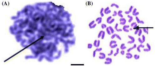
Table showed micronucleated cells after treatment with different doses of MNITMT. A significant difference between each treatment and the control was seen in all cases. The increase in number of micronuclei was associated with increasing doses 2, 5, 10 and 20 mg ml–1 (79, 125, 141, and 145% respectively).
Table 2. Micronucleated cells after treatment with different doses of MNITMT.
Cell survival
The statistical analysis of the data (ANOVA, p > 0.05) revealed that there is no significant difference between the treatment groups at different concentrations and exposure times and the control as shown in Table . This trend was noticed in almost all treatments. MNITMT induced cell proliferation at a concentration of 2, 5 and 10 mg ml–1; however, these effects did not significantly differ from the control. Such induction decreased with increased concentrations in most treatments, reaching an approximately 50% inhibition of proliferation at a 20 mg ml–1 dose.
Table 3. Proliferation index in bone marrow cells of mice treated with MNITMT for 24, 48 and 72 h.
Discussion
The traditional immunosuppressive agent azathioprine (AZA) is used to prevent rejection of organ transplants in recipients, particularly kidney allografts (MacPhee et al. Citation1998) and to treat various autoimmune disorders (Fraser et al. Citation2002). The clinical use of AZA is limited by potentially serious toxicity to the bone marrow (Connell et al. Citation1993; Kerstens et al. Citation1995). Crawford et al. (Citation1996) published their interesting results on a number of rationally designed analogs of AZA. The most effective and safest analog was MNITMT. It was more effective in inhibiting the human mixed lymphocyte reaction and in preventing skin allograft in mice than AZA (Crawford et al. Citation1996). Moreover, AZA had been reported to induce pancreatitis which is manifested by elevation of serum amylase (Weersma et al. Citation2004; Alexander and Dowling Citation2005). However, MNITMT did not induce any elevation in serum amylase although it was used at high dose (Tumah et al. Citation2006). Therefore, we decided to study the genotoxic and the cytotoxic effects of MNITMT.
According to our findings, a drastic reduction in mitotic index was observed in bone marrow cells of mice treated for different periods of time in all doses used. Two doses, 10 and 20 mg ml–1, showed significant differences compared to control. This result is consistent with previously reported results (Kabarity Citation1980). Moreover, such a reduction was also reported by Omari et al. (Citation1996) and Abderrahman (Citation1997, Citation2004). This inhibitory effect of MNITMT may result from either obstruction of the one set of prophases or from an arrest of one of the mitotic phases. In fact MNITMT can cause an arrest in metaphase due to its effect on spindle formation. Thus, the reduction in MI may be due to the inhibitory effect of MNITMT at the onset of mitosis, in spite of metaphase arrest (Kabarity Citation1980). On the other hand, the effect seen using high doses may be attributed to the inhibitory effect of MNITMT on DNA, RNA and protein synthesis (Al-Ahdal et al. Citation1988) or may be due to the decrease in the number of cells moving into prophase from G2 (Baszczynski et al. Citation1980). At 24 and 48 h after application of a low dose (2 mg ml–1), the mitodepression was lower than that at 72 h. This suggests that 48 h following application may be the threshold time in producing mitodepressive effect. After 48 h exposure, the effect of MNITMT started to decrease.
The micronucleus test (MN) is based on the observation that treatment of cells with certain chemicals or specific types of radiation will disturb chromatin distribution in anaphase. After anaphase, this disturbed chromatin can be excluded from the nuclei of the daughter cells and found in the cytoplasm as micronucleus (Schmid Citation1975). The purpose of this test is to screen chemicals for clastogenic (chromosome breaking) and aneugenic (loss of whole chromosome) effects (Seehy et al. Citation1989). We have observed a significant difference between each treatment and the control in all cases. This increase was associated with increasing the doses.
Genotoxic effects were reported with immunosuppressive drugs. Tacrolimus (FK-506), a potent immunosuppressive drug, induced chromosomal aberrations (CA) and MN frequency at all concentrations for 24 and 48 h (Kurtoglu and Yuksel Citation2012). Similar results were reported by others (Oliveira et al. Citation2004), who found that cultures supplemented with the immunosuppressive drugs mycophenolate mofetil and tacrolimus showed higher amounts of micronuclei compared to controls in all concentrations tested. Moreover, MN frequency rose significantly after 3 weeks of immunosuppressive therapy with cyclosporine A, mycophenolate mofetil and methylprednisolone (Rath and Oliveira-Frick Citation2009).
The effect of MNITMT on cell survival and proliferation index in bone marrow cells of mice has not been investigated previously. Thus our results are the first to be reported in this respect. Although the MTT assay is a way to determine the number of viable cells, more accurate measurement of proliferation can be achieved by BrdU labeling, or [3H] thymidine incorporation assays (Kermani et al. Citation2008).
The results in Table showed that MNITMT is not cytotoxic to the survival and proliferation of the bone marrow cells of mice when treated with 2, 5 and 10 mg ml–1 MINTMT in all periods of time. On the other hand, it significantly inhibits the proliferation index compared to the control when treated with the highest dose, 20 mg ml–1, at all time periods examined. These results are not consistent with Crawford et al. (Citation1996), who reported that MNITMT was devoid of any bone marrow toxicity.
Although our results obtained from this study indicated that MNITMT is not cytotoxic to survival and cell proliferation at the low dose treatments, 2, 5 and 10 mg ml–1, a significant inhibition was seen when treated with the highest dose, 20 mg ml–1, at all exposure time periods. The MNITMT concentration of 20 mg ml–1 resulted in approximately 50% reduction in the proliferation of bone marrow cells in treated mice at all time periods.
In addition to the better safety profile of MNITMT, drug efficacy was better than AZA in preventing skin allograft rejection (Crawford et al. Citation1996). Clearly, the toxicity of MNITMT is controversial especially when high doses of MNITMT are applied, but it has a better safety profile than AZA particularly when used at lower doses. The proliferation index showed that the effect of MNITMT on cell death is more active due to the linear decrease in proliferation index at the highest concentration.
More detailed and larger scale investigations are still required to gain more information on this promising novel immunosuppressive agent that may have various clinical applications, particularly in organ transplantation and autoimmune disorders, and especially when high doses of MNITMT are used.
Conclusions
The genotoxic effects of the novel immunosuppressive agent 3-[(1-methyl-4-nitro-1H-imidazol-5-yl)thio]-4-methyl-1,2,4-triazole (MNITMT) on cell division, chromosomal aberration and micronucleus formation in mice bone marrow cells were investigated. MNITMT induced a significant decrease in the mitotic index (MI) at all time periods and in almost all treatments. In contrast, a significant increase in percentages of micronuclei with increasing doses was noticed in all treatments. Moreover, few types of chromosomal aberration were noticed, those being mostly broken and sticky chromosomes. Administration of MNITMT at a dose of 2, 5 and 10 mg ml–1 did not induce any cytotoxicity. On the other hand MNITMT concentration of 20 mg ml–1 results in a reduction of approximately 50% in the proliferation bone marrow cells of mice in all time periods.
Nevertheless, the present investigation indicated that MNITMT has a better safety profile than other immunosuppressive agents such as AZA, particularly when lower doses are applied. As the toxicity of MNITMT is controversial especially when higher doses are applied, more detailed studies on this novel immunosuppressive agent are still needed to confirm its efficacy and safety.
Declaration of interest:
The author reports no conflicts of interest. The author alone is responsible for the content and writing of the paper.
Acknowledgment
The author sincerely thanks Prof. M. Hassan, Faculty of Pharmacy, Jordan University of Science and Technology (JUST), Irbid, Jordan for his generously supply of the immunosuppressive agent MNITMT.
References
- Abderrahman SM. 1997. Effect of Peganum harmala extract on root tips of Allium cepa. Cytobios. 90(362–363):171–174.
- Abderrahman SM. 2004. Mitodepressive effect of Rubia cordifolia extract on the bone marrow cells of mice. Cytologia. 69(3):307–311.
- Al-Ahdal MN, McGarry TJ, Hannan MA. 1988. Cytotoxicity of khat (Catha edulis) extract on cultured mammalian cells: effects on macromolecule biosynthesis. Mutation Research-Genetic Toxicology and Environmental Mutagenesis. 204(2):317–322.
- Alexander S, Dowling D. 2005. Azathioprine pancreatitis in inflammatory bowel disease and successful subsequent treatment with mercaptopurine. Internal Medicine Journal. 35(9):570–571.
- Al-Safi S, Hassan M, Tashtoush B. 2003. Comparison between the effects of azathioprine and its novel non-mercaptopurine analog on antibody response in rabbits. Polish Journal of Pharmacology. 55:239–243.
- Bakare AA, Adeyemi AO, Adeyemi A, Alabi OA, Osibanjo O. 2012. Cytogenotoxic effects of electronic waste leachate in Allium cepa. Caryologia. 65(2):94–100.
- Baszczynski CL, Walden DB, Atkinson BG. 1980. CYCLOHEXIMIDE-induced nuclear alterations in maize root tips. Canadian Journal of Genetics and Cytology. 22(3):319–331.
- Connell WR, Kamm MA, Ritchie JK, Lennard-Jones JE. 1993. Bone marrow toxicity caused by azathioprine in inflammatory bowel disease: 27 years of experience. Gut. 34(8):1081–1085.
- Crawford DJK, Maddocks JL, Jones DN, Szawlowski P. 1996. Rational design of novel immunosuppressive drugs: Analogues of azathioprine lacking the 6-mercaptopurine substituent retain or have enhanced immunosuppressive effects. Journal of Medicinal Chemistry. 39(14):2690–2695.
- Danesi R, Del Tacca M. 2004. Hematologic toxicity of immunosuppressive treatment. Transplantation Proceedings. 36(3):703–704.
- Erskine, IA, Mackay, JM, Fox, DP.1984. Monitoring patients on long-term drug therapy for genotoxic effects. Basic Life Sci. 29(Pt B):895–905.
- Fraser AG, Orchard TR, Jewell DP. 2002. The efficacy of azathioprine for the treatment of inflammatory bowel disease: a 30 year review. Gut. 50(4):485–489.
- Frescura VD-S, Laughinghouse HD, Tedesco SB. 2012. Antiproliferative effect of the tree and medicinal species Luehea divaricata on the Allium Cepa cell cycle. Caryologia. 65(1):27–33.
- Gearry RB, Barclay ML. 2005. Azathioprine and 6-mercaptopurine pharmacogenetics and metabolite monitoring in inflammatory bowel disease. Journal of Gastroenterology and Hepatology. 20(8):1149–1157.
- Kabarity A. 1980. Mitodepressive effect of khat extract in the meristematic region of Allium cepa root tips. Cytologia. 45:733–738.
- Kermani S, Karbalaie K, Madani SH, Jahangirnejad AA, Eslaminejad MB, Nasr-Esfahani MH, Baharvand H. 2008. Effect of lead on proliferation and neural differentiation of mouse bone marrow-mesenchymal stem cells. Toxicology in Vitro. 22(4):995–1001.
- Kerstens PJSM, Stolk JN, De Abreu RA, Lambooy LHJ, Van Putte LBAD, Boerbooms AAMT. 1995. Azathioprine-related bone marrow toxicity and low activities of purine enzymes in patients with rheumatoid arthritis. Arthritis & Rheumatism. 38(1):142–145.
- Kurtoglu EL, Yuksel S. 2012. Genotoxic effects of tacrolimus on human lymphocyte cells. Russ J Genet. 48(6):651–655.
- Luke CA, Tice RR. 1989. Effect of processing time on the quality of mouse bone-marrow metaphase preparations. Mutation Research Letters. 227(1):59–62.
- MacPhee IAM, Bradley JA, Briggs JD, Junor BJR, MacPherson SG, McMillan MA, Rodger RSC, Watson MA. 1998. Long-term outcome of a prospective randomized trial of conversion from cyclosporine to azathioprine treatment one year after renal transplantation. Transplantation. 66(9):1186–1192.
- Marder W, McCune WJ. 2004. Advances in immunosuppressive drug therapy for use in autoimmune disease and systemic vasculitis. Semin Respir Crit Care Med. 25(5):581–594.
- Marder W, McCune WJ. 2007. Advances in immunosuppressive therapy. Semin Respir Crit Care Med. 28(4):398–417.
- Mosmann T. 1983. Rapid colorimetric assay for cellular growth and survival: Application to proliferation and cytotoxicity assays. Journal of Immunological Methods. 65(1–2):55–63.
- Nagafuchi K, Miyazaki K. 1991. Modulation of genotoxicity of azathioprine by intracellular glutathione in hepatocytes. Journal of Cancer Research and Clinical Oncology. 117(4):321–325.
- Nath A, Berger J. 2012. Complications of immunosuppressive/immunomodulatory therapy in neurological diseases. Current Treatment Options in Neurology. 14(3):241–255.
- Oliveira VD, Zankl H, Rath T. 2004. Mutagenic and cytotoxic effects of immunosuppressive drugs on human lymphocyte cultures. Exp Clin Transplant. 2(2):273–279.
- Omari Y, Shuraideh Z, Abderrahman S. 1996. Mitodepressive effect of Khat (Catha edulis) on the bone marrow of mice. Biomedical Letters. 54:69–72.
- Patel AA, Swerlick RA, McCall CO. 2006. Azathioprine in dermatology: The past, the present, and the future. Journal of the American Academy of Dermatology. 55(3):369–389.
- Pollak R, Nishikawa RA, Mozes MF, Jonasson O. 1980. Azathioprine-induced leukopenia clinical significance in renal transplantation. Journal of Surgical Research. 29(3):258–264.
- Rath T, Oliveira-Frick V. 2009. Mutagenicity of immunosuppressive medications among renal transplant recipients. American Journal of Nephrology. 30(6):514–520.
- Rossi AM, Romano M, Zaccaro L, Pulci R, Salmona M. 1987. DNA synthesis, mitotic index, drug-metabolising systems and cytogenetic analysis in regenerating rat liver: Comparison with bone marrow test after in vivo treatment with cyclophosphamide. Mutat Res. 182(2):75–82.
- Schmid W. 1975. The micronucleus test. Mutation Research. 31(1):9–15.
- Seehy M, Badr E, Hafez A. 1989. Genotoxicity of pesticides. Egypt J Genetics Cytol. 18:59–76.
- Tjio JH, Whang J. 1962. Chromosome preparations of bone marrow cells without prior in vitro culture or in vivo colchicine administration. Biotechnic & Histochemistry. 37(1):17–20.
- Tumah H, Al-Safi S, Abdul-Razzak K. 2006. Biochemical, cytotoxic of a novel non-mercaptopurine immunosuppressant. Acta Pharmaceutica Sinica. 48:109–119.
- Weersma RK, Peters FTM, Oostenbrug LE, van den Berg AP, van Haastert M, Ploeg RJ, Posthumus MD, Homan van der Heide JJ, Jansen PLM, van Dullemen HM. 2004. Increased incidence of azathioprine-induced pancreatitis in Crohn’s disease compared with other diseases. Alimentary Pharmacology & Therapeutics. 20(8):843–850.