
 ?Mathematical formulae have been encoded as MathML and are displayed in this HTML version using MathJax in order to improve their display. Uncheck the box to turn MathJax off. This feature requires Javascript. Click on a formula to zoom.
ABSTRACT
B4C pellets used in the control rod of the experimental fast reactor ‘JOYO’ with different 10B burnups from lower than 10 × 1020 captures/cm3 to 80 × 1020 captures/cm3 and irradiated at less than 800 °C were examined by transmission electron microscopy (TEM). In a B4C pellet irradiated in an irradiation capsule of ‘JOYO’ at 800 °C up to 30 × 1020 captures/cm3, intragranular helium bubbles appeared in flat plate-shapes with the plane of the plate parallel to the (111) rhombohedral plane. However, in the other specimens that were taken from an actual control rod, the helium gas formed very tiny spherical intergranular bubbles with a diameter of a few nanometers . These tiny bubbles make wavy arrays roughly parallel to the (111) plane. The B4C specimens were heated on a TEM in situ heating holder up to 1040 °C for 10 min. Clustering of tiny bubbles was observed, but did not extend to the plate-shaped bubbles. In high burnup specimens, large bubbles/cracks were rarely found along the {100} planes, which may correspond to the amorphous bands caused by the slip. While heating the specimens in TEM over 800 °C, liquid phases of lithium-bearing compoundsappeared on the surface of specimen.
1. Introduction
Boron carbide (B4C) was assumed to be a promising candidate material to be used as the neutron absorber for fast reactors because of its high neutron absorption cross section of 10B—3840 barns for 25.3 meV neutrons for thermal neutrons and on the order of several barns for 1 keV neutrons [Citation1]; and good safety performance such as a very high melting point of around 2450 °C and chemical stability. However, the most significant flaw of a B4C control material is that the neutron absorption reaction of the 10B isotope produces helium coupled with lithium by the following neutron absorption reaction: (1)
(1) It is well known that the generated helium gathers to form bubbles both inside the grains and along the grain boundary. The more neutrons the B4C pellets absorb, the more severe swelling and cracking they will suffer. The risk of cladding failure of a control rod can restrict the lifetime of the B4C control rods in fast reactors. Therefore, the reactor needs more frequent downtime maintenance to replace the control rods, which is not economical.
In the experimental fast reactor ‘JOYO’ at Japan Atomic Energy Agency, B4C was selected as its neutron-absorbing material. In order to prolong the lifetime of the B4C control rods of JOYO, an advanced sodium-bonded(sodium-flowing)-type control rods were designed and applied in the Mark III core alignment. The evaluated 10B burnup limit of the sodium-bonded control rods with an inner shroud pipe was improved up to 120 × 1020 captures/cm3, three times higher than the previously used helium-bonded(helium-filled)-type control rods, which was used for JOYO Mark I and II core alignments. However, the lifetime of sodium-bonded-type control rods was still less than half that of the design target value (260 × 1020 captures/cm3) [Citation2]. Hence, it would be desirable to develop an improved B4C control material capable of reducing the swelling/cracking after neutron absorption.
Because the generated helium gathers to form bubbles that induce microcracking of the pellet, the formation of the bubbles directly affects the amount of swelling. Therefore, a good understanding of the nature of the helium bubbles in B4C would be very helpful in the development of high-performance B4C control-material pellets. The morphology of the helium bubbles in the B4C grains has been reported by some research studies since early 1970s by using TEM investigation [Citation2–12]. However, the specific direction of the parallel-arranged bubbles in a B4C crystal is still unclear. Ashbee [Citation3] observed small defects that were mostly parallel to the traces of the (111) plane in B4C irradiated in a thermal reactor (Oak Ridge Research Reactor, ORR) at 400 °C for 8% burnup (natural B4C). The shape of bubbles was unclear but appeared to be elongated ellipses. Ashbee et al. [Citation4], however, reported that the plate-shaped bubbles aligned parallel to the rhombohedral {110} planes of the B4C specimen after irradiation in the experimental breeder reactor (EBR-II) with up to 3.7% burnup and post-irradiation-annealing at 1900 °C for 1 h. Jostsons and DuBose [Citation8] reported black-spot defects in the as-irradiated natural B4C specimen up to 1.7% burnup and at an estimated temperature of 500 °C in EBR-II. The defects that possessed spherical strain field were deduced as small cavities with ribbon-like shape, with the largest face of the faceted cavity most frequently parallel to the trace of the rhombohedral (111) plane and less frequently parallel to the trace of {100} or {110}. The size of the cavities increased to 3–20 nm by annealing at 1150 °C. Furthermore, after further annealing at a higher temperature (1450 °C), the lattice strain around the cavities was eliminated, and size of the faceted cavity was further increased and the edges of bubbles were parallel to the traces of (111), {100} and, {011}. Jostson and DuBose [Citation8] observed natural and depleted B4C specimens irradiated both in thermal (Engineering Test Reactor (ETR)) and fast (EBR-II) reactors at 500–840 °C, and with burnups of 1.7%–81%. All the bubbles had disc-like morphology surrounded by strain fields, and the plane of the disc was aligned parallel to the (111) planes in the as-irradiated specimen at 800–840 °C in EBR-II. The larger bubbles were positioned along the dislocations, stacking faults, and grain boundaries. The grain boundary bubbles appeared more equiaxed. The disk-like morphology of the bubbles appeared less developed in the specimens irradiated at slightly lower temperature (710–735 °C). In this case, some bubbles were also observed with the {110} plane as the most prominent facet. The specimen irradiated in ETR was similar to the specimens irradiated in EBR-II. However, the specimen irradiated at 550 °C with up to 81% burnup (natural B4C) in the ETR contained only small bubbles without clear shape, and were aligned parallel to the trace of (111). Jostson et al. [Citation7] also mentioned that after annealing at 1400 °C, there was a significant increase in the bubbles growth and were more equiaxed; however, the (111) facet was still predominant. The grain boundaries were the preferred sites of bubble agglomeration. The strain fields around the bubbles disappeared. Hollenberg et al. [Citation9] reported the change in the morphology of the helium bubbles irradiated up to around 62 × 1020 capture/cm2 across a wide range of irradiation temperature. Further, they concluded that small (diameter less than 10 nm) and high-density lenticular bubbles were homogeneously created at temperatures lower than ∼900 °C. At temperatures higher than 1500 °C, low density, large equiaxed and faceted bubbles were formed heterogeneously. Copeland et al. [Citation10] also observed that the largest face of the plate-like bubbles is most frequently parallel to the trace of the (111) plane, and less frequently parallel to the trace of {100} or {110} rhombohedral planes in the specimen irradiated in EBR-II at 650 °C or 730 °C up to 10 and 7.7 × 10²⁰ capture/cm3 burnup, respectively. On the other hand, for the specimens irradiated up to 16 × 1020 capture/cm3 burnup in the ORR at 350°C–400 °C showed only black-spot defects. Cummings et al. [Citation11] found that almost all the bubbles were parallel to the trace of {110} plane. Basmajian et al. [Citation12] also mentioned that plate-like cavities were observed within a grain associated with both {110} and (111) crystallographic planes for the B4C specimen irradiated at EBR-II from 427 °C to 871 °C up to 2–20 × 1020 capture/cm3. Later, Stoto et al. [Citation6] observed B4C irradiated in the PHENIX fast reactor between 620 °C and 1500 °C, and they confirmed that the bubbles were parallel only to the (111) plane in specimens irradiated at 620–1000 °C. At 1500 °C, the shape of bubbles was changed to a more tridimensional form. It must be pointed out that the irradiation temperature of the B4C samples may have large ambiguity due to the difference in reactor types and measuring methods – that may cause a variety of observation results from similar reported irradiation temperatures and burnups.
In view of the different results in the past researches mainly based on the materials irradiated at constant temperature and flux, it is essential to clarify the exact state of the helium bubbles in B4C irradiated in the actual control rods of fast reactors. Furthermore, there is no report on the evidence of lithium which is concurrently created the same amount as that of the helium atoms by the nuclear reaction mentioned above.
This paper describes the TEM observation results of the B4C specimens obtained from different parts of an actual control rod of the experimental fast reactor ‘JOYO.’ Heat treatment up to a temperature of 1040 °C has been carried out at inside TEM using a high-temperature specimen holder to investigate the effect of temperature on the behaviors of helium and lithium in B4C polycrystals.
2. Experimental procedures
The fast-neutron-irradiated B4C materials examined in this paper were provided by Japan Atomic Energy Agency. Most of the specimens were taken from a retired control rod of the sodium-cooled JOYO fast reactor. The r001_33, r001c21, and r001b21 specimens were obtained from the top, middle, and bottom parts of the retired control rod, respectively. The appearance of the samples is shown in online Supplemental Figure 1. The control rods of JOYO were inserted from the top of the reactor; therefore, the r001b21 specimen from the bottom part stayed near the reactor core for the longest time, and was subjected to the largest fluence of neutron irradiation. The degree of enrichment of 10B of all specimens was 90 at.%. The 10B burnup and irradiation temperature were estimated at 80 × 1020 captures/cm3, the highest temperature during the reactor operation was approximately 800 °C. While the burnup of the r001_33 specimen from the top of the control rod was less than 10 × 1020 captures/cm3 and the highest irradiation temperature was estimated to be below 600 °C. It is noted that the temperature and the duration stay at the fuel-core region of each specimen obtained from the real control rod were not uniform and the uncertainty was large. In addition, the K711b41 specimen was separately irradiated in the materials irradiation rig of JOYO at a constant temperature. The parameters of the examined materials are listed in .
Table 1. Characteristics of the B4C specimens
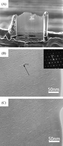
The K711b41 specimen for TEM was prepared using a focused-ion-beam-type micro-fabrication equipment (FIB) using accelerating Ga+ ion beam (Seiko Instruments/SMI9800, Japan). (A) is the scanning ion microscope (SIM) image of the specimen after sample preparation using the FIB. The grain boundaries were clearly visible. TEM observation was carried out with a JEM-2010F FE-TEM at 200 kV electron acceleration (JEOL, Japan). The other specimens were prepared by the ion-milling method using ∼3 kV argon beam (Ion Tech, England). The observations were carried out with a H-9000 TEM operated with 300 kV beam-acceleration (Hitachi, Japan).
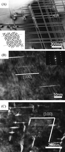
Figure 1. (A) SIM micrograph of the K711b41 specimen after thin foil preparation using FIB. (B) TEM micrograph of the as-received K711b41 specimen. (C) The same area with (B) but tilted 25°. The crowded dark spots were considered as the damages of FIB process.
In order to study the temperature effects on the helium microstructure, some thin foils of specimens were heated up to a maximum temperature of about 1040 °C and kept for 10–15 min on a single-tilt heating holder (GATAN, Model 628, USA). The heating and cooling rates were about 100 °C/min. During heating, it was generally difficult to obtain fine micrographs due to specimen drift. Therefore, the microstructure was observed just after cooling to room temperature, except for mentioned cases.
To examine the phase change of the irradiated B4C, the r001c21 specimen was crashed into powder and put in a Pt pan, followed by heat-treatment at 600 °C–800 °C for 1 h in Ar atmosphere. The heat-treated powder was identified using powder X-ray diffraction (XRD) technique on a PW-1710 diffractometer (Philips, Netherland), with CuKα radiation.
3. Results
In the K711b41 specimen irradiated at 800 °C up to a burnup of 30.1 × 10²⁰cap/cm3, the intragranular bubbles generally appeared in a flat-plate-like shape. When the specimen was adjusted to the proper angle that the bubbles were positioned parallel to the electron beam, many light-colored parallel straight lines were observed inside the B4C grains, as shown in (B). The typical lengths of these lines were in the range 10–50 nm, with most being approximately 30 nm in length. As shown in (C), when the sample was tilted, the fine lines gradually became oval-shaped with reduced contrast, which revealed the flat lenticular shape of the intragranular helium bubbles. The intragranular bubbles in any grain of K711b41 were proved to be parallel to the rhombohedral (111) planes of B4C by the selected area electron diffraction patterns.
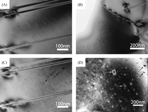
shows the transmission electron micrographs of the low-burnup specimen r001_33 from the control rod. Only a small amount of helium was produced; therefore, it was assumed that helium almost completely dissolved in the B4C lattice. (A) indicates the presence of parallel twin boundaries (stacking fault) that are frequently observed in as-received B4C crystals, as reported by Ashbee [Citation3] and Chen et al. [Citation13]. It is very difficult to determine the neutron-irradiation-induced helium bubbles in the grains. Only the strain fields along the grain boundaries indicate gathering of helium gas, as shown in (B). After the heat treatment up to 1040 °C for 15 min, as shown in (C), the invisible irradiation defects became visible by forming defect clusters (appearing as black dots). (C) is the same area as (A) after heating. (D) is taken from another grain after the heat treatment. Small helium bubbles nucleated and appeared along with the strain fields in this specimen after the heat treatment.
Figure 2. TEM micrographs of r001_33 specimen. (A) and (B) As-received specimen. (C) The same area with (A) after 1040 °C heat-treatment and observed at room temperature. (D) Another grain after heat-treatment and observed at room temperature.
In the r001c21 specimen, very small helium bubbles in the order of a few nanometers in diameter could be found. The bubbles looked approximately spherical in shape and heterogeneously nucleated, as shown in (A). They had a tendency to connect to form wavy strings approximately parallel to the (111) plane. Near the grain boundaries, there is a significant reduction in bubble density; the so-called denuded or depleted zone ((B)) as previously mentioned in many reports [Citation5–8,Citation10]. The formation of this zone is attributed to the strong defect-trapping effect of grain boundaries (i.e. sink). In contrast, the thickness of the grain boundary extended and the inner side of the grain boundary looked low density.
Figure 3. TEM micrographs of the as-received r001c21 specimen.
In the high-burnup specimen r001b21, there is no obvious difference in the size of the small helium bubbles when compared to the r001c21 specimen, as shown in (A,B). However, because more helium was produced, the bubbles had a very high density in each grain. The grain boundary bubbles are bigger and the grain boundary is thicker than in , as shown in (B).
Figure 4. TEM micrographs of the as-received r001b21 specimen.
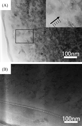
As can be seen in (A), a grid-like contrast is observed in the r001_33 specimen. The crystallographic orientation was examined using a selected area diffraction pattern, which showed that stacking faults lie on the rhombohedral {100} planes. As shown in (B), parallel to the trace of {100} planes, straight bubbles or cracks of lengths more than 200 nm are rarely observed in the as-received r001c21 specimen. As shown in (C), thick and large bubbles formed along the {100} planes in the r001b21 specimen after heat-treatment up to 1040 °C for approximately 10 min.
Figure 5. TEM micrographs of the B4C specimen. (A) As-received r001_33 specimen. Insert is structure model of B4C. (B) As-received r001c21 specimen. (C) The r001b21 specimen after heat-treatment at 1040 °C for 10 min, and observed at room temperature.
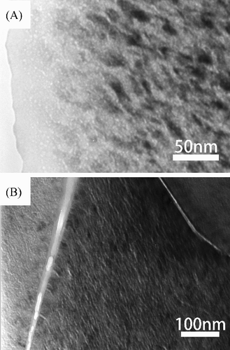
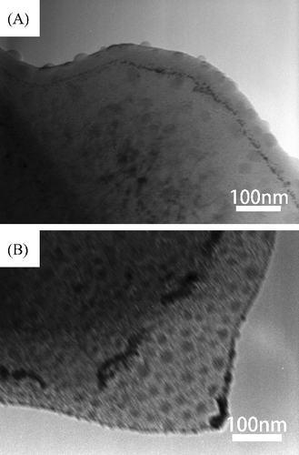
When the r001b21 specimen was heated to over 800 °C, a large number of liquid droplets suddenly appeared and quickly evaporated during TEM observation. (A) shows a micrograph of the r001b21 specimen during heating near 800 °C obtained using TEM. Hemisphere-shaped residues are visible on the specimen surface. (B) was taken at room temperature after heating up to 1040 °C; black traces remain after evaporation of the liquid drops. The formation of hemispherical drops on the surface of foils was observed for all irradiated B4C specimens, but more obviously for high burnup specimens. Because it may contaminate the inside of the TEM device, the in situ heating of the high burnup B4C specimens should be avoided, as they contain a large amount of lithium.
Figure 6. TEM micrographs of the r001b21 specimen during and after heating in TEM. (A) Observed at around 800 °C. (B) The edge of the same specimen after heating up to 1040 °C and then cooled down to room temperature.
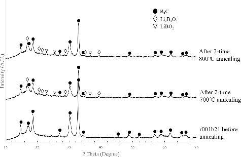
shows the XRD patterns of the r001b21 specimen after two-time heat-treatment at 700 °C and 800 °C in an Ar atmosphere for 1 h, respectively. In the XRD pattern for the 800 °C-annealed specimen, LiBO2 and Li2B4O7 are identified along with the B4C main phase. The peak heights of these compounds are slightly smaller for the specimen annealed at 700 °C for 1 h. However, after the first-time heat-treatment both at 800 °C or 700 °C, the peaks of LiBO2 and Li2B4O7 were not detected by XRD.
Figure 7. XRD patterns of the r001b21 specimen after 2-time heat-treatment at 700 °C and 800 °C for 1 h.
4. Discussion
Our TEM observations of the constant-temperature-irradiated specimen K711b41 showed that all the plate-shaped helium bubbles lie parallel to the (111) plane, which is in good agreement with previous reports for B4C specimens irradiated at temperatures lower than ∼1000 °C, presented by Jostsons et al. [Citation7], Ashbee [Citation3], and Stoto et al. [Citation6]. As Stoto et al. [Citation6] have already indicated, as several cuts in the reciprocal lattice of B4C are quite similar to each other, it is confusing to index the (110) and (111) plane, which might lead to some incorrect results for the orientation of helium bubbles in the B4C crystal. When the specimen K711b41 was capsule-irradiated in JOYO at a constant temperature of 800 °C, helium bubbles grew to a relatively large plate shape (10–50 nm in diameter). It has been reported that helium bubbles are usually accompanied by strain fields [Citation4,Citation6–9]. However, this phenomenon is not clearly observed for the K711b41 specimen in this study. It is reasonable to assume that this is because only this specimen was prepared by FIB technique, and a high-energy Ga+ beam used to fabricate thin films induces a local temperature rise of more than 1000 °C, same as for low thermal conductivity insulators such as SiO2 [Citation14]. After neutron irradiation, the thermal conductivity of ceramics generally decreases significantly due to the formation of crystalline defects [Citation15–17]. Because the local temperature rose during FIB fabrication, the local strain was mitigated in the K711b41 thin film. It has been reported that strain fields accompanying the bubbles disappeared after post-irradiation high temperature annealing at temperatures above 1400 °C [Citation4–8].
However, in the specimen irradiated in the actual control rod environment of JOYO which has a lower irradiation temperature than K711b41, the bubble size was much smaller (a few nanometers in diameter), and was mostly spherical rather than in a larger plate shape. The helium bubbles observed in the r001c21 and r001b21 specimens are tiny and spherical, with a tendency to align into wavy lines approximately parallel to the (111) plane; however, their sizes do not differ much. It is reasonable to infer that the effect of the 10B burnup mainly determines the bubble density. When comparing the K711b41 and r001b21 specimens, the shape of helium bubbles in the former (large plate-shaped) and latter (small spherical) is different, even though the 10B burnup and maximum temperature are relatively similar. Jostson et al. [Citation7] reported that small spherical bubbles were formed for the specimen irradiated at 550 °C with 81% burnup (natural B4C). However, for specimens irradiated at 700 °C and above, a flat plate-shape is developed [Citation7]. Copeland et al. [Citation10] also indicated that black-spot defects occur for lower temperature (350 °C–400 °C) irradiated specimens. Therefore, temperature seems to be the major factor affecting the helium bubble shape. A clear difference in irradiation conditions is ‘steady state irradiation’ for samples irradiated in materials testing rigs (such as K711b41 and Jostson's) and ‘cyclic or variable condition in actual control rod’ for the present specimens (such as r001_33, r001c21, and r001b21). In the actual control rod, the highest temperature was achieved near 800 °C, and the duration at that temperature was not so long; however, these conditions were experienced cyclically. This means that the generation of helium was cyclic, and there was no sufficient time for the helium to migrate and accumulate into large bubbles. However, there was a tendency to align with the (111) plane, the same plane as observed in the K711b41 specimen. In summary, in the actual control rod with a high burnup in the sodium-cooled fast reactor, small spherical bubbles are formed, and for high temperature steady state irradiation, lenticular or plate-shaped bubbles are produced. Bubble formation process should be dominated by the migration of helium atoms/vacancies in the crystal. Therefore, the large plate-shaped bubbles cannot be formed at low temperature where the helium/vacancy has poor mobility. Similarly, if the temperature is unstable, they also do not have enough time to form plate-shaped bubbles. The former aligns into a wavy plane and the latter forms onto a plane, both parallel to the (111) plane in the B4C crystal.
The heterogeneous distribution of helium bubbles in the r001c21 specimen may be caused by the migration of vacancies containing helium atoms (VHe) onto weakly-bonded interstices. Initially helium atoms are randomly generated by the neutron absorption reaction; therefore, distribution of helium is also random. Once helium bubbles are nucleated at proper sites such as the (111) planes, they grow by capturing free VHe. The difference in defect concentration generates the driving force for VHe migration. Hence, the bubbles can slowly grow. A study on helium gas release of neutron irradiated B4C pellets conducted by Suzuki et. al. [Citation18] determined that, the helium gas release rate is very low at temperatures lower than 700 °C. It is assumed that the migration of VHe in B4C at temperatures lower than 700 °C is slow; therefore, VHe are trapped by nearby helium bubbles to generate arrays of tiny bubbles. If the material is kept for a relatively long time at temperatures higher than 700 °C, these tiny bubbles will connect each other to make larger plate-shaped bubbles, as observed in the K711b41 specimen and other many reports [Citation2–12].
Other than on the (111) plane, it is confirmed that big helium bubbles or cracks are also generated on the {100} planes in some grains, possibly because the trace of slip is a good sink for VHe. These helium bubbles can be easily distinguished from the bubbles along the (111) plane by their longer length. According to Reddy et al. [Citation19] and An and Goddard [Citation20], slip in B4C can be induced by shock loading or indentation on the {100} planes by debonding of C-B-C chains under shear stress, followed by formation of amorphous bands. Chen and McCauley [Citation21] observed that mechanical scratching also induced amorphization of B4C. Therefore, one of a possible explanations for the formation of long bubbles or cracks along the {100} planes is due to the amorphous bands caused by slip or mechanical scratches during powder preparation are good sinks for irradiation-induced defects, and also are suitable places for helium bubble growth.
It was very difficult to find the trace of the lithium generated with the helium in neutron irradiated B4C, as it is generally invisible in TEM observation. The liquid phase that appeared during in situ heating of the high burnup specimen at over 800 °C () is suspected to be a consequence of the formation of lithium-bearing compounds on grain surfaces. From the XRD analysis shown in , two kinds of lithium borates are detected. The melting points of LiBO2 and Li2B4O7 are 845 °C and 917 °C, respectively. Because the Li2B4O7 have a higher melting point than that of LiBO2 and the former may only form under oxygen-rich condition, and the liquid phase was observed at around 800 C, the observed material on the specimen surface is more likely to be LiBO2. In considering the possible eutectic reaction of these two borates and the high vacuum inside TEM, the equilibrium melting points might be lower than the above value. The detection of Li-compounds in the heat-treated specimen is an evidence of the presence of Li on the surface of the specimens at that temperature. Considering the atomic size of lithium, a lot of it may remain in the interstitial sites of the B4C lattice. It can migrate to the surface easily under high temperatures, and subsequently form low melting-point compounds. Oxygen may be supplied from the oxidized surface layer, which generally exists on the surface of non-oxide ceramics [Citation22]. The reason why the borates detected only in the secondly heated specimens can be attributed for the high reactivity of metallic Li on the specimen surface. The specimen was kept in ambient atmosphere after the first heat-treatment, the lithium absorbs moisture easily from atmosphere to form lithium-H-O compounds. That compound will react with B4C during the second heat-treatment. If the temperature increases further or the surface is bombarded by the electron beam during TEM observation, lithium compounds will be evaporated away in a high vacuum.
5. Conclusions
The shape of helium bubbles in B4C, induced by fast neutron irradiation in an actual control rod of the fast reactor JOYO, was observed using TEM. The helium bubbles are tiny and spherical with a diameter of a few nanometers; the bubbles are produced inside crystals in the as-received specimens. If the burnup increases, the size of the bubbles is not affected, but the density increases. These tiny bubbles form arrays approximately parallel to the rhombohedral (111) plane. In the specimen irradiated in the materials testing rig under a constant temperature of 800 °C, the helium forms relatively large plate-shaped bubbles with a diameter of 10–50 nm, which lie parallel to the (111) plane of B4C. Other than the (111) plane, it is confirmed that large helium bubbles/cracks are sometimes generated on the {100} planes. It can be inferred that amorphous bands caused by slip or mechanical scratches during powder preparation are also good sinks for irradiation defects, and amorphous bands are a suitable place for helium bubble growth.
Finally, a liquid phase appeared during the in situ heating of the specimens at temperatures over 800 °C, which is believed to be caused by the melting of some lithium-bearing compounds. One of the possible candidates is LiBO2, which has a lower melting point of 845 °C. Considering the atomic size of lithium, it is possible that a lot of it may remain in the interstitial sites of the B4C lattice, and that it can migrate easily to the specimen surface at high temperatures.
Supplementary_Data.docx
Download MS Word (568.5 KB)Acknowledgments
The authors thank the Japan Atomic Energy Agency for their valuable aid during the course of this research for supplying the neutron-irradiated B4C pellets. This work was partially supported by the Ministry of Education, Culture, Sports, Science and Technology under the framework of the Innovative Nuclear Research and Development Program. (Research Project: Development of Highly Microstructure-Controlled Neutron Control Ceramics for Improving Safety of Fast Breeder Reactors).
Disclosure statement
No potential conflict of interest was reported by the authors.
Related Research Data
References
- Shibata K, Iwamoto O, Nakagawa T, et al. JENDL-4.0: a new library for nuclear science and engineering. J Nucl Sci Technol. 2011;48:1–30.
- Soga T, Tobita K, Mitsugi T, et al. The development of a sodium bonded type control rod in Joyo. O-arai, Higashi-ibaraki, Ibaraki (Japan): Saikuru Kiko Giho; 2000. p. 13–22 [In Japanese].
- Ashbee KHG. Defects in boron carbide before and after neutron irradiation. Acta Metall. 1971;19:1079–1085.
- Ashbee KHG, Frank FC, DuBose CKH. Voids in boron carbide. J Nucl Mater. 1973;48:193–198.
- Stoto T, Ardonceau J, Zuppiroli L, et al. Behaviour of implanted helium in boron carbide in the temperature range 750–1720 °C. Radiat Eff. 1987;105:17–30.
- Stoto T, Housseau N, Zuppiroli L, et al. Swelling and microcracking of boron carbide subjected to fast neutron irradiations. J Appl Phys. 1990;68:3198–3206.
- Jostsons A, DuBose CKH, Copeland GL, et al. Defect structure of neutron irradiated boron carbide. J Nucl Mater. 1973;49:136–150.
- Jostsons A, DuBose CKH. Microstructure of boron carbide after fast neutron irradiation. J Nucl Mater. 1972;44:91–95.
- Hollenberg GW, Mastel B, Basmajian JA. Effect of irradiation temperature on the growth of helium bubbles in boron carbide. J Am Ceram Soc. 1980;63:376–380.
- Copeland GL, Donnelly RG, Martin WR. Irradiation behavior of boron carbide. Richland, WA.USA: Conference on Reactor Materials Performance. 1972 Apr 24. CONF-720420-1.
- Cummings WV, Laidler JJ, Mahagin DE, et al. Microstructure of fast-reactor-irradiated boron carbide. Am Nucl Soc. 1972;15:742.
- Basmajian JA, Pitner AL, Mahagin DE, et al. Irradiation effects in boron carbide pellets irradiated in fast neutron spectra. Nucl Technol. 2017;16:238–248.
- Chen M, McCauley JW, Hemker KJ. Shock-induced localized amorphization in boron carbide. Am Assoc Adv Sci. 2003;299:1563–1566.
- Ishitani T, Kaga H. Calculation of local temperature rise in focused-ion-beam sample preparation. J Electron Microsc. 1995;44:331–336.
- Yano T, Miyazaki H, Iseki T. Effect of isochronal annealing on thermal diffusivity of neutron-irradiated AIN. J Nucl Mat. 1996;230:74–77.
- Akiyoshi M, Yano T, Tachi Y, et al. Saturation in degradation of thermal diffusivity of neutron-irradiated ceramics at 3 × 1026 n/m2. J Nucl Mater. 2007;367–370:1023–1027.
- Akiyoshi M, Tsuchida H, Takagi I, et al. Irradiation effects on thermal diffusivity and positron annihilation lifetime in ceramics induced by neutron and 30 MeV electron. J Nucl Sci Technol. 2012;49:595–601.
- Suzuki H, Maruyama T, Wakasa T. Postirradiation annealing of boron carbide pellet irradiated in fast breeder reactor. J Nucl Sci Technol. 1979;16:588–595.
- Reddy KM, Liu P, Hirata A, et al. Atomic structure of amorphous shear bands in boron carbide. Nat Commun. 2013;4:2483.
- An Q, Goddard WA. Atomistic origin of brittle failure of boron carbide from large-scale reactive dynamics simulations: suggestions toward improved ductility. Phys Rev Lett. 2015;115:105501–105505.
- Chen M, McCauley JW. Mechanical scratching induced phase transitions and reactions of boron carbide. J Appl Phys. 2006;100:123517.
- Park D-C, Yano T, Iseki T, et al. Effect of nitrate salts as sintering additives during the ball‐milling process of silicon nitride powders. J Am Ceram Soc. 2000;83:2967–2973.