
 ?Mathematical formulae have been encoded as MathML and are displayed in this HTML version using MathJax in order to improve their display. Uncheck the box to turn MathJax off. This feature requires Javascript. Click on a formula to zoom.
Abstract
Detection of O() atoms using one-photon resonant excitation to the
state at
nm followed by near-threshold ionisation, i.e. 1 + 1' resonance-enhanced multi-photon ionisation, has been investigated. The aim was to achieve low ion recoil, improved sensitivity, and reliable angular momentum polarisation information, with an as simple as possible laser setup. An efficient 1 + 1' scheme has been found where the VUV light for the first step is generated by difference frequency (
) VUV generation, and the ionisation step 1' uses
around 289 nm. The presented scheme induces 9 m/s recoil of the O
ion using a two-dye laser system, and zero recoil should be possible by generating 302 nm radiation with a third dye laser. While this approach is much more sensitive than a previous 1 + 1' scheme using 212.6 nm for the 1' step, we found that the relatively intense radiation of the
beam does not saturate the 1' step. To test the ability of this scheme to accurately determine branching ratios, fine structure yields, and angular distributions including polarisation information, it has been applied to O
photodissociation around 130 nm with subsequent O(
) fragment detection.
GRAPHICAL ABSTRACT
1. Introduction
Ground-state oxygen atoms, O(), which are highly reactive due to two unpaired electrons, play an important role in various chemical environments. In the atmosphere, photodissociation of O
leads to the formation of O(
) atoms, which can rapidly react with O
to form ozone, or with ozone to form two O
molecules [Citation1]. In the interstellar medium, collisions between O(
) and helium or atomic and molecular hydrogen have been identified as an important cooling process [Citation2]. In oxygen-containing plasmas, O(
) atoms are formed that play an important role in the chemistry in these complex systems [Citation3]. To better understand these processes, high-resolution lab-based experiments have to be performed.
For these experiments, it is crucial to be able to detect O() atoms in an efficient and sensitive way. Traditionally, laser induced fluorescence (LIF) was often used to detect O(
) by exciting the atoms to the
state using two 226 nm photons and detecting the
fluorescence at 845 nm [Citation4]. Later, 2 + 1 resonance enhanced multiphoton ionisation (REMPI) often became the preferred method of choice, especially in combination with detection techniques such as velocity-map imaging (VMI) [Citation5]. This 2 + 1 REMPI scheme (Figure a) uses 226 nm light to first excite O(
) to the
state by absorption of two photons, followed by the absorption of an additional photon to ionise the atoms [Citation6]. This REMPI scheme is easy to use, since only one dye laser is needed, with a wavelength that is commonly produced. It is moreover a very sensitive detection technique.
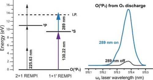
Figure 1. Overview of laser methods used in this study. a) Standard 2 + 1 (left) and threshold 1 + 1' (right) REMPI schemes for O() detection. b) Difference frequency four wave mixing scheme for generating 130.22 nm light used in the 1 + 1' scheme. Small changes in the
wavelength (see Figure ) are used for detection of O(
). c) Energetics of O
photodissociation around 130 nm. DL 1, 2, and 3 refer to the (O(
) + O(
)), (O(
) + O(
)), and (O(
) + O(
)) dissociation limits, respectively.
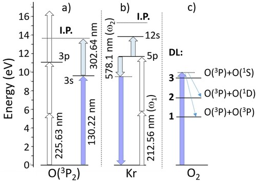
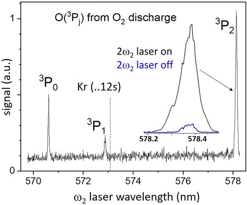
Figure 2. O total ion yield spectrum on scanning the
laser wavelength to generate tunable VUV in the vicinity of the
resonance of O(
) atoms.
However, in 2 + 1 REMPI, the three-photon energy is eV higher than the ionisation energy of the O(
) atoms, resulting in excess energy. Nearly, but not all, of this excess energy appears in the electron kinetic energy. Momentum conservation results in a slow recoil of the O
partner, which causes recoil of
eV, or 34 m/s [Citation7]. While small, this recoil velocity spoils the resolution of advanced molecular dynamics experiments, particularly those combining controlled molecular beams [Citation8] and final state-selective detection by VMI [Citation5].
In this article we focus on improving a previously used [Citation9] laser ionisation method for detection of O atoms using vacuum ultraviolet (VUV) resonant selection of O() with j = 2, 1, 0, followed by near-threshold ionisation to decrease the O
ion recoil. This 1 + 1' photoionisation scheme (Figure a) makes use of 130 nm VUV radiation to excite the O(
) atom to the
state, and
nm UV light to subsequently ionise the atoms slightly above the threshold at 302 nm. The greatly reduced recoil velocity of the O
ion paves the way towards high-resolution detection of O(
). Besides recoil, other critical aspects of laser ionisation methods include sensitivity, purity of state selection, product m state polarisation dependence, and ease or expense of use. All these aspects of the 1 + 1' detection scheme will be discussed here.
In the past, several studies have used 130 nm radiation for exciting O() atoms to the
state prior to detection. It is, for instance, possible to detect laser-induced fluorescence after this excitation step [Citation10]. Another method uses a second laser with a wavelength of 305 nm to excite the intermediate
state to high-lying Rydberg states which are then field ionised in a time-of-flight O-atom Rydberg tagging experiment [Citation11, Citation12]. This results in a high temporal and thereby kinetic energy resolution, and a negligible recoil velocity. In general, the lifetime of the Rydberg state used limits the signal levels and the combination of imaging with Rydberg tagging is not ideal due to conflicting experimental needs [Citation13]. However, the Rydberg tagging technique has been combined successfully with sliced VMI [Citation13] and this presents a suitable approach for high-resolution detection of O(
).
Lambert et al., on the other hand, employed a 1 + 1' REMPI method for photoionising O() atoms via the intermediate
state [Citation9]. In this pioneering study, two-photon resonance-enhanced difference frequency four-wave mixing (
) (Figure b) was used to generate tunable VUV radiation in the 120-132 nm region. Their work focused on photodissociation of O
(Figure c) via the
states at 124 and 120 nm, respectively, which have very large cross sections compared to excitation around 130 nm. By using two different
laser beams they generated two VUV wavelengths for dissociation via the E state, and O(
) excitation followed by ionisation at 212.56 nm (i.e. the radiation used for excitation at 2
). In the 130 nm region, the low signal to background ratio did not allow full analysis of their O(
) data.
Our work follows this study of Lambert et al. [Citation9]. We show that the use of a more intense laser beam with a longer wavelength for the ionisation step increases the sensitivity sufficiently, while at the same time creating less ion recoil. We also apply our low recoil 1 + 1' method to VUV photodissociation of O with O(
) fragment detection, and test the ability of our 1 + 1' scheme to accurately determine branching ratios, fine structure yields, and angular distributions including polarisation information.
2. Experimental methods
In this study, we used a standard molecular beam apparatus with time-sliced VMI detection. A supersonic molecular beam of neat O was generated by a Nijmegen pulsed valve [Citation14], using a backing pressure of 2.5 bar and a repetition rate of 10 Hz. A pulsed discharge in O
created O(
) atoms [Citation15] that we used for testing the overall detection sensitivity. For the O
photodissociation study, however, the discharge was switched off. The skimmed molecular beam was intersected by photolysis and probe lasers that were perpendicular to the molecular beam propagation direction. The O(
) atoms resulting from the discharge or photodissociation of O
were state-selectively ionised and the resulting ions were extracted perpendicular to the molecular beam propagation direction and focused onto a position-sensitive detector. This detector consisted of a microchannel plate (MCP) assembly coupled to a phosphor screen. A fast high-voltage switch with a pulse width of 100 ns was employed to gate the central slice of the atomic oxygen product's Newton sphere. Analysis of the images using the FINA program [Citation16] indicated a typical
degree of slicing. The VMI image was recorded by a CMOS camera and event counting was performed during the data acquisition. The final images were accumulated over
laser shots. Conversion from pixel to m/s was obtained from an O(
) image from O
photodissociation at 225.63 nm.
The 130 nm radiation used for exciting O() atoms to the
state was generated by difference frequency mixing (2
–
) in krypton (Kr) gas as the nonlinear medium, where the
and
frequencies were generated by two dye lasers, which were simultaneously pumped by a 532 nm laser beam from a single Nd:YAG laser operated at 10 Hz. The
and
laser beams were spatially and temporally overlapped and focused into a stainless-steel gas cell that was filled with 20 mbar of Kr. The VUV radiation, together with the
and
generation beams, passed through a MgF
lens at the output end of the Kr cell, which was positioned to create a collimated VUV beam of 4 mm diameter, and the
and
laser beams came to a focus outside the apparatus. The
laser beam was fixed at 212.55 nm to coincide with the two-photon resonance transition
of Kr [Citation17], and the
laser was tuned in the range of 570-579 nm to cover the wavelength range of the VUV for state-selective excitation of the O(
) photofragments to the
state. The O(
) products were probed using a two-colour resonance-enhanced multiphoton ionisation (1+1' VUV + UV REMPI) scheme in which the same VUV laser beam was used for the excitation step and a
nm laser was used to subsequently ionise the atoms. This 1' ionisation beam was produced by frequency-doubling a fraction of the
laser beam and was counter-propagating to the VUV beam. The size and flux of this
beam was adjusted for maximum signal using a 20 cm focal length lens positioned 25-30 cm from the crossing point. The
laser wavelength was scanned by
cm
over the nascent O-atom Doppler profile in order to uniformly detect all O
fragments with different velocities. A wavemeter was used to calibrate all reported wavelengths.
All lasers were linearly polarised and arranged such that the polarisation of the VUV was parallel to the plane of the MCP detector, which is the horizontal (H) plane in our setup. For the 1 + 1' REMPI detection scheme, the ionising laser polarisation was set at 54.7 (magic angle), M, relative to the exciting laser polarisation in order to negate any polarisation sensitivity of the ionisation step when measuring the angular momentum alignment of the nascent O(
) products. This set of polarisations is labelled from here on as HM. We also compared ionisation at the magic angle with the ionising laser polarisation set parallel and perpendicular (V) to the detector plane, thus HH and HV polarisations, respectively for our apparatus. The detector homogeneity was normalised using the geometry VM.
3. Results
A weak resonant O signal was obtained when intersecting the O(
) discharge beam [Citation15] with radiation from the Kr cell. Under these conditions the ionisation step of the 1 + 1' REMPI scheme is provided by 212 nm radiation, in accord with the results of reference [Citation9]. A significant increase in O
signal – up to 34 times at the highest laser fluence – was obtained on addition of the
laser beam. Figure shows the resulting O
signal on scanning the wavelength of the
laser from 569−579 nm. Three peaks are observed at the expected positions for resonant excitation of the O(
) states. The VUV wavelengths and energies correspond to O(
): 130.214 nm, 9.521 eV, O(
): 130.483 nm, 9.502 eV, and O(
): 130.600 nm, 9.493 eV, in accord with the literature values [Citation18]. Off-resonance, a background O
signal is observed due mainly to the 212 nm radiation, as seen by pumping the Kr out of the cell and thereby removing the VUV radiation.
The relative intensities of the O() peaks in Figure are quite different from those measured for a similar discharge beam [Citation15] with 2 + 1 REMPI at
nm, or for a microwave discharge beam with two-photon resonant degenerate four-wave mixing spectroscopy [Citation19], which show a regular decrease in signal on going from j = 2 to j = 1 to j = 0. The 1 + 1' REMPI spectrum in Figure , however, is quite irregular, with a particularly weak
peak, and a
peak that is stronger than
. VUV generation in this 130 nm region is affected by accidental resonances in Kr at 2
+
, see Figure b, which increase XUV frequency sum generation at the cost of the desired difference frequency VUV. The MgF
lens at the exit of the Kr cell blocks any XUV radiation from the detection chamber. In particular, the allowed
transition at
nm appears to most affect the sensitivity for O(
). The scan shown in Figure was taken at a Kr pressure of 20 mbar. VUV output at the O(
) wavelength can be increased by lowering the Kr pressure, but this leads to an overall decrease in signal across the full wavelength range of Figure . Similar effects most likely explain the
intensity ratio.
3.1. Photodissociation of O at 130 nm
The improved 1 + 1' REMPI method was applied to VUV photodissociation of O at the three VUV wavelengths nearby 130 nm used for resonant detection of O(
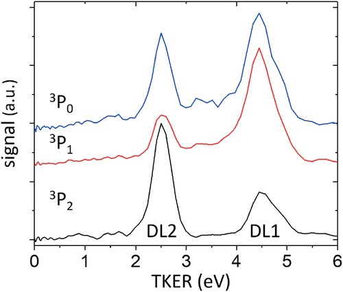
) fragments. While dissociation limit (DL) 3 (Figure c) is accessible below 133 nm, dissociation is found to take place only to limits DL 1 and 2 [Citation9], creating faster and slower O(
) atoms, respectively. Relative branching ratios for DL 2/DL 1, product channel angular distributions, and information on m state polarisation of the products are obtained. The results will be discussed in the following.
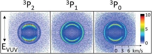
O images obtained by crossing an O
molecular beam with VUV radiation tuned to O(
) resonances are shown in Figure . The VUV causes both O
photodissociation and O(
) excitation as the first step in the 1 + 1' process, where 1' is driven by the counter-propagating
laser beam. The
field direction of the linearly polarised VUV (
) is indicated in the figure and the polarisation direction of the
ionisation laser is set at
(magic angle) relative to
. Two recoil rings are apparent in each image, corresponding to the fast (DL 1) and slow (DL 2) dissociation channels. Qualitatively, the fast ring peaks in the horizontal direction, i.e. perpendicular to
, which indicates a direct (axial) dissociation via a perpendicular (
) transition in O
. The slower ring peaks along the vertical direction, i.e. along
, indicating a parallel (
) transition.
Figure 3. Velocity mapped O images (HM geometry), after symmetrisation and correction for the detector inhomogeneity, from O
photodissociation with VUV radiation tuned to 1 + 1' REMPI of the O(
) resonances shown in Figure . The
field direction of the linearly polarised VUV radiation is shown on the left and the colour bar on the right codes the signal intensity.
A broad background, which is still partially present when the VUV wavelength is tuned outside the Doppler profile of the O() resonances, underlies the recoil rings in each image. It was not possible to reliably remove this background directly by any experimental ‘on-off’ subtraction method. Furthermore, by observing the regions in the image where the recoil signal is weak or not present, it was also apparent that the background varies slightly with the polarisation direction of the intense ionisation laser. For this reason, images taken with the ionisation laser polarisation set at the magic angle were fully analysed after subtracting a constant value at each pixel, typically
of the maximum intensity. The subtracted value was chosen so that no negative intensity appeared across the background-corrected or inverted image. The FINA inversion program [Citation16] was used to extract the DL 1 and DL 2 angular distributions and the relative branching ratios from the kinetic energy distributions shown in Figure , despite the possible effect of O-atom alignment on the cylindrical symmetry of the images. Integration of each peak in the TKER curves yielded the following relative branching ratios
at each dissociation wavelength:
for O(
) at 130.214 nm,
for O(
) at 130.483 nm, and
for O(
) at 130.600 nm.
Figure 4. Total kinetic energy release (TKER) distributions from the images shown in Figure , after inversion using FINA. The and
curves are given a vertical offset for clarity.
Angular distributions for the recoil rings in the inverted images are plotted in Figure . Angular distributions from O() DL 2 using the same background subtraction correction and HV, HM, and HH polarisations are shown in Figure . The observed angular distributions are described by the equation [Citation9]
(1)
(1) where
are Legendre polynomials. This equation was used for fitting the image data (scattered points) in Figures and . Best fitting coefficients for
and
are listed in Table . As discussed in more detail later, a non-zero value for
is an indication of alignment effects arising from the REMPI process for O(
) detection.
Figure 5. Angular data (scattered points) and fits (solid lines) for the DL 1 and DL 2 recoil rings obtained from the images with HM polarisation shown in Figure , after inversion using FINA. Values for and
recovered by the fitting routine are listed in Table .
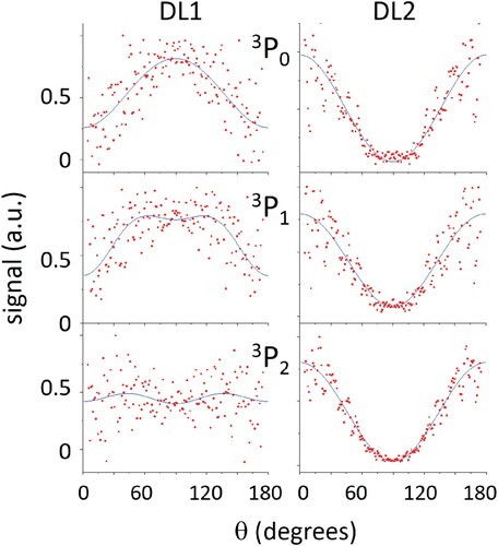
Figure 6. Angular data (scattered points) and fits (solid lines) for the DL 2 recoil ring obtained from the O() images with HV (red), HM (blue), and HH (black) polarisation. Values for
from the fitting routine are indicated for each curve. The signals for HV and HM polarisation are given a vertical offset for clarity.
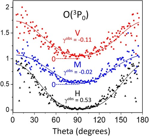
Table 1. Anisotropy parameters β and γ, and fractional populations of the different λ levels, where
, for the different O(
) states for the two different dissociation limits (DL).
3.2. Analysis
Molecular dissociation by polarised radiation can produce polarised atomic fragments, and analysis of this nascent atom polarisation affords unique insights into the dissociation process [Citation20, Citation21]. In the case of molecular oxygen, for example, photoexcitation from the upper state of the Schumann-Runge continuum leads to 89% of the produced O(
) atoms in the m = 0 state [Citation9, Citation22–24], which can be understood based on the long-range quadrupole-quadrupole interaction [Citation22]. Assuming that the electron spin is not polarised, the
-populations are the same for both atoms in a Σ state. The co-partner O(
) atoms are therefore expected to show similar m-state polarisation. The strong alignment of nascent O(
) atom products results in their ionisation probability depending significantly on the recoil direction in the laboratory (laser polarisation) frame, i.e. the expected angular distribution is perturbed by the atomic alignment. Unraveling nascent atom polarisation information from photofragment imaging data is a non-trivial task which is dependent on the details of the REMPI process used [Citation25]. A full polarisation study requires using linearly and circularly polarised laser beams in several pump-probe geometries. We use here only linear laser polarisation and a semi-classical analysis where coherence effects, which can be substantial for O(
) atoms [Citation26, Citation27], are not probed. Our method provides two-dimensional angle-speed distributions of O
ions, while the strategy is to determine alignment information based on the known alignment sensitivity of the O(
)+
O(
) transition.
In general, an experimentally observed photofragment angular distribution can be described by the following function [Citation9]
(2)
(2) Here, β is the spatial anisotropy parameter describing the angular distribution of the photodissociation fragments, θ is the angle between the dissociating laser polarisation and the recoil velocity,
is a Legendre polynomial of order 2, λ is the projection of the angular momentum j on the recoil direction,
is the fractional population of each λ level, and
is the probe frame angular detectivity function, which depends on the transition probed. When
is known, the alignment-free β parameter and the λ populations
can, in principle, be determined from
and
for each j-state. In reference [Citation28],
equations were derived for two-photon excitation of O(
) to the
state where the intermediate state was assumed to be only the
state. The same derivation becomes rigorous with our 1 + 1' REMPI scheme, where the upper state becomes the ion continuum with j = 0, 1, 2 allowed to be populated after ionisation. In reference [Citation28], the ionisation step was assumed to be saturated, i.e. only the resonant transition is polarisation sensitive. We can vary the polarisation direction of the
ionisation laser beam as a check of the ionisation step saturation. The H polarised VUV laser beam drives both photodissociation and the resonant excitation step of the O(
) ionisation scheme. In the O
photoexcitation step, a subset of the randomly oriented O
molecules that are pointing primarily along (for a parallel
transition) or perpendicular (for a
transition) to the
field direction of the linearly polarised VUV beam is photo-selected, and their subsequent direct (axial) dissociation creates a product angular distribution described by β. Photodissociation also creates O-atom fragments that are aligned in the molecular frame with respect to the interatomic bond axis. Only those molecules pointing directly along
create fragment atoms travelling in the
direction, and these atoms are excited to the
state following the selection rule
, with m the projection of j on the laser polarisation direction. Along this direction, the m = 2 state of
cannot be excited, and when starting from the
state, the intermediate state becomes aligned since only the m = 0 level of the
upper state is populated. When
as for the DL 2 channel, 2 + 1 REMPI at 226 nm and 1 + 1' REMPI via the
state are less sensitive for m = 2 detection, as pointed out by Lambert et al. [Citation9]. Most excited molecules are not pointing directly along
, and the recoil frame is thus rotated from the molecular frame. The projection of m onto the recoil frame results in mixing of m levels and thus excitation to the
of the
atom. Similar arguments provide an excitation pathway for the
state of O(
) via
to
in the ion continuum. In the recoil frame, all λ states can thus be detected.
Assuming that the nascent oxygen atoms are in a specific fine structure state, the probe frame angular detectivity functions for a two-photon transition are given by [Citation28]
(3)
(3) with
(4)
(4) Here,
is a geometrical factor [Citation28], and
are density matrix moments, which are given by
(5)
(5) with
the population of the
state. For a pure
state, this results in
(6)
(6) For the O(
) system we find that
,
and
for
,
and
for j = 1, and
for j = 0 for the
intermediate state [Citation28]. Altogether, this results in analytical expressions for
and
in terms of β,
, and
:
(7)
(7) From these expressions, in combination with
(8)
(8) we can analytically determine β and the
fractional populations from
and
.
Table contains the resulting β and values we found for all images. In the case of O(
) there are too many parameters to obtain one unique solution. We therefore determined the ranges of possible fractional populations. We obtain one solution with a positive β, and one with a negative β value. For DL 2, we only listed the solution for the positive β value, since β is also positive for the other two j states. For O(
) DL 1, for which the signal-to-noise ratio in the experimental data is low (Figure ), it is unclear which solution is more probable and we therefore listed both solutions.
General trends are that for DL 2, β is close to 2, indicating that the transition is close to parallel. The observed anisotropy parameter for DL 1 is smaller than 1 and even negative for O(), which indicates that this transition is more perpendicular. The most significant alignment effects, based on
, appear in the DL 1 channel where for O(
) DL 1 the most population is found in the
state.
4. Discussion
1 + 1' REMPI detection of O() atoms using resonant VUV radiation around 130 nm has been described here as an alternative to standard 2 + 1 REMPI at 226 nm. By applying intense
radiation for the ionising step, the sensitivity of 1 + 1' detection is greatly increased (see the inset of Figure ), and this approach provides much lower ion recoil, i.e. 9 m/s instead of 34 m/s for 2 + 1 REMPI. While recoil is not an issue in this or most other photodissociation experiments, the separation of the resonant excitation and the ionisation step does allow probing the polarisation sensitivity of the method. In terms of relative fine structure yields, the interfering resonances for VUV generation in the 130 nm region made a measurement of the total j-dependent O(
) relative yields unreliable in this study. This problem could be addressed by using a secondary calibration method such as 2 + 1 REMPI under the same conditions.
1 + 1' REMPI should be competitive with 2 + 1 REMPI for the following reasons. The resonant state of O(
) is the lowest optically allowed excited state, with a lifetime of
ns [Citation29] and, for excitation from the ground state, a maximum absorption cross section
, assuming the lineshape of a Doppler profile at 300 K [Citation30]. We estimate our VUV production at roughly
photons/pulse [Citation31] focused to
cm
, which for a pulse length
ns yields a rather large excitation probability of
, with
. For both 1 + 1' and 2 + 1 REMPI, the ionisation rate is usually smaller than the resonant excitation rate. However, when using strongly focused 226 nm radiation, the ionisation step is expected to ‘saturate’, and thus not affect the polarisation sensitivity. In this study with 1 + 1' REMPI, the intense (2.6 mJ/pulse)
pulse is also well-focused in order to drive ionisation during the 4 ns pulse length, but the O(
) polarisation dependences (Figure ) show that saturation is clearly not reached. While the VUV flux is relatively low, the high-energy photons create background at the mass/charge ratio of O
, as observed in Figure . The intense UV radiation used for O(
) 2 + 1 REMPI at 226 nm, however, can cause similar effects. For example, more O(
) signal usually arises from two-photon (VUV equivalent) dissociation of O
than from one-photon UV dissociation [Citation32].
In future studies, the use of a third dye laser for the 1' ionisation step could have several advantages. First of all, the ion recoil could be reduced even further by tuning the wavelength of this laser to ionise just above the threshold. Second, a common state for exciting and the same wavelength for ionising the different fine structure states could be used, making the REMPI spectra easier to interpret. Additionally, the 1' ionisation wavelength could then be tuned to reach an autoionising Rydberg state to possibly obtain a higher ionisation efficiency while maintaining the low ion recoil.
4.1. Photodissociation of O in the 130 nm region
O absorption in the range from 130 to 137 nm is most interesting as the only spectral region where two allowed transitions, i.e. the high energy tail of the
Schumann-Runge continuum (SRC), and
, are known to overlap with similar intensity [Citation33, Citation34]. Both upper states in these transitions undergo avoided crossings with Rydberg states of the same symmetry, causing shoulders in the repulsive walls of each potential energy curve, which are challenging to quantify accurately. Adiabatic correlation diagrams connect the B state to (O(
) + O(
)) of DL 2, and the
state to (O(
) + O(
)) of DL 1. Advanced theory [Citation35, Citation36] predicts interference effects resulting from overlap, where the maximal contributions by the weaker
state peak around 135.1 and 131.5 nm. Qualitatively, it can thus be expected that the DL 2 branching ratios will show a strong dip at these wavelengths. Our branching ratio data is quite limited in that single O(
) fine structure states are detected, each at a different dissociation wavelength near 130 nm. Furthermore, due to the varying strength of the generated VUV, it was not possible to directly calibrate the j-state yields. Measurements by Lambert et al. [Citation9] in this spectral region suggest that the relative O(
) fine structure yields are statistical, i.e. the population ratios for j = 2:1:0 are equal to 5:3:1, respectively, for the O(
) products from both DL 1 and DL 2. This can be expected given the large kinetic energy releases for both channels. Assuming this to be the case, the relative DL 2/total branching ratios we measure can be compared with the O(
) branching ratios measured previously, which are shown in Figure .
Figure 7. DL 2/total branching ratios in the 127–136 nm region. Closed circles are from the present work with O() detection (assuming a statistical j-state distribution), open triangles from [Citation9], open squares from reference [Citation37], and the continuous dashed-line is from reference [Citation38].
![Figure 7. DL 2/total branching ratios in the 127–136 nm region. Closed circles are from the present work with O(3Pj) detection (assuming a statistical j-state distribution), open triangles from [Citation9], open squares from reference [Citation37], and the continuous dashed-line is from reference [Citation38].](/cms/asset/68abede1-82e3-4075-ba07-fcc3939c88af/tmph_a_1979264_f0007_oc.jpg)
Our data and that of reference [Citation9] are from a cold molecular beam of O and narrow band lasers, while references [Citation37, Citation38] employed samples of O
in a gas cell. A lamp with monochromator light source was used in [Citation37], while data from Nee and Lee were obtained using a continuously tunable VUV synchrotron source [Citation38]. While the scatter in experimental data is significant, due to the different light source bandwidths and/or discrete points reported, the overall agreement is reasonable. It is apparent especially from our own data that the branching ratios oscillate rapidly in this wavelength region, on a very fine wavelength scale.
In general, the alignment-free angular anisotropy parameters β extracted from our data are also consistent with the states involved in the adiabatic correlation diagram, where excitation to the B-state leads to for DL 2 and excitation to the
state leads to
for O(
) DL 1, see Table . These measured values suggest that curve crossing after optical excitation (which would lead to less extreme values of β (
)), is present but minor. The anisotropy parameter for O(
) DL 1 suggests that curve crossing here has a larger effect. While Lambert et al. did not report β values at 130.2 nm or 132.7 nm, their O(
) image at 132.7 nm shows quite similar features as our images shown in Figure .
Our population values presented in Table are less certain than desired and will also be affected by the curve crossing mentioned above. Lambert et al. [Citation9] did not report alignment information at 130.2 nm or 132.7 nm, and their results for the strong absorption peaks around 124 and 120 nm could be affected by a polarisation dependence of the ionisation step (their laser polarisation corresponds to our HH in Figure ). In general, their analysis indicated a dominant
component for DL 2, which is not so clear from our data. A dominant
component was found for nascent O(
) fragments from O
photodissociation to DL 2 in the 120–125 nm region, as well as across the
Schumann-Runge continuum [Citation9, Citation22–24]. Previous measurements in the 140–165 nm region did not detect any significant alignment of the O(
) products for DL 2 [Citation22]. At present there are no simple predictions for alignment of DL 1 fragment atoms produced via
excitation. Our data for O(
) DL 1 suggests a dominant
component.
Our results can moreover be compared to predictions by adiabatic correlation diagrams. For DL 1, the states correlates with the production of O(
) + O(
), whereas for DL 2, the B-state is correlated with O(
) + O(
) [Citation39]. The adiabatic correlation diagrams therefore predict that no O(
) will be formed for both dissociation limits, and that no O(
) will be formed for DL 1. Since our results are not in agreement with these simple predictions, this is another indication that curve crossing after optical excitation and contributions from Rydberg states play a role at the high photon energies used in this study.
The combination of our 1 + 1' REMPI method with a second VUV source tuned in small steps across the 130-137 nm region should lead to a more quantitative understanding of the allowed VUV transitions of O.
5. Conclusions & outlook
We have presented a novel 1 + 1' near-threshold REMPI scheme for the detection of O() atoms. This scheme induces a recoil of only 9 m/s to the O
ion, instead of 34 m/s for the generally used 2 + 1 REMPI scheme. By using 302 nm radiation from a third dye laser for the 1' ionisation step, it should even be possible to approach zero recoil. Our scheme is more sensitive than the 1 + 1' scheme from Lambert et al. [Citation9], which uses 212 nm radiation for the 1' step, and therefore results in higher signal levels. It can moreover be used to extract polarisation information for the O(
) atoms, as demonstrated by our investigation of the photodissociation of O
around
nm.
The low ion recoil in combination with the good sensitivity of our scheme provides good prospects for applying this REMPI scheme to molecular scattering studies in crossed beam experiments combining controlled molecular beams and VMI. In these studies, the ion recoil is one of the limiting factors for the resolution that can be obtained. Moreover, signal levels are generally low, such that a high sensitivity is required. Scattering processes of interest include inelastic O() + He / H
and reactive C(
) + NO(
)
CN(
) + O(
) collisions, which are both considered as important processes for astrochemistry.
Acknowledgments
The authors thank Niek Janssen and André van Roij for expert technical support and Xingan Wang, Vikram Plomp, and Zhong-Fa Sun for fruitful discussions.
Disclosure statement
The authors declare no competing interests.
Additional information
Funding
References
- S. Chapman, Edinb Dublin Philos. Mag. J. Sci. 10, 369–383 (1930).
- F. Lique, J. Kłos, M.H. Alexander, S.D. Le Picard and P.J. Dagdigian, Mon. Not. R. Astron. Soc.474, 2313–2322 (2018).
- J.Y. Jeong, J. Park, I. Henins, S.E. Babayan, V.J. Tu, G.S. Selwyn, G. Ding and R.F. Hicks, J. Phys. Chem. A. 104, 8027–8032 (2000).
- W.K. Bischel, B.E. Perry and D.R. Crosley, Chem. Phys. Lett. 82, 85–88 (1981).
- A.T.J.B. Eppink and D.H. Parker, Rev. Sci. Instrum. 68, 3477–3484 (1997).
- D.J. Bamford, L.E. Jusinski and W.K. Bischel, Phys. Rev. A. 34, 185–198 (1986).
- A.G. Suits, Rev. Sci. Instrum. 89, 111101 (2018).
- S.Y.T. van de Meerakker, H.L. Bethlem, N. Vanhaecke and G. Meijer, Chem. Rev. 112, 4828–4878 (2012).
- H.M. Lambert, A.A. Dixit, E.W. Davis and P.L. Houston, J. Chem. Phys. 121, 10437–10446 (2004).
- H.F. Döbele, M. Hörl, M. Röwekamp and B. Reimann, Appl. Phys. B. 39, 91–95 (1986).
- C. Lin, M.F. Witinski and H.F. Davis, J. Chem. Phys. 119, 251–255 (2003).
- B. Jones, J. Zhou, L. Yang and C.Y. Ng, Rev. Sci. Instrum. 79, 123106 (2008).
- H.A. Cruse and T.P. Softley, J. Chem. Phys. 121, 4089–4096 (2004).
- B. Yan, P.F.H. Claus, B.G.M. van Oorschot, L. Gerritsen, A.T.J.B. Eppink, S.Y.T. van de Meerakker and D.H. Parker, Rev. Sci. Instrum. 84, 023102 (2013).
- Z. Farooq, D.A. Chestakov, B. Yan, G.C. Groenenboom, W.J. van der Zande and D.H. Parker, Phys. Chem. Chem. Phys. 16, 3305–3316 (2014).
- J.O.F. Thompson, C. Amarasinghe, C.D. Foley and A.G. Suits, J. Chem. Phys. 147, 013913 (2017).
- J.P. Marangos, N. Shen, H. Ma, M.H.R. Hutchinson and J.P. Connerade, J. Opt. Soc. Am. B. 7, 1254–1259 (1990).
- A. Kramida, Y. Ralchenko and J. Reader, NIST ASD Team: NIST Atomic Spectra Database (version 5.8) 2020. L<https://physics.nist.gov/asd>.
- E. Konz, G. Marowsky and H.G. Rubahn, Opt. Comm. 134, 75–79 (1997).
- R.J. Van Brunt and R.N. Zare, J. Chem. Phys. 48, 4304–4308 (1968).
- A.P. Clark, M. Brouard, F. Quadrini and C. Vallance, Phys. Chem. Chem. Phys. 8, 5591–5610 (2006).
- S.M. Wu, D. Chestakov, G.C. Groenenboom, W.J. van der Zande, D.H. Parker, G. Wu, X. Yang and C. Vallance, Mol. Phys. 108, 1145–1157 (2010).
- A.T.J.B. Eppink, D.H. Parker, M.H.M. Janssen, B. Buijsse and W.J. van der Zande, J. Chem. Phys.108, 1305–1308 (1998).
- B.R. Lewis, S.T. Gibson, T.G. Slanger and D.L. Huestis, J. Chem. Phys. 110, 11129–11132 (1999).
- A.G. Suits and O.S. Vasyutinskii, Chem. Rev. 108, 3706–3746 (2008).
- M. Brouard, R. Cireasa, A.P. Clark, T.J. Preston, C. Vallance, G.C. Groenenboom and O.S. Vasyutinskii, J. Phys. Chem. A. 108, 7965–7976 (2004).
- A.J. Gilchrist and G.A.D. Ritchie, J. Chem. Phys. 138, 214307 (2013).
- M.C.G.N. van Vroonhoven and G.C. Groenenboom, J. Chem. Phys. 116, 1965–1975 (2002).
- S.S. Tayal, Phys. Scr. 79, 015303 (2009).
- D.A. Parkes, L.F. Keyser and F. Kaufman, Astrophys. J. 149, 217–223 (1967).
- S.J. Hanna, P. Campuzano-Jost, E.A. Simpson, D.B. Robb, I. Burak, M.W. Blades, J.W. Hepburn and A.K. Bertram, Int. J. Mass Spectrom. 279, 134–146 (2009).
- B. Buijsse, W.J. van der Zande, A.T.J.B. Eppink, D.H. Parker, B.R. Lewis and S.T. Gibson, J. Chem. Phys. 108, 7229–7243 (1998).
- N. Balakrishnan, M.J. Jamieson, A. Dalgarno, Y. Li and R.J. Buenker, J. Chem. Phys. 112, 1255–1259 (2000). doi:https://doi.org/10.1063/1.480657.
- B.R. Lewis, S.T. Gibson, F.T. Hawes and L.W. Torop, Phys. Chem. Earth 26, 519–526 (2001). doi:https://doi.org/10.1016/S1464-1917(01)00040-X.
- A.C. Allison, S.L. Guberman and A. Dalgarno, J. Geophys. Res. 87, 923–925 (1982).
- S.T. Gibson and B.R. Lewis, J. Elec. Spec. Rel. Phen. 80, 9–12 (1996).
- L.C. Lee, T.G. Slanger, G. Black and R.L. Sharpless, J. Chem. Phys. 67, 5602–5606 (1977).
- J.B. Nee and P.C. Lee, J. Phys. Chem. A 101, 6653–6657 (1997).
- Y. Huang and R.J. Gordon, J. Chem. Phys. 94, 2640–2647 (1991).