
Abstract
Equine cutaneous fungal granulomas have been previously referred to in New Zealand (Fairley Citation1998), and are described in the veterinary literature from around the world, including North America and Australia (Pascoe and Summers Citation1981; Genovese et al. Citation2001; Valentine et al. Citation2006), but no peer-reviewed reports appear published in the literature in New Zealand. Described here is a case of multiple cutaneous fungal granulomas caused by Alternaria spp. in a horse in New Zealand.
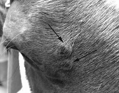
A 2-year-old chestnut Quarterhorse gelding was observed to have five non-painful and non-pruritic cutaneous nodules on the head and poll; it was unknown how long the nodules had been present. Two of the masses were adjacent to each other, medial to the right eye (), two were either side of the neck at the poll, and one was in the left frontal region. Clinical examination revealed the lesions were all ˜10 mm in diameter, and projected approximately 5 mm from the skin's surface. The masses were partially alopecic and darkly pigmented, and the surrounding skin appeared grossly normal.
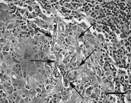
An excisional biopsy was performed on one of the masses near the poll. This mass was chosen as it allowed easy excision, with a good margin and simple closure. The excised nodule was fixed in 10% neutral buffered formalin, processed into paraffin blocks, cut at 5 μm, and stained with H&E, for histopathological examination. There was a well-demarcated nodular deep dermal infiltrate forming granulomas that consisted of a central core of cellular debris and neutrophils, surrounded by lymphocytes, epithelioid macrophages, and small numbers of multinucleate giant cells. Small numbers of negative-staining, non-pigmented fungal hyphae were present both within multinucleate giant cells and free within central areas of necrosis. Periodic acid-Schiff (PAS) stain showed that the fungal hyphae were septate, had non-parallel walls, and were 10–20 μm in diameter (). The histological appearance was consistent with a fungal granuloma.
Figure 1. Photograph of the area adjacent to the right eye of a 2-year-old chestnut Quarterhorse gelding with multiple cutaneous fungal granulomas caused by Alternaria spp. Two partially alopecic darkly pigmented nodules of about 10 mm diameter are visible (arrows).
The four remaining nodules were removed surgically, with wide margins. To confirm the initial diagnosis, one mass was fixed in formalin and again routinely processed for histological examination. As before, the mass was consistent with a fungal granuloma. Another of the excised masses was bisected, and the cut surface inoculated onto Sabouraud's dextrose agar, with antibiotics (100 mg chloramphenicol and 40 mg gentamicin per litre) (Fort Richard Laboratories Ltd, Auckland, NZ). The plates were sealed, and placed in an aerobic incubator for 3 weeks at 28°C. The plates were examined at least once a week during this time for evidence of fungal growth but none was observed. As fungal culture was unsuccessful, DNA was extracted from formalin-fixed sections of the second mass removed from the horse, and was amplified using PCR, as described previously (Munday et al. Citation2006). Amplicons were sequenced and compared with those within GenBank (http://www.ncbi.nlm.nih.gov/genbank), using the basic local alignment search tool (http://www.ncbi.nlm.nih.gov/blast). The amplified DNA sequences were found to be >99% similar to those of both Alternaria tenuissima and Alternaria alternata. Due to the high degree of conservation within these two fungi, the precise species of Alternaria within the granulomas could not be determined. Seven weeks after the surgery, no recurrence of lesions was detectible.
Equine cutaneous fungal granulomas have been reported in both humid and dry climates (Genovese et al. Citation2001; Valentine et al. Citation2006; Schwarz et al. Citation2009). Fungal granulomas comprised around 2% of all cutaneous nodular and proliferative lesions submitted for histology in the northwest of the United States of America (Valentine Citation2005). Similarly, fungal granulomas were reported to comprise 2.6% of all equine skin lesions submitted to a diagnostic laboratory in southeast Queensland, Australia (Pascoe and Summers Citation1981). As in the present case, cutaneous fungal granulomas caused by Alternaria spp. are reported to occur most frequently around the heads of younger horses (Coles et al Citation1978; Genovese et al. Citation2001). This is suggested to be due to the increased likelihood of skin trauma, allowing the fungi entry into the dermis (Genovese et al. Citation2001; Valentine et al. Citation2006).
Figure 2: Light photomicrograph of a section of fungal granuloma from the skin of a 2-year-old chestnut Quarterhorse gelding with multiple cutaneous fungal granulomas caused by Alternaria spp. Fungal hyphae (arrows) are septate, and have non-parallel walls (PAS, bar=35 μm).
Tissue biopsy is the most appropriate sample for fungal culture, and although it is reported that Alternaria spp. grow in most routine media used in laboratories, potato-carrot agar may be preferable (Pastor and Guarro Citation2008).
Different treatment options used for fungal granulomas are immunotherapy, anti-fungal chemotherapy, surgery (Scott and Miller Citation2003), or thermocautery (Knottenbelt Citation2009). Surgical excision is reported to have good success where wide margins can be achieved (Scott and Miller Citation2003; Valentine et al. Citation2006). A combination of oral fluconazole for 10 days (a loading dose of 14 mg/kg given once followed by 5 mg/kg every 24 h) and oral potassium iodide at a dose of 30 mg/kg every 24 h for 30 days was successful in treating one case (Schwarz et al. Citation2009). There have been reports of spontaneous remission of multiple lesions after a diagnosis was made on one lesion (Scott and Miller Citation2003), although another lesion persisted for 1.5 years before it was excised surgically (Valentine et al. Citation2006). In the case presented here, surgical treatment was considered the most appropriate therapy, and appears to have been curative.
In conclusion, Alternaria spp. fungi are present in New Zealand and can cause equine cutaneous granlomas. PCR appears to be a valuable diagnostic method in cases where culture is unsuccessful. Evidence from the present case suggests complete surgical excision can be curative.