

Abstract
Designing a temporary substrate to adhere, multiply and form new bone tissue for the bone-forming cells is a challenge in bone tissue engineering. In this article, a new biodegradable and composite scaffolding system was fabricated using an electrospinning technique to augment bone formation using poly-caprolactone (PCL), natural polymer gelatin (GL) and nano-hydroxyapatite (nHAp). The effect of fabrication parameters on nanofiber scaffolds (PCL, PCL + GL, PCL + GL + nHAp) production, as well as MC3T3-E1 proliferation and osteogenic differentiation was investigated. Physical properties of scaffolds, such as morphology, surface area, wettability, were examined using SEM, BET method, and contact angle measurements, respectively. A pre-osteoblastic cell line, MC3T3-E1 was seeded on these scaffolds to study the proliferation by DNA assay and cell viability using live-dead assay. After 21 days of in vitro culture, more than 89% of the MC3T3 cells remained viable within PCL + GL + nHAp scaffold, in contrast to 61.9% viability observed in the plain PCL scaffold. The differentiation of MC3T3 cells into the bone phenotype was determined quantitatively using alkaline phosphatase (ALP) activity and calcium deposition as well as qualitatively using alizarin red staining. At day 21, the DNA content of PCL + GL + nHAp scaffold was 4.5 times higher than its day 1 DNA contents. At day 21, the normalized intracellular ALP activity in the PCL + GL + nHAp group was 1.2 times higher than that in the PCL + GL group, which in turn was 1.7 times larger than that in the PCL group. Moreover, at day 21, PCL + GL + nHAp scaffold showed a 12.43-fold increase in ALP activity and deposited nearly 35-fold higher calcium relative to their respective values at day 1, thus indicating that the inclusion of gelatin and nHAp significantly enhanced cell binding, long-term cell survivability, proliferation, and stimulated osteogenesis in vitro. The findings from current work show that incorporation of nHAp into the nanofibrous scaffold of PCL + GL and the use of our optimized electrospinning process provides a favorable substrate to promote bone healing.
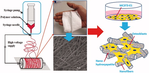
Graphical Abstract
Acknowledgment
The authors are thankful to Dr. Sudhir Arbuj, Center for Materials for Electronics Technology (C-MET), Pashan, Pune for his help during N2 adsorption analysis.
Disclosure statement
No potential conflict of interest was reported by the author(s).