

Abstract
Gallbladder torsion is a rare disease that requires immediate surgical intervention to avoid maternal and/or foetal sepsis and death. However, preoperative diagnosis is challenging because the disease has no specific symptoms. A 37-year-old pregnant woman at 34 weeks of gestation presented with severe epigastric pain. Ultrasonography and computed tomography scan findings showed a distended gallbladder without stones, floating from the hepatic bed, and laboratory examination demonstrated normal liver function; therefore, we made a diagnosis of gallbladder torsion and performed a caesarean section and an open cholecystectomy under general anaesthesia. This is the first report wherein gallbladder torsion in pregnancy was diagnosed preoperatively. Gallbladder torsion should be considered as a differential diagnosis in case of such imaging findings.
Introduction
Gallbladder torsion is a rare disease that causes acute abdominal pain. Patients are generally over 60 years; 79% are women, and the general mortality rate is 6% (Reilly et al. Citation2012). If cholecystectomy is delayed, there is a risk of gallbladder perforation, peritonitis, and sepsis (Kleiss et al. Citation2003). Since gallbladder torsion in pregnancy may cause maternal and/or foetal death, immediate diagnosis and surgery are necessary. However, it is challenging to diagnose preoperatively, and its characteristics in pregnancy remain unclear.
Case presentation
A 37-year-old pregnant woman, gravida 7, para 3, at 34 weeks of gestation, normal pregnancy course presented with severe epigastric pain and vomiting. She had a history of caesarean section (CS) for breech presentation after two vaginal deliveries and no significant medical history. The patient’s vital signs were: blood pressure, 116/64 mmHg; heart rate, 92 beats per minute and temperature 37.3 °C. There was no guarding or rebound tenderness. The laboratory data showed a white blood cell (WBC) count of 8100/mm3, a platelet count of 162,000/mm3, and C reactive protein (CRP) level of 0.16 mg/dL. Levels of aspartate aminotransferase (AST), alanine aminotransferase (ALT), lactate dehydrogenase (LDH), and total bilirubin (T-Bil) were within normal range. She was diagnosed with acute gastritis and started on intravenous fluids without antibiotics. However, 12 hours after admission, she became pyrexial (38.2 °C) and reported pain in the right upper quadrant. Acute appendicitis, small bowel obstruction, or ovarian torsion were considered. The patient’s vital signs were: blood pressure, 103/52 mmHg; heart rate, 86 beats per minute. Laboratory data showed a WBC count of 12,400/mm3, a platelet count of 164,000/mm3, and CRP level of 0.56 mg/dL; AST, ALT, LDH, and T-Bil levels were within normal range. Abdominal ultrasonography (US) revealed a distended gallbladder with thickened walls, containing no stones (). Abdominal computed tomography (CT) additionally demonstrated the body and fundus of the gallbladder were located outside the hepatic bed (). Gallbladder torsion was suspected based on the acalculous, distended, ‘floating’ gallbladder observed on imaging.
Figure 1. Imaging and intraoperative findings in this case. (A) Ultrasonography shows a distended gallbladder with thickened walls and debris storage without stones. (B) Coronal computed tomography shows a distended gallbladder (yellow arrow) with thickened walls, located outside the hepatic bed. (C) The yellow star indicates the cephalic side. Intraoperative findings show a distended, gangrenous gallbladder (yellow arrow) floating from the liver (blue star) with a 360° clockwise torsion around the cystic duct.
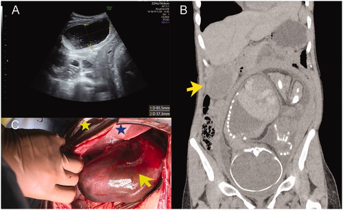
In preoperative cardiotocography, the foetal heart rate was tachycardic, and the biophysical profile was 6 points. Further, as insult to the foetus was possible due to gallbladder necrosis, we performed emergency CS and cholecystectomy under general anaesthesia. A female infant weighing 2510 g was delivered. The Apgar score was 8 at 1 minute and 9 at 5 minutes; umbilical artery pH was 7.309. The gallbladder was distended, gangrenous, and floating from the liver, with a 360° clockwise torsion around the cystic duct (). Pathological examination showed haemorrhagic infarction of gallbladder without any stones. Her postoperative course was uneventful, and she was discharged on postoperative day 6. The infant was admitted to the neonatal intensive care unit because of premature birth and discharged 22 days after birth with good progress.
Discussion
This report describes the first case of a radiological, preoperative diagnosis of gallbladder torsion in pregnancy. The diagnosis was made based on findings of a distended, acalculous gallbladder ‘floating’ from the hepatic bed and normal liver enzyme levels in our case. Moreover, the findings of Doppler-US (Boer et al. Citation2011) and magnetic resonance cholangiopancreatography (MRCP) (Bekki et al. Citation2017) are useful to diagnose and to avoid foetal radiation exposure. There are only two reports of gallbladder torsion in pregnancy (Kleiss et al. Citation2003, Lee et al. Citation2013); both cases were treated for acute cholecystitis and only diagnosed as gallbladder torsion intraoperatively. In both previous cases and the present one, the symptoms began at 17, 30, and 34 weeks of gestation, respectively, and included epigastric and right upper quadrant pain; liver enzymes were normal in all cases. Further, in all three cases, imaging showed a distended, acalculous gallbladder; however, gallbladder floatation was only confirmed preoperatively in this case. Preoperative diagnosis is associated with a good prognosis at 0% mortality (Reilly et al. Citation2012); however, diagnosis is challenging in clinical practice. Therefore, we should consider surgery when symptoms are exacerbated or antibiotics are ineffective as in other cases even if a precise preoperative diagnosis is impossible.
There are several congenital (anatomic abnormalities), acquired (severe emaciation), and physical (intraabdominal pressure changes) factors implicated in gallbladder torsion (Ohkura et al. Citation2013). In our case, the patient was thin (BMI 17 kg/m2) before pregnancy). Additionally, cholestasis due to relaxation of gallbladder smooth muscle by increased progesterone during pregnancy may be a risk factor for pregnant women.
In summary, if pregnant women present with acute-onset epigastric or right upper quadrant pain after the second trimester, US should be performed, and CT or MRCP should be considered if necessary. A diagnosis of gallbladder torsion should be considered when the gallbladder is distended without stones and ‘floating’ and/or when the liver enzymes levels are within normal ranges.
Acknowledgements
We would like to thank Editage (www.editage.com) for English language editing.
Disclosure statement
No potential conflict of interest was reported by the author(s).
References
- Bekki T, Abe T, Amano H, Fujikuni N, Okuda H, Sasada T, et al. 2017. Complete torsion of gallbladder following laparoscopic cholecystectomy: a case study. International Journal of Surgery Case Reports 37:257–260.
- Boer J, Boerma D, de Vries Reilingh TS. 2011. A gallbladder torsion presenting as acute cholecystitis in an elderly woman: a case report. Journal of Medical Case Reports 5:588.
- Kleiss K, Choy-Hee L, Fogle R, Lindsay M. 2003. Torsion of the gallbladder in pregnancy. a case report. The Journal of Reproductive Medicine 48: 206–208.
- Lee SE, Choi YS, Kim BJ. 2013. Torsion of the gallbladder in pregnancy. Journal of the Korean Surgical Society 85: 302–304.
- Ohkura Y, Hashimoto M, Sasaki K, Watanabe G. 2013. Complete acute gallbladder torsion diagnosed with abdominal ultrasonography and colour Doppler imaging. Case Reports 2013:bcr2012008460–bcr2012008460.
- Reilly DJ, Kalogeropoulos G, Thiruchelvam D. 2012. Torsion of the gallbladder: a systematic review. HPB 14:669–672.