A 4-day-old male kid of an indigenous Cretan breed was referred for evaluation because of excessive vocalizing despite normal suckling and milk intake. The kid was reported to be apparently healthy and presented in a good body condition and an adequate response to stimuli.
There was no history of congenital abnormalities in the flock or in previous parturitions of the same dam. The parturition was normal and the sibling of the referred kid did not present any deformity. There was no apparent difference in the body shape or size of the two kids. The dam was of a local polled breed. She was administered exogenous hormones (sponges impregnated with progestagen and prostaglandin F2-α) for estrus induction. After estrus detection, the goat was brought to a breeding male of the same local polled breed and returned to the flock a week later after mating. Regarding the disease control, organophosphate spray (1000 mg phoxim/L solution) was applied for the control of ectoparasites before breeding, albendazole (15 mg/kg body weight) was orally administered for deworming in the fourth month of pregnancy, and vaccination against enterotoxinemia was given in the last month of pregnancy. Furthermore, an oxytetracycline spray (2.2 g/can) was topically applied at the umbilical site of the neonates soon after their birth. The site was sprayed from a distance of 15 cm and for a minimum of 4 s, according to the manufacturer's instructions.
In addition to grass feeding and grazing in clover, almond and olive tree leaves and other feedstuff, including maize, barley, wheat and soya, were supplied during pregnancy according to the availability and the need.
Physical examination revealed a large (5 cm in diameter) bladder-like mass protruding from the perineal midline and it was difficult for urination. Observation of voiding behavior of the kid against its sibling showed the absence of normal voiding position and stranguria.
The animal showed no other clinical signs or anatomic abnormalities except for discomfort, which coincided with the filling of bladder with urine. The urethral process and the free part of penis were apparently normal except for an opening of the dorsal side of the penis. Testicles could be palpated in the scrotal area. After inserting a urethral catheter for feline use, a solution of chlorexidin was flushed through the catheter and the urethral lumen to bladder, and then bladder was emptied employing slight pressure ( and ).
Figure 1. Epispadias exstrophy complex in a goat-kid. Observe the protruding bladder (arrow) and the catheterized urethra (arrowhead).
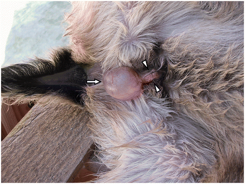
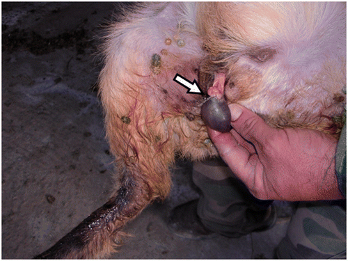
Figure 2. Epispadias exstrophy complex in a goat-kid. The bladder is easily emptied (arrow) by employing slight pressure.
The farmer declined any surgical correction of the deformity. He was given instructions on palpating the bladder and emptying it once daily, if needed. Whenever the bladder was full, the kid showed discomfort with intense vocalizing and the owner assisted the emptying of the bladder. No pain was reported either on physical examination or during daily observations of the farmer.
Hematological and biochemistry parameters, including renal function tests and urinalysis, remained within the reference intervals. A urine sample was submitted for culture, which was also negative. The animal was euthanized three months later. Necropsy revealed no other abnormality. Differential diagnoses included hernia, cutaneous cyst, ectopic urinary bladder, hypospadias, epispadias, and exstrophy–epispadias complex. The diagnosis of the latter abnormality was based on the visible defect of the lower abdominal wall with the protruding bladder and the opening of the dorsal side of the penis.
Most occurrences of congenital abnormalities are sporadic (Al-Ani Citation1998) in animals are attributed either to genetic factors despite the absence of family recurrence (Kalfa et al. Citation2009) or to prenatal exposure to chemicals or plants (Panter, Bunch, et al. Citation1990; Panter, Keeler, et al. Citation1990; Soto-Blanco and Gorniak Citation2004). The key of the congenital malformation is usually the interaction between the genome and the microenvironment of the embryo during the prenatal development (Magras Citation2002).
Congenital defects are relatively uncommon in goats including the congenital abnormalities of the genitourinary system (Sonfada et al. Citation2010). The exstrophy–epispadias complex is a complex congenital anomaly that, although rare, remains the largest genitourinary birth defect that is surgically correctable. The primary defect in exstrophy is a derangement in the midline development that shows a spectrum of severity. In its mildest form, epispadias, the dorsal urethral unit is not fused and has failed to form into a tube. Next, patients with classic bladder exstrophy have a bladder and urethra open and continuous with the abdominal wall. Also associated is a failure of the abdominal muscles, pelvic ring, and pelvic floor musculature to fuse in the midline. Cloacal exstrophy, the most severe variant, includes exstrophied hindgut tube and a more severe degree of concomitant congenital derangements of musculoskeletal, genitourinary, gastrointestinal, and neurological systems. Bony pelvic and pelvic floor muscular defects are characteristics of exstrophy (Stec et al. Citation2002; Stec Citation2011).
The etiopathogenesis of the abnormality has been experimentally studied, mainly in chicks and pigs. A premature rupture of the cloacal membrane was evidenced as the mechanism leading to this abnormality (Ebert et al. Citation2009). A sheep model was also created for the study of bladder exstrophy (Slaughenhoupt et al. Citation1996).
In reported abnormalities of the urogenital system of goats, mainly hypospadias, concurrent malformations (such as retained testicles, kidney agenesis, bone, or anorectal defects), umbilical hernia, hydrocephalus, and urinary incontinence were reported (Otiang’a-Owiti et al. Citation1997; King et al. Citation2002; Smith et al. Citation2006), but they were not detected in this case.
No corrective surgery is indicated for goats with minimal defects, even in humans, while moderate urethral defects can be treated with a two-layer closure, and major defects with excision of the external genitalia and urethrostomy (Kramer and Kelalis Citation1981; Karras et al. Citation1992; Sakhaee and Azari Citation2009). Surgery was declined in this case. In spite of the deformity, the animal did not show related health problems and did not show urine scalding of the skin. Although patients with this defect are susceptible to ascending urinary tract infections (Watson Citation1994), urinalysis including urine culture was without any pathological findings and urine culture was negative. The abnormality was not incompatible with satisfactory growth and development, since it did not affect the ability of the kid to suckle.
Despite investigation in human cases, the etiology of the abnormality is not clear. Polled ancestry, which existed in the present case, has been connected with congenital abnormalities without strong scientific evidence (King et al. Citation2002). As for more of the congenital abnormalities, a combination of environmental factors including exposure to either natural chemicals, such as certain plants, or to synthetic pharmaceutical substances and genetic alterations remains the basic incriminated factor (Sakhaee and Azari Citation2009; Kalfa et al. Citation2009). Benzimidazoles have been reported to cause developmental abnormalities in sheep fetuses (Navarro et al. Citation1998). Organophosphates have been reported to have various toxicological effects on pregnant animals and their offspring. However, malformation of the genitourinary system is not reported among them (Elmazoudy et al. Citation2011). As oxytetracycline was applied postnatally, it is less likely to have had any effect on this developmental anomaly.
Identification and evaluation of the various etiologies in animal cases remains a challenge for the future, which will contribute to the better understanding of the impact of chemical exposure in animal models for the study of pathogenesis in humans. To the authors’ knowledge, this is the first reported case of exstrophy–epispadias complex in a goat-kid.
References
- Al-Ani , FKKW , Al-Qudah , KM and AI-Rawashdeh , O . 1998 . Occurence of congenital anomalies in Shami breed goats: 211 cases investigated in 19 herds . Small Rum Res , 28 : 225 – 232 .
- Ebert , AK , Reutter , H , Ludwig , M and Rosch , WH . 2009 . The exstrophy–epispadias complex . Orphanet J Rare Dis , 4 : 23
- Elmazoudy , RH , Attia , AA and Abdelgawad , HS . 2011 . Evaluation of develpmental toxicity induced by anticholinesterase insecticide diazinon in female rats . Birth Defects Res B Dev Reprod Toxicol , 926 : 534 – 542 .
- Kalfa , N , Philibert , P and Sultan , C . 2009 . Is hypospadias a genetic, endocrine or environmental disease, or still an unexplained malformation? . Int J Androl , 32 : 187 – 197 .
- Karras , S , Modransky , P and Welker , B . 1992 . Surgical correction of urethral dilatation in an intersex goat . J Am Vet Med Assoc , 201 : 1584 – 1586 .
- King , WW , Young , ME and Fox , ME . 2002 . Multiple congenital genitourinary anomalies in a polled goat . Contemp Top Lab Anim Sci , 41 : 39 – 42 .
- Kramer , S and Kelalis , P . 1981 . Correction of total incontinence in male and female epispadias . J Pediatr Surg , 16 : 812 – 816 .
- Magras , I and Tsiligianni , ThC . 2002 . Congenital malformations and environment . JHVMS , 54 : 154 – 164 .
- Navarro , M , Cristofol , C , Carretero , A , Arboix , M and Ruberte , J . 1998 . Anthelmintic induced congenital malformations in sheep embryos using netobimin . Vet Rec , 142 : 86 – 90 .
- Otiang’a-Owiti , GE , Oduor-Okelo , D , Kamau , GK , Makori , N and Hendrickx , AG . 1997 . Morphology of a six-legged goat with duplication of the intestinal, lower urinary, and genital tracts . Anat Rec , 247 : 432 – 438 .
- Panter , KE , Bunch , TD , Keeler , RF , Sisson , DV and Callan , RJ . 1990 . Multiple congenital contractures (MCC) and cleft palate induced in goats by ingestion of piperidine alkaloid-containing plants: reduction in fetal movement as the probable cause . J. Toxicol Clin Toxicol , 28 : 69 – 83 .
- Panter , KE , Keeler , RF , Bunch , TD and Callan , RJ . 1990 . Congenital skeletal malformations and cleft palate induced in goats by ingestion of Lupinus, Conium and Nicotiana species . Toxicon , 28 : 1377 – 1385 .
- Sakhaee , E and Azari , O . 2009 . Hypospadias in goats . Iran J Vet Res Shir Uni , 10 : 298 – 301 .
- Slaughenhoupt , B , Chen , C and Gearhart , J . 1996 . Creation of a model of bladder exstrophy in the fetal lamb . J Urol , 156 : 816 – 818 .
- Smith , KC , Brown , P and Parkinson , TJ . 2006 . Hypospadias in rams . Vet Rec , 158 : 789 – 795 .
- Sonfada , ML , Sivachelvan , MN , Haruna , Y , Wiam , IM and Yahaya , A . 2010 . Incidence of congenital malformations in ruminants in the north eastern region of Nigeria . Int J Anim Vet Adv , 2 : 1 – 4 .
- Stec , AA . 2011 . Embryology and bony and pelvic floor anatomy in the bladder exstrophy–epispadias complex . Seminars in Pediatric Surgery , 20 : 66 – 70 .
- Soto-Blanco , B and Gorniak , SL . 2004 . Prenatal toxicity of cyanide in goats – a model for teratological studies in ruminants . Theriogenology , 62 : 1012 – 1026 .
- Stec , AA , Hommer , R , Walker , LC , Pannu , HK , Fishman , EK and Gearhart , JP . 2002 . Classic bladder exstrophy in a nonhuman primate: a comparative analysis . Urology , 59 : 180 – 183 .
- Watson , A . 1994 . Urinary tract infection in early childhood . J Antimicrob Chemother , 34 : 53 – 60 .