
ABSTRACT
Purpose: To observe (1) changes in meibomian gland (MG) after exposure to intense pulsed light (IPL) and (2) to understand the mechanism by which IPL treats meibomian gland dysfunction (MGD) in patients.
Methods: A cohort study, including 35 MGD patients, was conducted. IPL treatment was administered in one group (IPL group; n = 18), and eyelid hygiene in another (control group; n = 17) for 3 months. All patients were given artificial tears during the treatment period. Associated ocular-surface indexes (ocular surface disease index, OSDI; tear breakup time, TBUT, Schirmer 1Test, corneal staining, and conjunctival staining), MG function, MG macro-morphology, and MG micro-morphology were examined before and after treatment. The relationships between the change in symptom score and the change in the other indexes (related ocular-surface indexes, MG functional indexes, and MG morphological indexes) were evaluated.
Results: There was no statistical difference in pretreatment between the IPL and the control groups in terms of age, gender, related medical history, MGD stage, and all examined indexes, with the exception of conjunctival staining. OSDI, TBUT, meibum quality, MG expressibility, and MG dropout improved after treatment in both of the two groups (all P < 0.05). The MG microstructure indexes, including the MG acinar longest diameter (ALD), MG acinar unit density (AUD), and the positive rate of inflammatory cells (ICs) around glandular structures were significantly improved in the IPL group. No improvements of microstructure were found in the control group.
Conclusion: IPL treatment improves the symptom score of patients, associated ocular-surface indexes, MG function, and MG macrostructure as well as eyelid hygiene. And IPL treatment particularly improves MG microstructure and decreases MG inflammation in MGD patients.
Introduction
Meibomian gland dysfunction (MGD) is a chronic, diffuse abnormality of the meibomian gland that is characterized by terminal duct obstruction and/or qualitative/quantitative changes in glandular secretion.Citation1 Intense pulsed light (IPL) treatment is an emerging therapy for MGD. IPL is a noncoherent polychromatic light source with a broad wavelength spectrum of 500–1200 nm.Citation2 As an established commercial technology, IPL treatment is broadly used in diseases involving facial sebaceous glandsCitation3, and it has been proven that IPL treatment is effective for treatment of the eyelid sebaceous gland, also known as the meibomian gland (MG).Citation2,Citation4–Citation7 Compared to routine physical therapy (such as eyelid hygiene) for MGD, IPL treatment is more time-efficient and has better efficacy, lasting more than 6 months.Citation2 Thus, IPL is a promising new therapy for MGD, though the mechanism by which IPL works in the MG and improves MGD is still unclear. The photothermal effect, a decrease in inflammation, and MG activity stimulated by photomodulation are all the hypotheses under discussion.Citation2,Citation4–Citation7
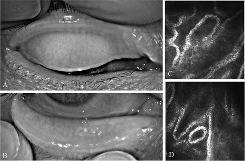
Although MGD is commonly characterized by the dysfunction of the MGCitation1, patients with MGD suffer from both abnormalities of MG function and morphology.Citation8 MG functional and morphological abnormalities are closely related to each other. MG dysfunction induces MG atrophy, and severe MG atrophy leads to a complete loss of MG function.Citation8 In fact, in MGD patients, remarkable gland dropout can be observed via noncontact infrared meibographyCitation8 (). In addition, changes of microstructure, such as an enlarged MG acinar diameter and a decreased MG acinar unit density, have also been discovered via in vivo laser scanning confocal microscopyCitation9,Citation10 (). Evaluation of MG morphology is as important as evaluation of MG function, and the potential reversibility of MG morphology has recently attracted attention.Citation11–Citation13
Figure 1. A 50-year-old male patient with severe obstructive MGD. The meibography examination (A: meibography image of upper eyelid; B: meibography image of lower eyelid) showed that the MGs of this patient were vague and difficult to identify in both the upper and lower eyelids; the acinar units were extremely enlarged as seen with confocal microscopy (C, D: confocal microscope images of meibomian acinar structure in upper eyelid).
Eyelid hygiene is a routine physical MGD treatment conducted by doctors or patients themselves, including warming and massage.Citation1 In this study, a comprehensive evaluation of MG function and morphology was conducted in MGD patients after exposure to IPL. A comparison of MG function and morphology was also done between patients treated with IPL and those treated using eyelid hygiene. The results obtained in this study will be helpful for understanding the mechanisms by which IPL works to treat MGD and will provide data for future studies involving IPL treatment for MGD.
Material and methods
Subjects
Adult Asian subjects (IPL group, n = 18; control group, n = 17), who were diagnosed with MGD (>stage1, according to the 2011 International Workshop on MGDCitation1) and had not conducted eyelid hygiene or undergone any alternative treatments for at least 3 months, were enrolled consecutively in the study. The study was conducted in the ophthalmology clinic of the Eye & ENT Hospital of Fudan University. The eyes with MGD (in cases where only one eye was affected) or the eyes with more severe MGD (according to stage) were assessed in the study. Diagnostic criteria: Citation1 symptoms of ocular discomfort, such as eye irritation that limited activities; Citation2 clinical signs: meibum quality grade ≥4(1) or MG expressibility ≥ 1(1). Exclusion criteria: Citation1 previous ocular surgery or trauma (excluding chalazion section); Citation2 blepharal dysraphism; Citation3 a history of blepharal and periorbital skin disease in 1 month; Citation4 acute inflammation; Citation5 rheumatic immune systemic diseases. Patients with excessive sun exposure in 1 month, a history of herpes zoster infection, pregnancy, use of photosensitive drugs/foods, or skin Fitzpatrick scale V/VI were excluded from the IPL group. Informed consent was obtained from all subjects after explanation of the nature and possible consequences of the study. The sample size was sufficient for statistical calculation. This study was approved by the Institutional Review Board of the Eye and ENT Hospital of Fudan University and was registered with Chinese Clinical Trial Registry prior to the first subject being enrolled. This study adhered to the tenets of the Declaration of Helsinki. All examiners were blinded to the treatment group.
Treatment
(1) Drug
All patients were given artificial lubricant four times a day for 3 months (Tears Naturale, Alcon, America).
(2) Eyelid hygiene
Control group subjects were required to perform an eyelid hygiene regimen at home once daily for 3 months as follows: (1) warming: closed eyelids were warmed for 10 min at about 40°C; (2) massage: traction was applied on the lateral canthus to immobilize the upper and lower eyelids, and then the eyelids were mildly compressed downward or upward with fingers (5 times per hygiene regimen). Warming and massage were performed consecutively.
(3) IPL treatment
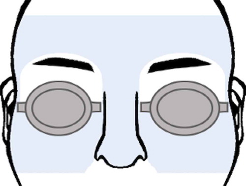
Three IPL treatments were administered once a month for 3 months. A modular laser multi-application platform (M22, Lumenis, America) was used to administer treatment to the periorbital area (). IPL treatment intensity was chosen based on the Fitzpatrick scale as follows: Fitzpatrick scale III, 17 J/cm2 with a 560-nm filter; and Fitzpatrick scale IV, 16 J/cm2 with a 590-nm filter. Patients were required to wear opaque goggles during the IPL procedure. Makeup and contact lenses were removed before treatment. To prevent facial pigmentation secondary to IPL, patients were urged to avoid sun exposure for 1 month after each IPL treatment.
Figure 2. IPL treatment area (marked in blue). To avoid hair loss and eye injury, eyebrow and eyelid were excluded from the treatment area.
Assessments
(1) Associated ocular-surface indexes
Ocular surface disease index (OSDI), tear breakup time (TBUT), Schirmer 1Test (S1T), corneal staining, and conjunctival staining were assessed. (1) OSDI: a self-administered questionnaire containing 12 items, gives a range of zero (no symptoms) to 100 (severe symptoms) points. (2) TBUT: TBUT was measured three times consecutively after fluorescein delivery, and the median value was recorded. (3) The S1T was performed for 5 min without topical anesthesia, using a sterile Schirmer test strip. (4) Corneal stainingCitation14: Five areas (upper, lower, nasal, temporal, and optical-diameter) were evaluated after the instillation of fluorescein. Superficial punctate keratopathy of the cornea was scored between 0 and 3 in each area. (4) Conjunctival stainingCitation14: Four areas (upper, lower, nasal, temporal) were evaluated after the instillation of lissamine green and scored between 0 and 3 in each area.
(2) MG function indexes
Meibum quality and MG expressibility of the upper eyelid were assessed. (1) Meibum qualityCitation1: Eight MG glands in the nasal and middle parts of the eyelid were assessed using a scale of 0–3 for each gland: 0, clear; 1, cloudy; 2, cloudy with debris (granular); and 3, thick, like toothpaste. The scores were added to calculate the total score. (2) ExpressibilityCitation1: Five MG glands in the nasal part were evaluated on a scale of 0–3: 0, all glands expressible; 1, 3–4 glands expressible; 2, 1–2 glands expressible; and 3, no glands expressible.
(3) MG morphological indexes
MG dropout and MG acini parameters of the upper eyelids were assessed. (1) MG macrostructure index: MG dropout. After the upper eyelids were everted, the MG dropouts were observed via a noncontact infrared meibography system (Keratograph, OCULUS, German), according to a published method.Citation15 The whole area of the tarsal plate was limited to the four boundariesCitation15: the proximal border, the distal border, the nasal border (tear punctum), and the temporal border (the most visible tarsal conjunctiva of everted eyelid). The examiner defined the array of “string-like” structures traversing palpebral surface vertically as MGs.Citation15 Partial loss or truncation of these structures was regarded as MG dropout.Citation15 With ImageJ V1.49 software (provided in the public domain by Bethesda, MD, USA, http://imagej.nih.gov), the MG dropouts were calculated. (2) MG microstructure indexes: confocal microscopy parameters. An in vivo laser scanning confocal microscope (HRT II Corneal Rostock Module, Heidelberg Engineering GmbH, Germany) was used to observe MG histological structure. The examiner first everted the upper eyelid and moved the center of the Tomo-Cap onto the palpebral conjunctiva.Citation9 After the first superficial conjunctival cells were visualized, the focal plane was gradually moved to the subconjunctival tissue until the glandular structures were visualized.Citation9 MGs were scanned with vertical movements. Images (in a 400 × 400-μm frame) of the nasal, middle, and temporal parts were obtained and used to calculate the confocal microscopy parameters (MG microstructure indexes): MG acinar longest diameter (ALD), MG acinar shortest diameter (ASD), and MG acinar unit density (AUD). Inflammatory cells (ICs) around the glandular structures were also noted.
Statistical methods
Data were analyzed using SPSS 22.0 (IBM Corp, America). Continuous intergroup variables were analyzed using an independent t-test, and pretreatment and continuous intragroup variables were tested with a paired t-test. Categorical intergroup variables were analyzed with the nonparametric Kruskal–Wallis test, and categorical variables intragroup were analyzed with the nonparametric Wilcoxon signed-rank test. Correlations between normally distributed values and non-normally distributed values were analyzed with the linear Pearson correlation coefficient and the Spearman correlation coefficient respectively. Statistical significance level was <0.05.
Results
Population characteristics
As shown in , no intergroup differences were found in age, gender, and related medical history (dry eye, blepharokeratoconjunctivitis (BKC), chalazion, chalazion section, and disease duration). MGD stages in the IPL group (age 41.56 ± 9.7 years, 9 female and 9 male) were not statistically different compared with the control group (age 40.76 ± 13.93 years, 8 female and 9 male).
Table 1. Characteristics of MGD patients.
Associated ocular-surface indexes
There was no statistical difference in pretreatment regarding OSDI, S1T, TBUT, and corneal staining between the IPL and control groups (all P > 0.05). As shown in , OSDI and TBUT improved significantly after treatment in both the IPL and control groups. There was no significant change in S1T or corneal staining after treatment in either IPL or control group (all P > 0.05, ). Pretreatment conjunctival staining in the IPL group was slightly higher than that in the control group (P = 0.040) and accordingly decreased in the IPL group after treatment (P = 0.001, ).
Table 2. Clinical indexes of IPL group and control group before and after treatment.
MG function indexes
There was no statistical difference in MG quality or expressibility between the IPL and the control groups prior to treatment (all P > 0.05). Meibum quality and MG expressibility improved in two groups with statistical significance after treatment (all P < 0.05, ).
MG morphological indexes
There was no statistical difference between the IPL and the control groups in MG dropout, MGALD, MGASD, MGAUD, and IC (all P > 0.05). As shown in , there was mild improvement in MG dropout in both the IPL (5.44 ± 6.18%, P = 0.002) and the control groups (4.05 ± 5.04%, P = 0.008). Pretreatment MGALD (101.89 ± 21.44 μm to 84.67 ± 20.25 μm), MGAUD (91.50 ± 37.42/mm2 to 113.11 ± 40.12/mm2), and the positive rate of IC (44.44% to 16.67%) significantly improved after treatment (all P < 0.05) in the IPL group, but not in the control group (all P > 0.05). MGASD in both the IPL and the control groups had no statistical change after treatment.
Factors related to the change in OSDI after treatment
Relationships between the change in OSDI and the change in other indexes (related ocular-surface indexes, MG functional indexes, and MG morphological indexes) were evaluated in the IPL and the control groups. In the IPL group, the improvement in OSDI was positively related to the improvement in MGAUD. In the control group, the improvement in OSDI had no correlation with the other indexes.
Discussion
IPL treating MGD was first reported in an article in. 2015Citation4 Since then, four studies have been published that confirm the efficacy of IPL for the treatment of MGD.Citation2,Citation5–Citation7 Although IPL treatment had already been used to treat MGD patients in some regions, the specific mechanisms by which IPL affects MGD are yet to be elucidated. Many proposed hypotheses are based on the effects of IPL when treating facial sebaceous abnormalitiesCitation2,Citation4–Citation7, though there is no strong evidence to support the idea that the mechanism is the same when treating MGD. Therefore, in order to clarify the specific effects of IPL on MG, examination of the changes in the MG after exposure to IPL was conducted, and these changes were compared to changes in the MG after treatment with eyelid hygiene. Since there were no significant differences in population characteristics, MGD stages, or in most pretreatment indexes between the two groups, the posttreatment differences between the IPL and control groups in the current study appear to be the result of IPL.
Results of this study support those found by Toyos and several doctors.Citation2,Citation4–Citation7 In addition, the current study confirmed improvements in the symptom score of patients (OSDI), ocular surface injury in patients (conjunctival staining), TBUT, and MG function (meibum quality and MG expressibility) after 3 months of IPL treatment. Except for conjunctival staining, these improvements were also seen in patients undergoing eyelid hygiene treatment. For safety reasons, patients with acute inflammation at the beginning of the study were excluded, since it was improper for those patients to accept eyelid hygiene or IPL treatment immediately. Therefore, the corneal staining in two groups and the conjunctiva staining in the control group was mild. This is the likely reason why these indexes were not statistically different posttreatment.
What interested us most were the differences in MG morphological change before and after treatment between the two groups. As for the MG macrostructure, the patients in both groups had remarkable MG dropout before treatment (IPL group, 45.72 ± 12.93%; control group 40.28 ± 13.15%). This level of dropout is much higher than that in healthy individuals of a similar age (14.7 ± 5.7%).Citation15 After treatment, MG dropout decreased by 4–5% in both groups with statistical significances, which is in accordance with the decreasing degree of MG dropout reported in previous studies.Citation12,Citation13
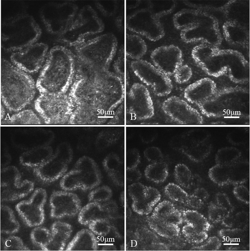
When it comes to the microstructure change, differences emerged. Both the IPL and the control groups had significant enlarged MGALDs (IPL group, 101.89 ± 21.44 μm; control group, 98.00 ± 29.01 μm), which were much higher than the cutoff value of 65 μm.Citation10 Nevertheless, only the IPL group showed improvement in MGALD after 3 months of treatment (). MGAUD in IPL group also increased accordingly. Considering the positive relationship between the change of OSDI and the change of MDAUD, it is suggested that IPL treated MGD condition through improving MG microstructure, and we further speculated that the particular improvement in MG microstructure was induced by the photomodulation effect of IPL. Photomodulation was the photobiostimulatory effect originally developed for NASA plant growth experiments 300 in space, and was later discovered efficacy of promoting cell activity like wound healing and photorejuvenation.Citation16,Citation17 NASA found that the optimal light wavelengths (proven in prior studies of laser and LED light) for photobiostimulation included 680, 730, and 880 nmCitation16, which are all included in the IPL wavelength spectrum used for treatment. We presumed that the photomodulation stimulates acinar cell activity, thus improving MG microstructure. And this is also the likely reason that one procedure of IPL treatment can last between 6 and 12 months.Citation2 Furthermore, the positive rate of IC around glandular structures decreased after treatment in only the IPL group. The anti-inflammation effect of IPL has been broadly reported in dermatology studies.Citation18 Although strong evidence is still lacking, previous ophthalmological studies also considered decreasing inflammation as a possible mechanism of IPL treating MGD.Citation2,Citation4,Citation7 This study provided primary evidence supporting this hypothesis.
Figure 3. The MG figure under confocal microscopy from a 66-year-old female MGD patient before and after three simple IPL treatments. Enlarged acinar diameter was decreased and AUD was increased after treatment. A, B: before treatment. C, D: after three simple IPL treatments.
According to the results, IPL not only improved the MG macrostructure, but also improved the MG microstructure, in particular, and decreased the MG inflammation. Consequently, we presumed that photomodualtion and anti-inflammatory effect are two working mechanisms of IPL treating MGD. It is likely that the photothermal effect also plays a role in the mechanism; however, it is beyond the discussion of this study. One limitation of this study is that only primary evidence was provided and the possible mechanisms were only verified on a histological level. Further cytological and molecular studies are required to fully elucidate the mechanisms involved in IPL treating MGD.
In conclusion, IPL treatment improves MG function, MG macrostructure as well as eyelid hygiene, and IPL treatment particularly improves MG microstructure and decreases MG inflammation in MGD patients.
Acknowledgments
This study was supported by the research grant number [81670819] from National Natural Science Foundation of China. and the research grant number [17411961800] from Science and Technology Commission of Shanghai Municipality.
Disclosure statement
The authors report no conflicts of interest. The authors alone are responsible for the content and writing of the paper.
References
- Nichols KK, Foulks GN, Bron AJ, Glasgow BJ, Dogru M, Tsubota K, et al. The international workshop on meibomian gland dysfunction: executive summary. Invest Ophthalmol Vis Sci. 2011;52(4):1922–29. doi:10.1167/iovs.10-6997a.
- Vora GK, Gupta PK. Intense pulsed light therapy for the treatment of evaporative dry eye disease. Curr Opin Ophthalmol. 2015;26(4):314–18. doi:10.1097/ICU.0000000000000166.
- Babilas P, Schreml S, Szeimies RM, Landthaler M. Intense pulsed light (IPL): a review. Lasers Surg Med. 2010;42(2):93–104. doi:10.1002/lsm.20877.
- Toyos R, McGill W, Briscoe D. Intense pulsed light treatment for dry eye disease due to meibomian gland dysfunction; a 3-year retrospective study. Photomed Laser Surg. 2015;33(1):41–46. doi:10.1089/pho.2014.3819.
- Vegunta S, Patel D, Shen JF. Combination therapy of intense pulsed light therapy and meibomian gland expression (IPL/MGX) can improve dry eye symptoms and meibomian gland function in patients with refractory dry eye: a retrospective analysis. Cornea. 2016;35(3):318–22. doi:10.1097/ICO.0000000000000735.
- Jiang X, Lv H, Song H, Zhang M, Liu Y, Hu X, et al. Evaluation of the safety and effectiveness of intense pulsed light in the treatment of meibomian gland dysfunction. J Ophthalmol. 2016;2016:1910694. doi:10.1155/2016/1910694.
- Craig JP, Chen YH, Turnbull PR. Prospective trial of intense pulsed light for the treatment of meibomian gland dysfunction. Invest Ophthalmol Vis Sci. 2015;56(3):1965–70. doi:10.1167/iovs.14-15764.
- Tomlinson A, Bron AJ, Korb DR, Amano S, Paugh JR, Pearce EI, et al. The international workshop on meibomian gland dysfunction: report of the diagnosis subcommittee. Invest Ophthalmol Vis Sci. 2011;52(4):2006–49. doi:10.1167/iovs.10-6997f.
- Matsumoto Y, Sato EA, Ibrahim OM, Dogru M, Tsubota K. The application of in vivo laser confocal microscopy to the diagnosis and evaluation of meibomian gland dysfunction. Mol Vis. 2008;14:1263–71.
- Ibrahim OM, Matsumoto Y, Dogru M, Adan ES, Wakamatsu TH, Goto T, et al. The efficacy, sensitivity, and specificity of in vivo laser confocal microscopy in the diagnosis of meibomian gland dysfunction. Ophthalmology. 2010;117(4):665–72. doi:10.1016/j.ophtha.2009.12.029.
- Guillon M, Maissa C, Wong S. Eyelid margin modification associated with eyelid hygiene in anterior blepharitis and meibomian gland dysfunction. Eye Contact Lens. 2012;38(5):319–25. doi:10.1097/ICL.0b013e318268305a.
- Yin Y, Gong L. Reversibility of gland dropout and significance of eyelid hygiene treatment in meibomian gland dysfunction. Cornea. 2017;36(3):332–37.
- Arita R, Suehiro J, Haraguchi T, Maeda S, Maeda K, Tokoro H, et al. Topical diquafosol for patients with obstructive meibomian gland dysfunction. Br J Ophthalmol. 2013;97(6):725–29. doi:10.1136/bjophthalmol-2012-302668.
- DEWS. Methodologies to diagnose and monitor dry eye disease: report of the diagnostic methodology subcommittee of the international dry eye workshop (2007). Ocul Surf. 2007;5(2):108–52. doi:10.1016/S1542-0124(12)70083-6.
- Yin Y, Gong L. Uneven meibomian gland dropout over the tarsal plate and its correlation with meibomian gland dysfunction. Cornea. 2015;34(10):1200–05. doi:10.1097/ICO.0000000000000533.
- Whelan HT, Smits RL Jr., Buchman EV, Whelan NT, Turner SG, Margolis DA, et al. Effect of NASA light-emitting diode irradiation on wound healing. J Clin Laser Med Surg. 2001;19(6):305–14. doi:10.1089/PLT.2001.19.issue-6.
- Helbig D, Simon JC, Paasch U. Epidermal and dermal changes in response to various skin rejuvenation methods. Int J Cosmet Sci. 2010;32(6):458–69. doi:10.1111/j.1468-2494.2010.00573.x.
- Wat H, Wu DC, Rao J, Goldman MP. Application of intense pulsed light in the treatment of dermatologic disease: a systematic review. Dermatol Surg. 2014;40(4):359–77. doi:10.1111/dsu.12424.