Abstract
Although diffuse thymic hyperplasia after therapy has been reported in lymphomas, no report is found in the medical literature about hyperplasia of other lymphoid tissues following chemotherapy in malignancies as far as we could reach. So, we planned to investigate the nasopharyngeal region of the lymphoma cases during their follow-up while at remission. Children who were in follow-up after cessation of oncological therapy with the diagnosis of lymphoma were evaluated for their nasopharyngeal lymphoid tissues by means of computed tomography. When a mass lesion was diagnosed, biopsies were taken. Among 23 lymphoma cases in follow-up, at a median of eight months (range 1–13 months), eight patients (six Hodgkin's and two Non-Hodgkin's malignant lymphoma) developed nasopharyngeal lymphoid hyperplasia (34.78%) that were proven to be benign by means of biopsies. Two of the patients also developed thymic hyperplasia. Nasopharyngeal hyperplasia regressed in four patients at a median of four months. Nasopharyngeal lymphoid hyperplasia may be diagnosed after the cessation of therapy in lymphomas and whatever the cause, it seems to be a benign process.
Diffuse thymic hyperplasia following chemotherapy has been reported in Hodgkin's disease and it was found to be a benign process Citation[1–14]. But in the literature, as far as we could find, there is no information about the hyperplasia of other lymphoid tissues following cancer therapy. When we realized, by chance, that there was a benign nasopharyngeal lymphoid hyperplasia in a patient following his treatment, we planned to investigate the nasopharyngeal region of the lymphoma cases during their follow-up while at remission.
Reported here are eight cases of lymphoma (six Hodgkin's, two Non-Hodgkin's malignant lymphoma) cases that developed nasopharyngeal lymphoid hyperplasia after the cessation of their therapies during complete remission. As far as we can tell, this is the first report of diffuse lymphoid hyperplasia of nasopharyngeal region following chemotherapy.
Materials and methods
In this prospective study patients who were in follow-up after cessation of oncological therapy with the diagnosis of lymphoma were evaluated consecutively for their nasopharyngeal lymphoid tissue by means of computed tomography (CT). There were 13 Hodgkin's and 10 Non-Hodgkin's malignant lymphoma cases, whose median age was 11 years (range: 2–14 years) and there were 15 males and 8 females (M/F = 15/8). All of the patients were investigated with a nasopharyngeal computed tomography at diagnosis. None of them had a nasopharyngeal hyperplasia at diagnosis nor did they have nasopharyngeal involvement during treatment of their cancer. After completion of oncological treatment, nasopharyngeal tomographies were taken at every three months in addition to their routine laboratory and radiological controls. Nasopharyngeal biopsies were done when hyperplasia was seen at CT.
Treatment protocol of the patients with Hodgkin disease were ABVD or MOPP/ABV hybrid chemotheraphy protocol chosen according to the stage and involved field radiotheraphy. In NHL patients BFM-90 chemotheraphy protocol was used.
Results
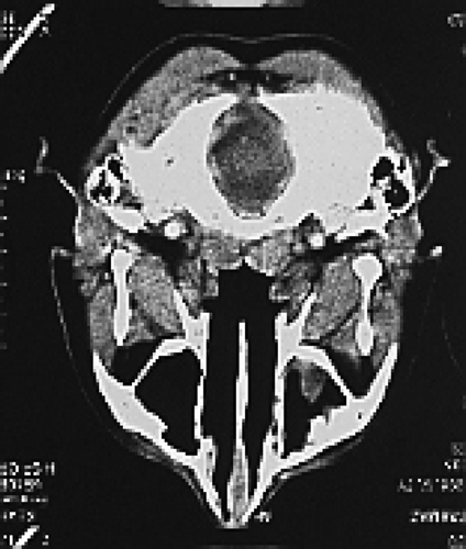
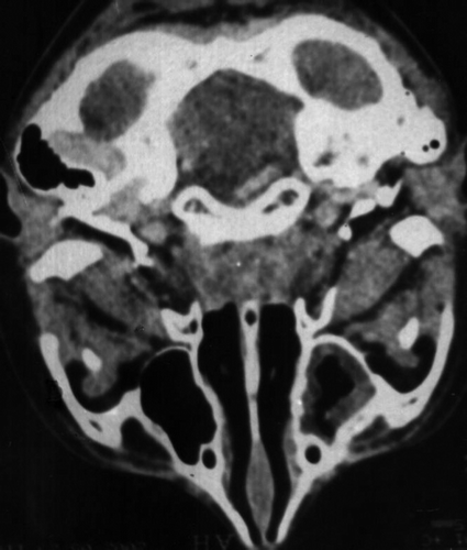
Among 23 lymphoma cases in follow-up, eight patients developed nasopharyngeal lymphoid hyperplasia (34.78%). Their median age was seven years (range: 2.5–13 years) and female:male ratio was 2:6. There were six Hodgkin's disease and two non-Hodgkin's malignant lymphoma cases. During their follow-up, a mass lesion was diagnosed at nasopharyngeal tomographies at a median of eight months (range: 1–13 months) after cessation of their therapies although they were normal at the beginning ( and , ). None of the patients had any complaints about their nasopharyngeal region and they were all in complete remission. Nasopharyngeal biopsies were taken after the diagnosis of the mass. The histopathological diagnosis was “Benign lymphoid hyperplasia” in all. Two of the patients also developed thymic hyperplasia respectively six and eight months after the completion of treatment. Nasopharyngeal hyperplasia regressed in four patients at a median of four months (range: 2–13 months). The other four of the patients still have the same appearance of their nasopharyngeal CT with a follow-up interval of median 13 months (range: 6–16 months) and they all are still in complete remission.
Table I. Summary of the patients with postherapy nasopharyngeal mass
Discussion
Evaluation of a mass during follow-up of a cancer patient is of critical importance because prompt confirmation of active disease may allow institution of effective salvage therapy. In recent years there have been some reports observing thymic hyperplasia in patients recovering by combination chemotherapies for cancer without obvious explanation, mostly young patients with lymphomas Citation[1–14].
In a series of 65 patients with mediastinal masses following therapy for Hodgkin's Disease, Jochelson et al. Citation[4] noted that 88% (57 patients) had some form of residual abnormality in the mediastinum; approximately half of these patients had mediastinal widening greater than 6 cm and they recommended that additional therapy should not be used to treat residual mediastinal widening Citation[4].
To our knowledge, the hyperplasia of other lymphatic organs has never been established after therapy of cancer patients. In our series of 23 pediatric patients with the diagnosis of lymphoma, eight cases (34.5%) developed nasopharyngeal masses during remission after therapy while they had no complaints. All of them underwent biopsy and pathology revealed only benign lymphoid hyperplasia.
In different series, it is reported that, thymic enlargement may last from 60 days to one year Citation[3], Citation[6] and some authors concluded that the masses which fail to regress completely by 9 to 12 months should be evaluated further to determine their nature Citation[3].
Among our patients who developed nasopharyngeal hyperplasia, four had complete resolution at a median of four months. Two of them also developed post therapy thymic enlargement. Among these two patients neither thymic hyperplasia nor nasopharyngeal hyperplasia had regressed in ten and 16 months respectively after the diagnosis of hyperplasia.
Thymic hyperplasia was reported in a patient after chemotherapy for acute myeloid leukemia and suggested to be a rebound enlargement after initial atrophy caused by the drugs Citation[15]. This hypothesis can also be used for nasopharyngeal hyperplasia. Whatever the cause, it seems that it is a benign process and does not appear to correlate with survival as thymic hyperplasia. But there wasn't any clue to distinguish benign hyperplasia from relapse in computed tomographies and nasopharyngeal examination. So, we recommend taking biopsies when a mass lesion was diagnosed at CT. A study with MRI or PET scan or Galium scan for this investigation and differential diagnoses can be planned in the future.
References
- Stolar CJ, Garvin JH, Rustad DG, et al. Residual or recurrent chest mass in pediatric Hodgkin's disease. Am J Pediatr Hematol Oncol 1987; 9: 289–94
- Durkin W, Durant J. Benign mass lesion after therapy for Hodgkin's disease. Arch Intern Med 1979; 139: 333–6
- Chen JL, Osborne BM, Butler JJ. Residual fibrous masses in treated Hodgkin's disease. Cancer 1987; 60: 407–13
- Jochelson M, Mauch P, Balikian J, et al. The significance of the residual mediastinal mass in treated Hodgkin's disease. J Clin Oncol 1985; 3: 637–40
- Radford JA, Cowan RA, Flanagan M, et al. The significance of the residual mediastinal abnormality on the chest radiograph following treatment for Hodgkin's disease. J Clin Oncol 1988; 6: 940–6
- Tartas NE, Korin J, Dengra CS, et al. Diffuse thymic enlargement in Hodgkin's disease. JAMA 1985; 254: 406
- Shin MS, Kang-Jey H. Diffuse thymic hyperplasia following chemotherapy for nodular sclerosing Hodgkin's disease. Cancer 1983; 51: 30–3
- Grisson JR, Durant JR, Whitley RJ, Flint AF. Thymic hyperplasia in a case of Hodgkin's disease. South Med J 1983; 76: 1189–92
- Canellos G. Residual mass in lymphoma may not be residual disease. J Clin Oncol 1988; 6: 931–3
- Cohen M, Hill CA, Cangir A, Sullivan MP. Thymic rebound after treatment of childhood tumors. AJR 1980; 135: 151–6
- Carmossino L, Dibenedetto A, Feffer S. Thymic hyperplasia following successful chemotherapy. Cancer 1985; 56: 1526–8
- Bangerter M, Behnisch W, Griesshammer M. Mediastinal masses diagnosed as thymus hyperplasia by fine needle aspiration cytology. Acta Cytol 2000; 44(5)743–7
- Shimizu H, Sakakibara Y, Fujimoto T. Mediastinal widening simulating relapse in a case of Hodgkin's disease. Rinsho Ketsueki 1993; 34(7)865–9
- Feldges A, Wagner HP, Bubeck B, et al. Recurrent mediastinal mass in a child with Hodgkin's disease following successful therapy: A diagnostic challenge. Pediatr Surg Int 1997; 12(8)613–7
- Mishra SK, Melinkeri SR, Dabadgha S. Benign thymic hyperplasia after chemotherapy for acute myeloid leukemia. Eur J Haematol 2001; 67(4)252–4

