Abstract
Background. The purpose of this study was to quantify the set-up errors of patient positioning during IGRT and to correlate set-up errors to patient-specific factors such as weight, height, BMI, and weight loss. Patients and methods. Thirty four consecutively treated head-and-neck cancer patients (H&N) and 20 lung cancer patients were investigated. Patients were positioned using customized immobilization devices consisting of vacuum cushions and thermoplastic shells. Treatment was given on an Elekta Synergy accelerator. Cone-beam acquisitions were obtained according to a standardized Action Limit protocol and compared to pre-treatment CT images. The average 3D deviation from three initial cone beam scans was compared to deviations at the 10th and 20th treatment session and correlated by linear regression analysis to height, weight, and BMI, and in H&N to weight loss as expressed by the relative weight change over time. Results. The SD of the translational and rotational random set-up errors during the first three sessions for H&N were 0.9mm (Left-Right), 1.1mm (Anterior-Posterior), 0.7mm (Cranio-Caudal) and 0.7° (LR-axis), 0.5° (AP-axis), and 0.7° (CC-axis). The equivalent data for lung cancer patients were 1.1mm (LR), 1.1mm (AP), 1.5 mm (CC) and 0.5° (LR-axis), 0.6° (AP-axis), and 0.4° (CC-axis). The median BMI for H&N and lung was 25.8 (17.6–39.7) and 23.7 (17.4–38.8), respectively. The median weekly weight change for H&N was −0.3% (−2.0 to 1.1%). With H&N and lung cancer analyzed separately, no statistically significant correlation was observed between set-up errors and height, weight, BMI, or weight change during treatment, irrespectively whether the 3D deviations from the initial three cone beam scans or scans from the 10th or 20th treatment sessions were used. Conclusion. This IGRT study did not support the hypothesis that set-up errors during radiotherapy are correlated to patient height, weight, BMI, or weight loss.
Radiation treatment of head and neck cancer (H&N) is associated with considerable toxicity. Several normal tissues may be affected by ionizing radiation, and both acute and late radiation-induced reactions may be dose-limiting for radical treatment. This regards particularly neurological structures such as the brain stem or spinal cord where the tolerance levels to radiation are considerable lower than the prescribed tumor doses for curative radiation treatment of head and neck cancer.
Lhermitte's syndrome is a distressing, sub-acute reaction which may occur a few months after the finish of radiotherapy presumably owing to radiation-induced lesions of the dorsal columns of the cervical cord Citation[1], Citation[2]. Per definition, the syndrome is reversible but may proceed to an irreversible spinal cord injury in case of localized spinal cord over-dosage.
The risk of chronic progressive myelopathy after radiation treatment of the head and neck is small after conventional fractionation without concurrent chemotherapy Citation[1–3], however, because of the devastating consequences of spinal cord injury, it must be prevented.
During 2006, our department observed an excess number of patients with Lhermitte's sign compared to previously. No changes in fractionation schedules or treatment indications had been performed and there were no obvious modifications of treatment techniques or verification procedures that could explain the incidents. While randomness was a plausible explanation to the incidents, it was essential to exclude any factors which would impact on the risk of spinal cord injury. Set-up errors during radiotherapy had to be eliminated. The fixation procedure was changed immediately and cut-outs in the immobilization face masks were abandoned to optimize patient immobilization and reduce set-up errors which potentially could reduce the risk of spinal cord injury.
It has been indicated that weight loss is associated with increased set-up errors (random and systematic) in patients during image guided radiotherapy (IGRT) for nasopharyngeal cancer Citation[4]. We also hypothesized that overweight or obesity could lead to destabilization as well. Therefore, after the introduction of cone-beam CT in our department these questions were investigated by a prospective study of head and neck patients to assess the set-up errors during radiotherapy and compare the variations to patient-specific factors such as weight, height, BMI, and weight loss during treatment. The study was extended to investigate whether such patient-related factors would impact on treatment set-up errors in lung cancer patients as well.
Patients and methods
Radiation treatment
From November 2007 to February 2008, 34 consecutively treated H&N cancer patients and 20 lung cancer patients were investigated. The H&N group consisted of 6 females and 28 males with a median age of 60.2 years (range 45.3–88.8). The diagnostic groups were oropharynx 11, larynx 9, hypopharynx 3, oral cavity 5, nasal cavity/paranasal sinuses 3, and carcinoma of unknown primary 3.
Three patients received postoperative radiation after radical surgery. Of the 31 patients undergoing primary radiotherapy, 58% (18/31) had stage III–IV disease according to UICC 2002 and 42% (13/31) had stage I-II disease. Fifteen H&N patients (44%) had nodal metastases at the beginning of treatment while 19 (56%) had no neck disease. The size of neck metastases ranged from 1 to 5cm (median 2.4 cm).
H&N patients were treated with a slightly accelerated schedule of 66–68 Gy in 33–34 fractions, 6 fractions per week during 5 and a half weeks Citation[5]. Two patient were treated with palliative intent, 52 Gy/13 fractions during six and a half week. Patients were followed weekly at the head and neck clinic. This consisted of a clinical examination with blood tests and a recording of patient-related problems including pain scores, analgetic consumption, nutritional status, and weight changes.
The lung cancer group consisted of 8 females and 12 males, slightly older that H&N patients with a median age of 68 years. The patients received 60–66 Gy in 30–33 fractions, 5 fractions per week, the total dose dependent on whether concomitant docetaxel was given or not. As part of the pre-treatment nutritional status and counselling for these patients, base line weight and height were obtained. Weight was measured under standardized conditions. No weekly weight data was obtained for this group.
Treatment planning was performed as 3D conformal or IMRT on a Pinnacle3® based dose planning system with 3 mm slice thickness on CT.
Immobilization
The patients were immobilized in supine position using customized vacuum cushions (VacFix™). H&N patients had a full thermoplastic face mask (AquaPlast®) covering and fixating the shoulders as well and lung cancer patients were immobilized with arms up in a thermoplastic shell from the chin to the umbilicus. Reference lines were marked on the immobilization device during simulation defined by laser beams, and equivalent alignment was performed at treatment.
Image acquisition, analysis, and set-up variation
Treatment was given on an Elekta Synergy accelerator. Cone-beam kV CT acquisitions (CBCT) were obtained at fractions number 1, 2, and 3 as well as at the 10th and 20th. The CBCT scans for H&N patients were done as a half rotation 70 kV CBCT of 60 s and 1.1 cGy per scan while lung cancer patients had a full rotation 120kV CBCT scan of 120 s and 2.1 cGy per scan.
Patient position at treatment was assessed from the CBCT using an automatic grey scale matching algorithm in a predefined volume (clip box) according to the set-up protocols for H&N and lung cancer patients in our department. The CBCT images were compared to pre-treatment CT images from the dose planning system. This provided three translational displacements and three rotations. Patients were repositioned if the measured displacements were larger than the levels according to a standardized Action Level protocol.
The average 3D deviation from three initial cone beam scans were estimated and compared to deviations at the 10th and 20th treatment session and correlated by linear regression analysis to height, weight, and BMI. In H&N, a correlation analysis was also performed between the 3D deviations and weight loss as expressed by the relative weight change per week. BMI (kg/m2) was calculated as weight (kg) divided by the square height (m2).
Statistics
Correlation analysis was done by linear regression or Spearman's non-parametric regression analysis. ANOVA was used for comparison between individual subgroups. To obtain a parameter of weekly weight changes over the whole treatment course, the weekly percentage weight change (%) for H&N patients was fitted by linear regression for the correlation analyses with the set-up errors.
Patients who for technical reasons (tracheostomy, moved to another accelerator) and who did not have a full data set were excluded from the analysis. This left 31 H&N and 19 lung cancer patients for the correlation analyses. All tests were performed on SPSS version 15.0. Reported p-values describe the two-tailed probabilities of the observations.
Results
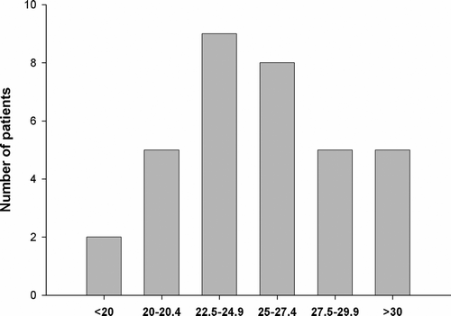
A considerable variation in weight was observed, varying between 51.3–133.0 kg (median 74.3) and 51.9–130.0 kg (median 70.0) in H&N and lung cancer patients, respectively. The median BMI values for H&N and lung cancer patients at the beginning of treatment were 25.8 (17.6–39.7) and 23.7 (17.4–38.8), respectively. Overweight (BMI > 25) was observed in 46% (25/54) of H&N ().
Figure 1. BMI distribution of 34 H&N cancer patients at the beginning of radiotherapy.
Weight changes over the full course of treatment differed for the H&N group between a gain of 3.8% to a weight loss of 12.7%. The median weight loss over the whole treatment course was 1.6%. This corresponded to a weekly weight change of −0.3% (−2.0 to 1.1%).
The SD of the translational and rotational random set-up errors during the first three treatment sessions for H&N patients were 0.9 mm (Left-Right), 1.1 mm (Anterior-Posterior), 0.7 mm (Cranio-Caudal) and 0.7° (LR-axis), 0.5° (AP-axis), and 0.7° (CC-axis). The equivalent data for lung cancer patients were 1.1 mm (LR), 1.1 mm (AP), 1.5 mm (CC) and 0.5° (LR-axis), 0.6° (AP-axis), and 0.4° (CC-axis).
The H&N population standard error of the overall variation (systematic and random) at the 10th session was 2.3 mm (AP), 2.1 mm (LR), and 2.0 mm (CC), while the equivalently data for the 20th session were 2.6 mm, 2.2 mm, and 2.2 mm which from a clinical point of view showed that the present fixation provided a satisfactory immobilization of H&N patients.
The equivalent results from the lung cancer group were 1.6 mm (AP), 1.9 mm (LR), and 3.2 mm (CC), however, the magnitude of the standard error of the overall variation increased for this group at the 20th session: 2.8 mm (AP), 3.1 mm (LR), and 3.2 mm (CC).
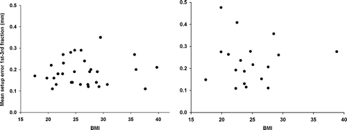
The individual mean set-up errors from session 1, 2 and 3 were analyzed with respect to height, BMI, weight, and weight change. The H&N and lung cancer groups were analyzed separately. No statistically significant correlations were observed between the set-up errors and any of the patient-related factors for either H&N or the lung cancer group. This is illustrated in which shows the relationship between BMI and the initial mean set-up error for both H&N and the lung cancer group. The lack of correlation was found irrespectively whether the 3D deviations from the initial three cone beam scans or the deviations from the 10th or 20th treatment sessions were used.
Figure 2. Average 3D deviations in H&N patients (left panel, n = 31) and lung cancer patients (right panel, n = 19) from three initial CBCTs as a function of BMI. No significant correlation was found to different patient characteristics (rs=0.05, p = 0.80, rs=0.01, p = 0.96, respectively).
An exploratory analysis was performed with patients divided into groups defined by the median or quartiles of the BMI, height, weight, and weight change and compared to the set-up variations. These tests also failed to demonstrate a relationship between the patient-related factors on set-up deviations.
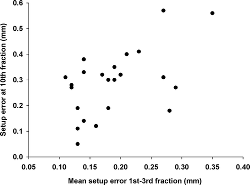
A significant correlation was found between the translational and rotational errors from the initial three cone beam scans (p = 0.009) indicating that a poor alignment in three planes is associated with considerable rotational deviations as well. Also, a correlation between the set-up error at the beginning of the treatment course and at the 10th fraction could be detected for the H&N group as shown in (rs=0.43, p = 0.037), a finding also observed for the rotational variation (p = 0.038). However, the initial set-up variation was not shown to be related to the variation at the 20th fraction indicating that other factors than the patient-related factors occurring during radiotherapy seem to impact on set-up deviations.
Figure 3. Correlation between the average set-up error in H&N at the beginning of radiotherapy and at the 10th fraction (rs=0.43, p = 0.037).
Discussion
Set-up reproducibility during fractionated radiotherapy of head-and-neck cancer may be affected by factors such as tumor shrinkage, soft tissue edema, and body weight loss. We quantified the set-up errors of patient positioning by CBCT and correlated the set-up errors to patient-specific factors such as weight, height, BMI, and weight loss. The hypothesis that obesity or weight loss during radiotherapy produce larger set-up errors could not be confirmed in this study. No correlation was observed between the magnitude of set-up errors during IGRT and patient-related factors such as height, weight, BMI, and weight loss during radiotherapy despite of a large proportion of overweight patients in the study.
Set-up error rather than organ motion has been shown to be the dominant force in positioning errors in obese men undergoing radiation treatment for prostate cancer Citation[6], and weight loss has been associated with LR and AP displacement in a study of 16 nasopharynx patients Citation[4]. A fairly large proportion of this study group was considered overweight according to international definitions and the variation of weight in the study population and weight changes was of a magnitude making it possible to discern a significant impact on set-up deviations for both H&N and lung cancer patients. However, the present immobilization techniques produced such small set-up variations that the population size may have been too small to pick up a significant difference. This calls for an extension of our study to include additional patients.
We included H&N patients with large TNM stages, i.e., 44% had nodal metastases, but node shrinkage could hardly have had an interactive effect on the set-up errors after the 10th or 20th session since the neck nodes were just 1–5 cm in the largest diameter.
The significant correlation between the average 3D translation deviation and the 3D rotational deviations is logical. A proper fixation with small 3D translational deviations evidently restricts rotational movements. We also found a good correlation between the average 3D deviation at the three initial treatment fractions and the translational and rotational error at the 10th fraction. However, this was not observed for the set-up variation at the 20th fraction which indicates that a good mobilization initially does not predict positioning later. Unfortunately, this study was not able to identify those patient-factors which may affect positioning during radiotherapy.
The present immobilization systems showed a reliable day-to-day set-up of H&N and lung cancer patients as assessed from our CBCT scans and within the margins of our general action level of 4 mm. The patient positioning has shown to be reliable in the context of reducing the risk of radiation-induced Lhermitte.
CBCT has demonstrated improved accuracy compared to other verification methods Citation[7], Citation[8], and a daily set-up correction using CBCT would be ideal Citation[9]. However, daily IGRT is time consuming and pose a heavy work load in a busy treatment facility. The set-up deviations in the initial phase of treatment in our study as assessed from CBCTs showed a high reproducibility of the patient immobilization. In addition, we found a good correlation between the initial average set-up and the set-up error at the 10th fraction for both H&N and lung cancer patients but no correlation to later sessions. The inference from this finding is that if we want to reduce workload during IGRT, one initial CBCT would suffice with the present immobilization devices and repeat CBCTs should be performed later on during the treatment course Citation[10].
Conclusion
The present immobilization has ensured a reliable patient positioning during radiotherapy of head and neck and lung cancer despite weight loss and a large proportion of patients with overweight. Deviations as assessed from CBCT were within the limits of our planning margins with a correction strategy of 4 mm. The next step in our prospective investigation is to repeat the present study but allowing for cut-outs in the face masks of head and neck patients to reduce skin doses during treatment. Declaration of interest: The authors report no conflicts of interest. The authors alone are responsible for the content and writing of the paper.
References
- Grau C. Stråleskader i medulla spinalis [Damage to the spinal medulla caused by radiation]. Ugeskr Laeger 1993; 155: 208–11
- St Clair WH, Arnold SM, Sloan AE, Regine WF. Spinal cord and peripheral nerve injury: Current management and investigations. Semin Radiat Oncol 2003; 13: 322–32
- Marcus RB, Jr, Million RR. The incidence of myelitis after irradiation of the cervical spinal cord. Int J Radiat Oncol Biol Phys 1990; 19: 3–8
- Wang C, Chong F, Wu J, Lai M, Cheng J. Body weight loss associates with set-up error in nasopharyngeal cancer patients undergoing image guided radiotherapy. Int J Radiat Oncol Biol Phys 2007; 69: S203
- Overgaard J, Hansen HS, Specht L, Overgaard M, Grau C, Andersen E, et al. Five compared with six fractions per week of conventional radiotherapy of squamous-cell carcinoma of head and neck: DAHANCA 6 and 7 randomised controlled trial. Lancet 2003; 362: 933–40
- Hansen EK, Bucci MK, Quivey JM, Weinberg V, Xia P. Repeat CT imaging and replanning during the course of IMRT for head-and-neck cancer. Int J Radiat Oncol Biol Phys 2006; 64: 355–62
- White EA, Cho J, Vallis KA, Sharpe MB, Lee G, Blackburn H, et al. Cone beam computed tomography guidance for set-up of patients receiving accelerated partial breast irradiation. Int J Radiat Oncol Biol Phys 2007; 68: 547–54
- Borst GR, Sonke JJ, Betgen A, Remeijer P, van HM, Lebesque JV. Kilo-voltage cone-beam computed tomography set-up measurements for lung cancer patients; first clinical results and comparison with electronic portal-imaging device. Int J Radiat Oncol Biol Phys 2007; 68: 555–61
- Zeidan OA, Langen KM, Meeks SL, Manon RR, Wagner TH, Willoughby TR, et al. Evaluation of image-guidance protocols in the treatment of head and neck cancers. Int J Radiat Oncol Biol Phys 2007; 67: 670–7
- Bertelsen A, Nielsen M, Jones W, Jensen HR, Brink C. The representitativeness of patient position during the first treatment fraction. Acta Oncol 2008 ( in press).