Abstract
Background. The number of CT examinations performed in Denmark increased from 14 500 examinations in 1979 to 301 617 in 2005. This implies increased radiation dose to the population. On this background, an analysis of the practice for CT examinations including potential limitations of radiation exposure and the associated risk is needed. Purposes. To analyse 1) the current use of CT in a university department compared to 1996, 2) the radiation dose and risk associated with the examinations and 3) the use of CT in Denmark since 1979. Material and methods. The administrative data of CT examinations performed in the Department of Radiology, Aarhus Sygehus, during 2005 and 1996, respectively, were obtained. Additionally national CT data were obtained from the database at the National Board of Health. Results. In 1996 1 840 patients obtained 5 538 CT examinations at Aarhus Sygehus. Their mean age was 46.7 years (0–88). The most frequent referring speciality was oncology followed by abdominal surgery and orthopaedic surgery. In 2005 3 769 patients obtained 11 216 CT examinations. They were generally older with a mean age of 56.9 years (0–97). The most frequent referring speciality was oncology followed by chest medicine and abdominal surgery. In 2005 the total effective dose was 71 043 mSv (mean 18.9 mSv/per patient). According to the BEIR VII model this radiation level corresponded to a risk for inducing a cancer in 7 patients, being fatal in half of them. The national data showed a gradual increase of the number of CT examinations from 1979 to 2005, most pronounced after year 2000 coinciding with the introduction of multi-slice CT (MSCT). Conclusion. The number of CT examinations at Aarhus Sygehus doubled during a 9 year period. The increase occured especially in middle and high age groups.
The use of Computed Tomography (CT) has increased rapidly worldwide as CT has transformed from a specialized diagnostic examinations to a more routinely used method. In the United States (USA) nearly 60 million CT examinations are performed annually. They probably account for more than 10% of all examinations, but contributes to about 67% of the total radiation dose to the population Citation[1]. In the United Kingdom (UK), CT has been reported to account for 8.6% of all X-ray examinations and contribute to nearly 50% of the total radiation Citation[2]. Unfortunately the total collective dose from diagnostic radiology is unknown in Denmark, but CT comprised 13% Citation[3] of all X-ray examinations performed in 2005.
In 1981, Doll and Peto Citation[4] estimated that about 0.5% of the cancer deaths in the USA were attributable to diagnostic X-rays. In a recent report Berrington de Gonzalez and Darby Citation[5] estimated that diagnostic X-rays cause 0.9% of the cumulative risk of cancer to age 75 years in the USA, which is equivalent to 5 695 cases per year. In the same report it was concluded that the corresponding cumulative cancer risk was 0.6% in UK, 0.9% in Sweden, 1.5% in Germany and 3.2% in Japan. These values correspond to 700 cancer cases annually in UK, 162 in Sweden, 2 049 in Germany and 7 587 cases in Japan. In the UK, the estimated annual radiation-induced cancer risk started to rise about age 40 and was still rising at the age of 70 years. The number of cancer cases depended on X-ray modality, frequency of radiation, irradiated organs and patient age. CT examinations were responsible for the largest number of radiation induced cancers, followed by barium enemas and hip or pelvic radiography.
There are diagnostic benefits from the increasing use of CT as a diagnostic tool, but the increased radiation exposure to the patients has to be taken into account. The recent introduction of multi-slice CT (MSCT) has enabled radiologist to obtain images in shorter time and view the data in multiple planes. This has improved the diagnostic abilities, but has also contributed to the increased use of CT Citation[6]. Depending on the technique, MSCT can imply a higher radiation dose than serial CT. Thus, in a recent analysis the effective doses given by 10 routine MSCT examinations were found on average 35% higher than for similar single slice CTs Citation[7].
The basic principle of radiation protection for medical exposure as recommended by the International Commission of Radiological Protection (ICRP) Citation[8] is justification of practice. Justification of a practice means that exposure to radiation should only be adopted if it produces sufficient benefit to the exposed individual or the society. Justification is also emphasised in the EURATOM directive (EURATOM 97/43) Citation[9]: ”Medical exposure shall show a sufficient net benefit, weighting the total potential diagnostic or therapeutic benefits it produces, including the direct health benefits to an individual and the benefits to society, against the individual detriment that the exposure might cause, taking into account the efficacy, benefits and risks of available alternative techniques having the same objective but involving no or less exposure to ionizing radiation”. Justification of CT implies weighting the diagnostic benefit against the adverse effects induced by the accompanying radiation dose.
As recommended by ICRP and mentioned in the EURATOM Directive justification of practice is important. It is therefore necessary to analyse current practice to put focus on relevant patient groups based on the number of CT examinations.
The objectives for this study were to analyse 1) the current use of CT in a university department compared to 1996, 2) the radiation dose and risk associated with the examinations and 3) the use of CT in Denmark since 1979.
Materials and methods
The administrative data of CT examinations performed in the Department of Radiology, Aarhus Sygehus, Aarhus University Hospital, were systematically registered in a Radiology Information System (RIS) since 1996. Based on data extract the CT practice in the year 2005 when the department had 2 MSCT scanners (A Philips Brilliance 40 and a Philips MX8000) was compared to that of 1996 when only one single slice Somatom Plus CT scanner was used. For every patient examined during the year 2005 and 1996, respectively, the data included age and gender, year of birth, examination date, anatomic region and examination protocol, referring medical speciality and in- or outpatient, respectively.
The department of radiology served inhabitants locally and offers specialist treatment for the western part of Denmark. The CT patient mix reflected the organisation and included mainly top specialities, complex patients, general medicine and emergency. The CT scanners were predominantly used for patients with malignancies, thoracic, abdominal, traumatic and skeletal disorders. The Department of Neuroradiology had their own CT scanner, both in 1996 and 2005. In 2005 there were two CT scanners in hospitals nearby, which was not the case in 1996. Working hours for CT were office hours for out-patients on appointment and 24-hour for in-patients at short notice and emergency service.
Descriptive analyses of all data were performed.
Radiation dose
The radiation dose was measured for all standard examination protocols used in 2005. The scanner used in 1996 was not available for dose measurements.
The radiation dose measurements were performed with a Capintec PC-4P pencil shaped ionization chamber and two perspex phantoms with diameters of 16 cm (head phantom) and 32 cm (body phantom), respectively. The weighted Computer Tomography Dose Index (CTDIw) was obtained based on the CTDI values measured centrally (c) and peripherally (p), respectively, in the phantoms according to the following formula
For helical scanning, the absorbed radiation dose is inversely proportional to the pitch and the volume CTDI (CTDIvol) is then used. Its definition is according to the formula:
To take the scan length into consideration the Dose Length Product (DLP) Citation[10] were estimated for all protocols in accordance with its definition:
where L is the scan length between the start and end position.
The effective dose was calculated from DLP using conversion factors (EDLP) depending on age, scanned region and CT geometry Citation[10].
The radiation risk
The radiation risk was estimated according to risk calculation model proposed by the US Food and Drug Administration (FDA) Citation[11] and the BEIR VII lifetime risk model Citation[12], respectively.
In these models the death risk rate is estimated after exposure to ionising radiation at ages 0, 5, 10, 15, 20, 30, … and 80 years. According to both models the risk of death for patients exposed to ionising radiation at age 5 years is two times higher than for patient exposed at age 20 years and five times higher than for patient exposed at age 40 years, whereas the risk declines in older age groups.
National survey
The national use of CT was analysed for the years 1979–2005 based on data collected by the National Board of Health Citation[3].
Results
lists the medical specialities referring to CT and the number of examinations in 1996 and 2005, respectively. For this purpose, the specialities were coded in accordance with the standard International Classification of Diseases, ICD 10.
Table I. List of medical specialities referring to CT and the number of examinations.
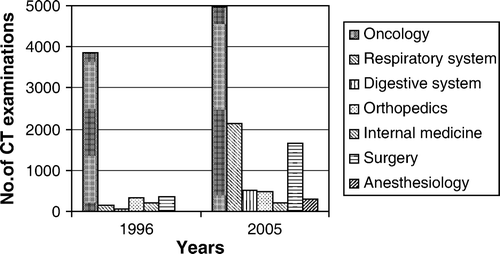
In 1996 the most frequent referring speciality was oncology followed by orthopaedics and surgery. Oncology was also the most frequent speciality in 2005 followed by chest medicine, abdominal surgery, digestive system specialities and orthopaedics. shows the number of examinations performed for the main referring specialities.
Figure 1. Number of examinations distributed by main referring speciality.
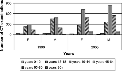
The age distribution was analysed according to the recommended general groups in MedLine: 0–12 years, 13–18 years, 19–44 years, 45–64 years, 65–80 years and more than 81 years. shows the patient populations in 1996 and 2005, respectively. In 1996, the average age of the patients was 46.7 years, in 2005 56.9 years. In 1996 60.1% of the patients were males and in 2005 54.0%. shows gender distribution of the patients divided according age for 1996 and 2005, respectively.
Figure 2. Age distribution of the patient population in 1996 and 2005.
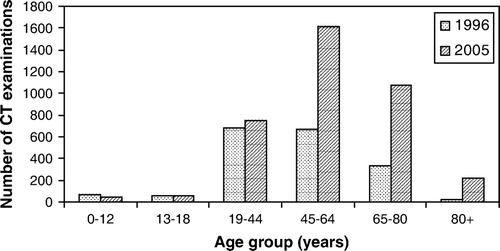
Figure 3. Gender distribution of the patients examined by CT in 1996 and 2005.
In 1996, 1 840 patients (719 females and 1 121 males) were examined. Their age range was 0–88 years (mean 46.7 years). shows further details of the patients examined in 1996. One hundred and twenty-two patients were children (53 girls and 69 boys; mean age 10.2 years). The average number of examination for this patients group was 3.0 (min. 1; max. 12). Orthopaedics was the most frequent referring speciality (35 children), followed by paediatrics (33 children), oncology (32 children), digestive system specialties (6 children), surgery (5 children), haematology (3 children), internal medicine and neurology (2 children from each speciality) and specialties of the genitourinary system (1 children). There were three children without specified referring speciality.
Table II. Some characteristics of the patients examined in 1996 divided according to age.
One thousand seven hundred and eighteen patients were between 19 – 88 years old, with a mean age of 49.2 years. Their average number of examinations was 3.0 (min. 1; max. 12). In this age group the most frequent referring speciality was oncology (981 patients) followed by orthopaedics (184 patients) and surgery (127 patients).
In 1996 there were 142 patients obtaining more than six CT examinations during the one-year study. They constituted 8% of the patients and accounted for 24% of all examinations. The highest number of examinations for a patient was 12. This number occurred only in patients referred from the Department of Oncology. The high number of examinations can partly be explained by the data collection. A simultaneous CT of the chest, abdomen and pelvis was registered as three examination.
In 2005 3 767 patients (1 731 females and 2 036 males) were examined. Their mean age was 56.9 years (0–98 years). shows further details of the patients. One hundred and five patients were children (50 girls, 55 boys; mean age 12.3 years). The average number of examinations in this group was 2.1 examinations (min. 1; max. 11). Only one patient obtained 11 examinations, a 15-years-old boy with post-traumatic liver lesion. The referring specialities were orthopaedics (53 children), traumatology (26 children), abdominal surgery (10 children), oncology (8 children) and chest medicine (6 children). Two children were registered without referring speciality.
Table III. Some characteristics of the patients examined in 2005 divided according to age.
Three thousand six hundred and sixty-two patients (1 681 females, 1 981 males) were between 19 and 98 years with a mean age of 57.7 years. Their average number of examinations was 3.0 (min. 1; max. 20). For patients older than 18 the most frequent referring speciality was oncology (1 113 patients) followed by chest medicine (958 patients) and abdominal surgery (523 patients). Three hundred and thirty-one patients were registered without specified referring speciality.
There were 300 patients with more than six CT examinations in 2005. They constituted 8% of the patients and accounted for 27% of all examinations. The highest number of CT examinations (20) occurred in a 60-year-old man with post-traumatic splenic vein thrombosis.
The total radiation doses given by the examinations in 2005 appear from . The total effective dose was 71 043 mSv for all examinations with a mean average of 18.9 mSv per patient. The average total patient dose was highest in the age group 45–64 years, being 21.3 mSv. This group therefore had the highest risk of radiation-induced cancer among adults. The calculated risks for radiation-induced cancer according to the FDA and BEIR VII model appear from .
Table IV. Effective dose and risk for patients obtaining CT examinations in 2005 according to FDA and BEIR VII, respectively.
National Survey
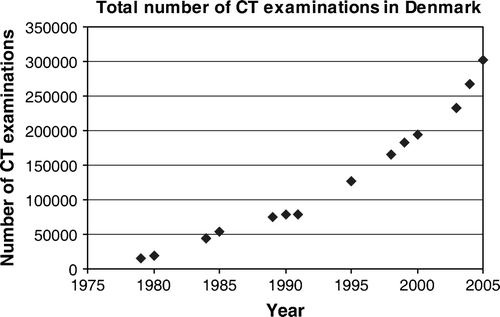
In Denmark, the number of CT examinations increased from 14 500 in 1979 to 301 617 (females 144 517, males 157 045) in 2005 Citation[3]. shows the gradual increase of the number of examination, most pronounced after year 2000 coinciding with the introduction of MSCT. Compared with other examinations the number of CTs was high in 2005. During this year there were performed 2 269 154 conventional X-ray examinations, 1 141 607 ultrasonographies, 50 682 angiographies, and 146 086 magnetic resonance examinations (MRIs).
Figure 4. The total number of CT examinations performed in Denmark from 1979 to 2005.
DISCUSSION
The present analysis showed that the number of CT examinations performed in our department doubled in less than 10 years. This correlated with a doubling of the number of CT scanners during the same period. Thus, the efficiency has not been improved with new and faster types of scanners. This can partly be explained by similar time consumption for patient positioning in 1996 and 2005, and the scanning time is short compared with this.
The age distribution of the patients shifted 10 years upwards from 1996 to 2005. This was mainly due to a substantially increase in the middle and older age groups, whereas the number of CT examinations in the age-groups from 19–44 years increased only moderately and there was a decrease in the youngest age group (0–12 years) (). Thus the increased number of CT examinations occurred predominantly in patients where the risk by radiation exposure is less important.
The increasing number of CT examinations at Aarhus Sygehus may to some extend be defined locally by the organisation. It is partly due to more comprehensive treatment of malignancies using new drugs or other therapies. The increased use for this purpose was, however, exceeded by CT for diagnosing diverse chest disorders (chronic obstructive pulmonary disease etc.) and in relation to abdominal surgery. The use of CT contributes to an adequate therapy, but the indications for CT have to be valid to omit unnecessary low dose radiation contributing to the total public risk of cancer. To ensure valid indications for CT examinations, they should be approved by the examining radiologist, and the clinician and the radiologist must be alert when patients obtain many CT examinations in a short period. Both in 1996 and in 2005 8% of the patients obtained more than six CT examinations during one year and this accounted for 24% and 27% of all examinations in 1996 and 2005, respectively. Further analysis of these patients with many CT examinations and thereby an increased risk has been performed and will be published separately.
The risk by low dose radiation is still controversial and many publications have encompassed this item. The ICRP, the United Nations Scientific Committee on the Effects of Atomic Radiation (UNSCEAR), the FDA and the BEIR VII report do not recommend the use of threshold doses for cancer induction. Investigations which described mortality from the Life Span Study cohort of atom-bomb survivors showed that excess solid cancer risk appears to be linear in the range 0–150 mSv with no evidence for threshold Citation[13]. In US the National Council of Radiation Protection and Measurements concluded that there is no evidence to reject the assumption of a linear non-threshold dose-response to low level radiation. This assumption was supported by the National Academy of Science in the BEIR VII report Citation[12]. In this report a “low dose” ranged from 0 to 100 mSv and the calculations were based on a linear dose response function, which imply that the risks increase linearly with dose. The dose was also considered cumulative. Thus, the doses of regular imaging such as in follow-up examinations correspond to the total dose attained.
The FDA Citation[11] assumed that the risk for adverse health effects from cancer is proportional to the amount of radiation dose absorbed, which depends on the type of X-ray examination. The FDA estimated that CT examinations with effective doses of 10 mSv may provide an increased chance for developing fatal cancer in approximately one patient out of 2000, whereas the BEIR VII lifetime risk model implies that one out of 1000 patients will develop cancer Citation[14]. Irrespectively the calculation model used it is important to be aware of the carcinogenic potential of radiation at doses encountered in CT.
The calculated radiation dose and risk for the 3 767 patients undergoing CT examination in our department in 2005 implies that 3.6 patients will have a risk for developing a radiation induced fatal cancer according to the FDA estimation and the total number of patients in risk of cancer will be 7.1 according to the BEIR VII estimation.
The doses by CT are much higher than by conventional radiography. The effective dose is often 10 mSv (e.g. a chest CT), implying a risk of cancer in one to two out of 2000 patients examined Citation[11], Citation[12]. From a public health risk perspective this small individual cancer risk must be multiplied by the large and increasing number of CT examinations. In Denmark the number of chest CTs increased from 36 739 in 2003 to 53 689 in 2005 Citation[3]. This implies an increased public dose of 169 500 mSv if the average examination dose was 10 mSv. During the same period abdominal CT increased from 66 687 to 92 142. With an average dose of 10 mSv for abdominal CT this implies an increased public dose of about 254 450 mSv. Unfortunately, there are no data available regarding the radiation dose given by medical exposure in Denmark. It would have given more perspective for the whole study. Thus, we do not know to which extend CT contribute to the total collective radiation dose, but CT is (as in other countries) a main contributor to the radiation dose received by the population from medical imaging. Therefore, it is important that the expected benefits of the CTs performed outweigh the risk of radiation exposure.
Reduction of radiation exposure can be obtained if image modalities without radiation can substitute CT. In this respect, studies analysing the diagnostic accuracy of MSCT compared with other diagnostic modality are generally needed. Supplied with sufficient clinical information, the radiologist should be able to suggest an acceptable alternative method that does not use ionizing radiation, such as MRI or ultrasonography, if they are diagnostically valid Citation[14]. Dialogue between the referring physician and the radiologist is essential, especially when knowledge about radiation exposure is inadequate. A recent study from a large hospital in the UK tested the knowledge of primary care and specialist physicians concerning understanding of radiation exposure, radiation doses and risks Citation[15]. The results showed that only 27% of doctors attained the general cut off of a 45% passing mark, and only 57% of the radiologists and radiology-related sub-specialists passed the test.
Acknowledgements
The study was part of the 6th Framework Research Project no. FP6/002388, “Safety and efficacy of computed tomography (CT): a broad perspective”.
References
- Mansson LG, Bath M, Mattsson S. Priorities in optimisation of medical X-ray imaging-a contribution to the debate. Radiat Prot Dosimetry 2005; 114(1–3)298–302
- Adam E.J. Changes in the computed tomography patient population. Eur Radiol Suppl 2006;16(Suppl 4):D38–D41.
- The Danish National Board of Health. http://www.sst.dk/Informatik_og_sundhedsdata/Download_sundhedsstatistik/Behandling_ved_sygehuse/DSNM.aspx. 2008.
- Doll R, Peto R. The causes of cancer: quantitative estimates of avoidable risks of cancer in the United States today. J Natl Cancer Inst 1981; 66(6)1191–308
- Berrington de GA, Darby S. Risk of cancer from diagnostic X-rays: estimates for the UK and 14 other countries. Lancet 2004 31;363(9406):345–51.
- Lee CI, Haims AH, Monico EP, Brink JA, Forman HP. Diagnostic CT scans: assessment of patient, physician, and radiologist awareness of radiation dose and possible risks. Radiology 2004; 231(2)393–8
- Yates SJ, Pike LC, Goldstone KE. Effect of multislice scanners on patient dose from routine CT examinations in East Anglia. Br J Radiol 2004; 77(918)472–8
- ICRP 103. Recommendations of the International Commission on Radiological protection. Ann ICRP. 2007;37(2–4):1–332.
- Council D. Council Directive 97/43/EURATOM of 30 June 1997 on health protection of individuals against the dangers of ionizing radiation in relation to medical exposure, and repealing Directive 84/466/EURATOM. Official Journal L180. 1997.
- EUR 16262: European Guidelines on Quality Criteria for Computed Tomography (2003) , 2000. Radiologists (in alphabetical order): G. Bongartz, S.J. Golding, A.G. Jurik, M. Leonardi, E. van Persijn van Meerten; Physicists (in alphabetical order): J. Geleijns, K.A. Jessen, W. Panzer, P. C. Shrimpton, G. Tosi. Office for Official Publications of the European Communities, .
- U.S.Food and Drug Administration. What are the radiation risks from CT?, http://www.fda.gov/cdrh/ct/risks.html. 2005.
- BEIR VII. National Academy of Sciences Committe on Biological Effect of Ionizing Radiation. Report VII: health effect of exposure to low levels ionizing radiations. The National Academies Press, Washington DC, 2006.
- Preston DL, Shimizu Y, Pierce DA, Suyama A, Mabuchi K. Studies of mortality of atomic bomb survivors. Report 13: Solid cancer and noncancer disease mortality: 1950–1997. Radiat Res 2003; 160(4)381–407
- Semelka RC, Armao DM, Elias J, Jr, Huda W. Imaging strategies to reduce the risk of radiation in CT studies, including selective substitution with MRI. J Magn Reson Imaging 2007; 25(5)900–9
- Jacob K, Steel JR. X-ray dose training: are we exposed to enough? A reply to Dr McCoubrie's letter published in Vol 60 No 6 (2005) on p. 730. Clin Radiol 2005;60(8):936.