
Abstract
Objective: Our aim was to study whether recovery from a Raynaud’s attack and involvement of the thumb are differentiators for systemic sclerosis (SSc) in patients with Raynaud’s phenomenon (RP).
Method: A stepwise cooling and recovery procedure was performed, provoking an RP attack, in patients with primary Raynaud’s phenomenon (PRP, n = 68) and SSc (n = 18). During the procedure, the perfusion of all five fingers during cooling and recovery was assessed by photoelectric plethysmography.
Results: In SSc patients, perfusion after 10 min in one or more fingers was more frequently not restored than in PRP patients (p = 0.001), with a negative predictive value of 98%. The thumb was more frequently involved in SSc patients (p = 0.036), with a negative predictive value of 95%. Positive predictive values were low.
Conclusions: In patients with RP, when there is restoration of perfusion in all fingers after 10 min or when the thumb is spared, the presence of an underlying SSc is very unlikely. Although these results need to be validated in a clinical setting in a larger prospective study, these signs can help physicians to select additional testing for SSc in RP patients.
Distinguishing primary Raynaud’s phenomenon (PRP) from Raynaud’s phenomenon secondary (SRP) to systemic sclerosis (SSc) is crucial in the early detection of SSc (Citation1). Although serology and nailfold capillary microscopy (NCM) are predictive of SSc with high sensitivity and specificity, these tools are generally unavailable to primary care givers (Citation2). Identifying clinical methods to rule out SSc would facilitate early separation of those at risk from those who are not.
We have previously reported that patients with more severe vasculopathy suffer from prolonged ischaemic time during an Raynaud’s attack (Citation3). In addition, it appears that the thumb is more frequently involved in SSc, whereas it seems to be spared in PRP (Citation4–Citation6). These two characteristics could possibly be recognized by patients and physicians, and can help to increase awareness of SSc.
In the current study, the ischaemic time of each finger of one hand, including the thumb, was assessed by a stepwise cooling and recovery procedure, provoking a Raynaud’s attack, in patients with PRP or SSc. We hypothesize that the ischaemic time during recovery after a Raynaud’s attack and involvement of the thumb are possible signs that can help to differentiate between PRP and SSc.
Method
Patients
Patients with PRP or SSc, selected when referred to the vascular laboratory between November 2008 and August 2013, were included when a complete cooling and recovery fingertip photoelectric plethysmography (PPG, as described below) was performed (n = 69), or when they underwent a complete cooling and recovery fingertip PPG, according to the same protocol, as part of another study (n = 17) (Citation7).
Raynaud’s phenomenon (RP) was defined as biphasic or triphasic discoloration of the hands and confirmed by the cooling and recovery PPG. Antinuclear antibodies (ANAs) were measured by indirect immunofluorescence, and were defined as positive for an ANA titre ≥ 1:80. NCM was assessed by wide-field videocapillaroscopy, as described previously (Citation3). Patients were classified as PRP (n = 68) if NCM was normal, serology was negative, and no secondary cause was diagnosed during at least 2 years of follow-up. Patients with SSc (n = 18) were classified according to the American College of Rheumatology/European League Against Rheumatism criteria (Citation8).
The study was conducted in accordance with the ethical standards of the Declaration of Helsinki. The study was approved by the local ethics committee (Groningen, The Netherlands, approval numbers 2016.305 and 2015.219), which provided exemption from written informed consent, given that the study does not fall under the Dutch law on medical research in humans.
Cooling and recovery procedure
Cooling and recovery fingertip PPG was performed as described previously (Citation3). In brief, one hand (the side with the most severe complaints or, when equal, the left hand) was submerged in water up to the radiocarpal joint. The water temperature was lowered in steps of 3°C from 33°C, and the procedure was complete when the water temperature was cooled to at least 9°C, or when all fingers lost perfusion on PPG and the pain was intolerable. After a stabilization period of 4 min for each temperature, 15 s of PPG signals were analysed to assess perfusion of all individual fingers. After all cooling steps, the hand was taken out of the water and perfusion was analysed every minute during 10 min of recovery in room air of 23–24°C. The vascular technician performing the procedure was unaware of clinical diagnosis or antibody status.
The procedure was considered positive for RP when two or more fingers lost perfusion and stayed abnormal during two or more subsequent steps. The time-point of loss of perfusion of the separate fingers was assessed, as well as the time-point of recovery of perfusion during rewarming. The time between those points is called the ischaemic time, and the mean ischaemic time of the five fingers was calculated. If the pain was intolerable and all fingers lost perfusion, the ischaemic time was calculated as if the procedure had been completed, as perfusion is never restored during further cooling. If perfusion was not restored within 10 min, then the recovery time was counted as 10 min.
Statistical analysis
Statistical analysis was carried out using SPSS Statistics version 23 (IBM Corp., Armonk, NY, USA). Non-normally distributed data were compared between the groups with the Mann–Whitney U-test, and binary data with the chi-squared test.
Results
Patients
Patient characteristics per group are given in . None of the patients had active ulcers during the cooling and recovery procedure. Twenty-six patients had intolerable pain at a temperature higher than 9°C, of whom 15 patients had PRP and 11 SSc. All these patients had loss of perfusion in all fingers prior to termination of the procedure.
Table 1. Patient characteristics.
Cooling and recovery procedure
The mean ischaemic time of the five fingers was over twice as long in patients with SSc compared to PRP (). The number of fingers with abnormal perfusion during cooling was higher in SSc patients, as was the number of fingers without restoration of perfusion during 10 min of recovery (mean number of fingers per patient with abnormal perfusion during cooling 4.72 vs 3.97, p = 0.007; without restoration of perfusion 3.71 vs 1.61, p < 0.001).
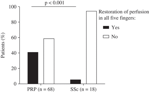
Seventeen SSc patients (94%) had no restoration of perfusion after 10 min in one or more fingers, compared to 28 PRP patients (41%) (). The SSc patient who had complete restoration of perfusion did have puffy fingers. For perfusion remaining absent in one or more fingers (which is a positive test), the negative predictive value (NPV) for SSc is 98%, with a positive predictive value (PPV) of 38%. Combining this with signs of skin involvement (e.g. puffy fingers or sclerodactyly), the NPV goes up to 100%.
Figure 1. Percentage of patients with and without restoration of perfusion in all fingers during 10 min of recovery, measured in one hand by photoplethysmography. PRP, primary Raynaud’s phenomenon; SSc, systemic sclerosis.
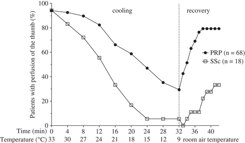
During cooling, 17 SSc patients (94%) developed abnormal perfusion in the thumb compared to 48 PRP patients (71%) (p = 0.036) (), while there was no difference in involvement of the other fingers during cooling (all p > 0.05). The SSc patient without involvement of the thumb did have sclerodactyly. The NPV for SSc of involvement of the thumb during cooling is 95%, with a PPV of 26%.
Figure 2. Cooling and recovery fingertip photoelectric plethysmography of the thumb. PRP, primary Raynaud’s phenomenon; SSc, systemic sclerosis.
When combining restoration of perfusion and involvement of the thumb, the NPV for SSc goes up to 100% and the PPV is 41%.
Discussion
This study shows that for patients with RP with quick restoration of perfusion in all fingers (within 10 min) and no involvement of the thumb, as objectively measured by a cooling and recovery procedure, underlying SSc is very unlikely. In addition to the well-known red flags of early SSc, these findings could potentially contribute to the differentiation between PRP and SRP in daily practice, especially when serology and NCM are not easily accessible. The combination of a long recovery period with skin involvement or involvement of the thumb gives an NPV for SSc of 100%; therefore, physicians could take this into account for ruling out SSc, combined with thorough assessment of the skin.
In our study, SSc patients more frequently had no restoration of perfusion, and a longer mean ischaemic time during recovery. The difference in recovery duration of perfusion is more pronounced than in our previous study (Citation3). This is because in the previous study we compared PRP with SRP, the latter also including patients with no classifiable underlying disease and other connective tissue diseases. The difference in recovery duration is also in line with previous studies, which also showed a difference in recovery when comparing PRP and SSc (Citation9, Citation10). The SSc patient who did have restoration of perfusion in all fingers had puffy fingers upon presentation, and therefore additional tests would have been recommended, even though recovery was quick.
Previously, Chikura et al suggested that involvement of the thumb could be a sign of underlying disease, as they found that the thumb was less affected in PRP than in SRP (Citation5). A more recent study by Ingegnoli et al also described sparing of the thumb, although they did not find a difference between PRP and SRP in this smaller cohort (Citation6). In agreement with the results of Chikura et al (Citation5), we confirmed that there is more frequently involvement of the thumb in SSc patients. However, as the PPV was low in our study, involvement of the thumb should not be considered a red flag. Conversely, SSc is very unlikely if the thumb is spared, with an NPV of 95%.
A limitation of this study is that patient reports of observations during their RP attacks in daily life were not obtained using a questionnaire. Although the cooling and recovery procedure does provoke an RP attack, it is not verified whether loss of perfusion and duration of recovery during the procedure is related to daily life complaints. In addition, SSc patients were not all assessed in an early stage of disease. It would be of special interest to study these patients in an early phase of SSc. In future studies, our results should be verified in an larger (early) patient cohort with a prospective setting, including patients’ reports of their complaints.
Conclusion
Patients with RP who have restoration of perfusion in all fingers within 10 min after a standardized cooling procedure and/or in whom the thumb is spared are unlikely to have SSc. This is an indication that a specific patient’s complaints are of additional value in differentiating between PRP and SRP. Although this objective measurement needs to be verified with patients’ reports, these results suggest that a physician could specifically ask the patient whether recovery after a Raynaud’s attack takes less than 10 min, and whether the thumb is uninvolved. Combined with the absence of skin involvement, these simple signs can help physicians to assess whether the patient needs to be referred for additional tests.
Disclosure statement
No potential conflict of interest was reported by the authors.
References
- Herrick AL. The pathogenesis, diagnosis and treatment of Raynaud phenomenon. Nat Rev Rheumatol 2012;8:469–79.
- Koenig M, Joyal F, Fritzler MJ, Roussin A, Abrahamowicz M, Boire G, et al. Autoantibodies and microvascular damage are independent predictive factors for the progression of Raynaud’s phenomenon to systemic sclerosis: a twenty-year prospective study of 586 patients, with validation of proposed criteria for early systemic sclerosis. Arthritis Rheum 2008;58:3902–12.
- van Roon AM, Smit AJ, van Roon AM, Bootsma H, Mulder DJ. Digital ischaemia during cooling is independently related to nailfold capillaroscopic pattern in patients with Raynaud’s phenomenon. Rheumatology (Oxford) 2016;55:1083–90.
- Chikura B, TL M, JB M, Vail A, Herrick AL. Sparing of the thumb in Raynaud’s phenomenon. Rheumatology (Oxford) 2008;47:219–21.
- Chikura B, Moore T, Manning J, Vail A, Herrick AL. Thumb involvement in Raynaud’s phenomenon as an indicator of underlying connective tissue disease. J Rheumatol 2010;37:783–6.
- Ingegnoli F, Gualtierotti R, Orenti A, Schioppo T, Marfia G, Campanella R, et al. Uniphasic blanching of the fingers, abnormal capillaroscopy in nonsymptomatic digits, and autoantibodies: expanding options to increase the level of suspicion of connective tissue diseases beyond the classification of Raynaud’s phenomenon. J Immunol Res 2015;2015:371960.
- Abdulle AE, van Roon AM, Smit AJ, Pasch A, van Meurs M, Bootsma H, et al. Rapid free thiol rebound is a physiological response following cold-induced vasoconstriction in healthy humans, primary Raynaud and systemic sclerosis. Physiol Rep 2019;7:e14017.
- van Den Hoogen F, Khanna D, Fransen J, Johnson SR, Baron M, Tyndall A, et al. Classification criteria for systemic sclerosis: an American College of Rheumatology/European League against Rheumatism collaborative initiative. Arthritis Rheum 2013;65:2737–47.
- Rosato E, Rossi C, Molinaro I, Giovannetti A, Pisarri S, Salsano F. Laser Doppler perfusion imaging in systemic sclerosis impaired response to cold stimulation involves digits and hand dorsum. Rheumatology (Oxford) 2011;50:1654–8.
- Grattagliano V, Iannone F, Praino E, De Zio A, Riccardi MT, Carrozzo N, et al. Digital laser doppler flowmetry may discriminate ‘limited’ from ‘diffuse’ systemic sclerosis. Microvasc Res 2010;80:221–6.