Abstract
The aim of study was to evaluate a case of granulomatous conjunctivitis, clinically and pathologically, in the right eye of a 2-year-old, female ostrich. A mass measuring 5 cm×3 cm×4 cm was removed surgically from the eye of the ostrich. Morexella phenylpyruvica was recovered from the mass. On histopathological examination, hyperplasia or squamous metaplasia in some area of conjunctival palpebra, and a granulomatous inflammation in the submucosa were observed. The lesion was described as a granulomatous conjunctivitis caused by M. phenylpyruvica. The lesion was located in the lower eyelid conjunctiva and was not only restricted to the gl. lacrimalis, but also present in the connective tissue. After excision of the mass, the ostrich was treated with topical and systemic antibiotics and corticosteroid. The ostrich recovered fully and the function of the eye appeared to be normal.
Résumé
Granulamateuse conjuctivitise lié à Morexella phenylpyrisvica chez une autruche ( Struthio canelus )
Dans ce travail on a pour but d'analyser la patologie et le clinique de lipogranulamateuse conjuctivitise semblable a chalation qui developpe sur l’œil droit d'une femelle autruche de deux ans et gui empeche la vision d'autruche. On a retire une masse de 5×3×4 cm. par l'intervention chirurgicale, on a identifie Morexella phenylpyrisvica. Par le controle microscopique on a remarque hiperplazi et squamoz metaplazi sur l'epitel de palpebral conjuctiva et l'inflamation granulamateuse qui se compose de plazma cellule, epiteloid, geant cellule sur submukoza. Cette lesion donne une description de granulamateuse conjuctivitis qui se compose de Morexella phenylpyrisvica. La lesion a ete localise sur la palpebra inferieure et elle n'etait pas seulement limitè just à glandula lacrimalis mais en même temps elle se trouve sur la connective tissu. Apres avoir retirer cette masse avec l'operation et apres avoir utiliser l'antibiotique et la korticosteroid topical et systemique la vision d'autruche a rèpris son cours normal.
Zusammenfassung
Granulamateuse conjuctivitise lié à Morexella phenylpyrisvica chez une autruche ( Struthio canelus )
Bei dieser Untersuchung war Ziel, bei einem 2 Jährigen weiblichem Strauß, mit einer granulomatosen Konjunktivitis am rechten Auge, eine klinisch-pathologische Bewertung zu machen. Die 5×3×3 cm große Masse wurde durch eine chirurgischen Eingriff entfernt. Aus der Masse wurde Moraxella phenylepyruvica identifiziert. Bei der mikroskopischen Untersuchung wurde bei dem palpeteralen konjunktiva-ephitel platzweise Hyperplasie, bei der Submukoza eine aus ephiteloide-,plasma-und gigantische Zelle entstehenden granulomatose Entzündung festgestellt. Es wurde festgestellt das die pathologische Veranderungen icht nur mit den gl. lacrimalis begränzt ist sondern sich auch im Umfang befindende konektive Gewebe ausbreitet. Im Zusammenhang wurde die pathologische Veranderungen an der unteren palpebra als sich eine gegen Moraxella phenylepyruvica bildende konjunktivitis bestimmt. Nach entfernung der Masse wurde der Strauß topisch-systemisch mit Antibiotika und Kortikosteroiden behandelt. Der Strauß wurde geheilt und das Auge gewann seine normale Funktion wieder.
Resumen
Conjuntivitis granulomatosa asociada a Morexella phenylpyruvica en un avestruz ( Struthio camelus )
El objetivo de este estudio fue el de evaluar clínica y patológicamente un caso de conjuntivitis granulomatosa en el ojo derecho de un avestruz hembra de dos años de edad. Una masa de 5×3×4 cm fue reseccionada quirúrgicamente del ojo de un avestruz. Se aisló Morexella phenylpyruvica a partir de la masa. Al examen histopatológico se observó hiperplasia o metaplasia escamosa en algún área de la conjuntiva palpebral, e inflamación granulomatosa de la submucosa. La lesión fue descrita como una conjuntivitis granulomatosa causada por M. phenylpyruvica. La lesión fue localizada en la conjuntiva del párpado inferior y no sólo se restringió a la gl. lacrimalis, sino que también estaba presente en el tejido conjuntivo. Tras la excisión de la masa, el avestruz fue tratado con antibióticos tópicos y sistémicos y corticosteroides. El avestruz se recuperó totalmente y la función del ojo parecía ser normal.
Introduction
In animals a number of pathological changes can occur in the conjunctiva, with different causes (Haziroğlu & Milli, Citation2001). In the eye, conjunctivitis is the most commonly seen pathology besides rarely occurring tumours. Allergy, dryness, physical, chemical and infectious factors are among the reasons for conjunctivitis (Jones & Hunt, Citation1983; Haziroğlu & Milli, Citation2001). Chalazion is a rare, non-infectious lipogranulomatous conjunctivitis that is seen in domestic mammals (Khurana et al., Citation1986; Prasad & Gupta, Citation1992; Read & Lucas, Citation2001). Clinically, it is recognized by nodules of various sizes (Procope & Kidwell, Citation1994). In addition, inflammation of gl. lacrimalis (dacryoadenitis) that is characterized by hyperaemia, oedema and mild leucocyte infiltration can occur (Jones & Hunt, Citation1983; Scroggs & Klintworth, Citation1996; Haziroğlu & Milli, Citation2001).
The mammalian meibomian gland (gl. tarsale) does not exist in avian species. Rather in these species there are gl. membranae nictitantis, which is located medial to bulbus oculi and secretes its content to the medial eye angle, and gl. lacrimalis, which is located in the lateral eye angle (King & McLelland, Citation1984; McLelland, Citation1990).
In the ostrich, eye complications due to avian influenza and poxviruses (Allwright et al., Citation1993; Clavijo et al., Citation2001), spontaneously occurring cataract (Bruning & Dolensek, Citation1986; Ofri & Horowitz, Citation1995), and laceration, abrasion and ulceration due to direct contact of objects have been reported. However, there are no reports describing granulomatous conjunctivitis in avians including the ostrich. To the best of our knowledge, this is the first report describing granulomatous conjunctivitis in ostriches. In this report, we examined the clinical and pathological features of a case of granulomatous conjunctivitis that microscopically resembled chalazion.
Case history
A 2-year-old ostrich (Struthio camelus) with a mass measuring 5 cm×3 cm×4 cm in diameter, located between the internal surface of the right eyelid and cornea, was submitted to the department of surgery. The ocular surface was completely covered by this mass; the cornea and sclera were not visible. In this location, a mucopurulent eye tear and occasionally caseous substances were noticed. When the lower eyelid was pulled in the ventral orientation, it was recognized that the mass was hyperaemic with the appearance of a cauliflower (). The mass originated from the lower palpebral conjunctiva and completely obstructed the puncta lacrimalis. The mass was removed surgically.
Figure 1. Macroscopical view of the mass in the right eye of the ostrich.

Materials and methods
A piece from the mass was kept in our department for histopathological examination while the rest was sent for bacteriological analysis. The tissue was fixed in 10% formalin and embedded in paraffin. From the block, 5 μm sections were cut and stained with haematoxylin and eosin, Ziehl–Nielseen, and Grocott (Luna, Citation1968). In addition, we used Sudan IV stain for our tissues that were formalin-fixed and kept in running top water for 2 to 3 h and cut using a frozen microtome. Finally, the sections were examined under a light microscope. For the bacteriological examination, swabs collected from the lacrimal secretion and the masses were inoculated onto blood agar and MacConkey agar. The cultures were incubated in aerobic conditions at 37°C for 48 to 72 h. Gram-stained preparations were prepared from the isolated colonies. In addition, to obtain pure cultures, inoculations in blood agar were performed. For the identification of the infectious agent, inoculations into biochemical test media (nitrate, urease, phenylalanine deaminase, gelatinase, growth in the presence of 6.5% NaCl) were performed. Catalase and oxidase tests were also carried out (Holt et al., Citation1994).
Results
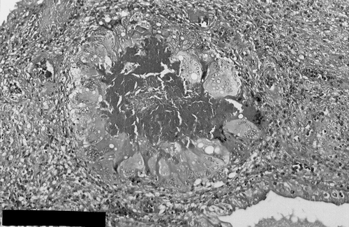
On histopathological examination, palpebral conjunctival epithelia was partially hyperplastic and papilliform growths were recognizable, while in some places squamous metaplasia was seen. In the submucosa, many lacrimal glands and duct lumens were filled with eosinophilic and vacuolated substances. These lesions were surrounded with a granulomatous cell infitration composed of many giant cells, lymphocytes, plasma cells, epitheloid cells, histiocytes, heterophils, fibrocytes and fibroblasts. In addition to these lesions, again in the submucosa and connective tissue, similar cell infiltrations with varying sizes were present (). In some lacrimal glands, gland epithelia showed papilliform growth into the lumen and these cells completely or partially filled the gland lumen. In sections stained with Ziehl–Nielseen and Grocot, no signs of tuberculosis and mycotic infections were seen. We have concluded that the lesion was a granulomatous conjunctivitis after we observed that the vacuoles that we previously thought as being lipid vacuoles were not stained by the lipid stains.
Figure 2. Granulomatous inflammation composed of numerous giant cell, inflammatory cells and macrophages in submucosa. Haemotoxylin and eosin stain: Bar=100 μm.
On microbiological examination, small (1 mm diameter) and non-haemolytic colonies grew in blood agar and MacConkey agar. In the Gram stain, Gram-negative short rods, occurring as pairs, were seen. In biochemical tests the bacterium was positive for catalase, oxidase, nitrate reduction, urease and phenylalanine deaminase, and negative for gelatinase and beta haemolysis. From this it was deduced that the infectious agent was Morexella phenylpyruvica ().
Table 1. Results of biochemical tests
Discussion
Our investigation of a lesion collected from the right lower eyelid from an ostrich was typical for a granulomatous inflammation. This lesion resembles the microscopic findings in chalazion described by Scroggs & Klinworth (Citation1996). Chalazion is a lesion seen in human and other mammalian species (Bowman et al., Citation1996; Read & Lucas, Citation2001). However, in this case, the presence of the lesion in an ostrich and the absence of the meiobomian gland in these animals led us to consider that the lesion was not chalazion and that it might be a dacryoadenitis originating from the gl. lacrimalis. However, in the literature review, no reports of dacryoadenitis with similar lesions were found in either mammals or birds, including the ostrich. We have described the lesion as a granulomateous conjunctivitis, caused by M. phenylpyruvica. The lesion was located in the lower eyelid conjunctiva and was not restricted to the gl. lacrimalis, but was also present in the connective tissue.
Members of the genus Morexella — M. bovis, M. nonliquefaciens, M. osloensis, M. phenylpyruvica and M. atlantae — can localize in the eye membrane and nasopharynx and cause infections in human and animals (Arda et al., Citation1997). It was reported that M. phenylpyruvica was isolated from genital tracts and brains of cows, sheep and pigs, and the intestinal tracts of goats (Quinn et al., Citation1994). It is known that Morexella spp. cause conjunctivitis in humans and many animal species, especially the cows (Quinn et al., Citation1994). The granulomatous inflammation and the giant cells seen in the conjunctivitis observed in our case supports the view that the lesions are associated with M. phenylpyruvica. Other bacteria, namely Staphylococcus, Enterobacteriacea, and Haemophilus, and occasionally Morexella ecinobacter, Branhamella cataralis and Streptococcus, are among the most commonly isolated infectious agents in conjunctivitis of the ostrich (Toka, Citation2001). Therefore, isolation of M. phenylpyruvica in this case is an important finding.
The lesion in this report resembles those seen in tuberculosis and mycotic infections. However, the Ziehl–Nielseen and Grocot stainings did not reveal infection with either of these infectious agents.
The treatment of the ostrich was similar to that described for humans (Khurana et al., Citation1986; Prasad & Gupta, Citation1992) and cats (Read & Lucas, Citation2001). First, the mass was surgically removed and then sefazolin (1 g sefazol) was intramuscularly injected every 12 h for 6 days. In addition, thiocilline was applied locally three times a day for 7 days. Thereafter, local treatment was continued with cortimicin for 15 days. At the end of this period the patient was able to recover her vision. There was no recurrence of the lesion when the patient was examined 4 months later.
Related Research Data
References
- Allwright , DM , Burger , WP , Geyer , A and Terblanche , AW . 1993 . Isolation of an influenza A virus from ostriches (Struthio camelus) . Avian Pathology , 22 : 59 – 65 .
- Allwright , DM , Burger , WP , Geyer , A and Wessels , J . 1994 . Avian pox in ostriches . Journal of the South African Veterinary Association , 65 : 23 – 25 .
- Arda M Minbay A Leloğlu N Kahraman M Akay Ö. Ilgaz A (zgür M Diker KS 1997 Morexella ve Morexella infeksiyonları In Arda M. (Ed.) Özel Mikrobiyoloji 4th edn pp. 103–105 Ankara Medisan Yayınevi
- Bowman RW McCulley JP Jester JV 1996 Meibomian gland dysfunction and Rrosacea In Pepose J.S. (Ed.) Ocular Infection & Immunity pp. 335–336 Boston Mosby-Year Book
- Bruning DF Dolensek EP 1986 Ratites In Fowwler M.E. (Ed.) Zoo & Wild Animal Medicine 7th edn p. 286 Philadelphia PA W.B. Saunders Company
- Clavijo , A , Riva , J , Copps , J , Robinson , Y and Zhou , E-M . 2001 . Assessment of the pathogenicity of an emu-origin influenza A H5 virus in ostriches (Struthio camelus) . Avian Pathology , 30 : 83 – 89 .
- Hazıroğlu R Milli Ü.H 2001 Veteriner Patoloji Cilt II 2nd edn Ankara Medipres
- Holt JG Krieg NR Sneath PHA Staley JT Williams ST 1994 Bergeys Manual of Determinative Bacteriology 9th edn Baltimore MD Williams & Wilkins
- Jones TC Hunt RD 1983 Veterinary Pathology 5th edn Philadelphia PA Lea & Febiger
- Khurana , AK , Ahluwillia , BK and Rajan , C . 1986 . Chalazion theraphy. Intralesional steroids versus incision and curettage . Acta Ophthalmology , 66 : 352 – 354 .
- King AS McLelland J 1984 Birds Their Structure and Function 2nd edn London Bailliere Tindall
- Luna LG 1968 Manual of Histologic Staining Methods of the Armed Forces Institute of Pathology 3rd edn New York McGraw-Hill Book Company
- McLelland J 1990 A Colour Atlas of Avian Anatomy London Wolfe Publications
- Ofri , R and Horowitz , I . 1995 . Spontaneous cataract resorption in an ostrich . Veterinary Record , 136 : 276
- Prasad , S and Gupta , AK . 1992 . Subconjunctival total excision in the treatment of choronic chalazia . Indiana Journal Ophthalmology , 40 : 103 – 105 .
- Procope JA Kidwell ED Jr. Delayed postoperative hemorrhage complicationg chalazion surgery Journal of National Medical Association 86 865 866 1994
- Quinn PJ Carter M Markey BK Carter GR 1994 Clinical Veterinary Microbiology London Wolfe Publications
- Read , RA and Lucas , J . 2001 . Lipogranulomatous conjunctivitis: clinical findings from 21 eyes in 13 cats . Veterinary Ophthalmology , 4 : 93 – 98 .
- Scroggs MW Klintworth KG 1996 The eye and ocular adnexa In Stenberg S.S. (Ed.) Diagnostic Surgical Pathology 2nd edn Philadelphia PA Lippincott-Raven Publishers
- Toka S 2001 Devekuşu Hastalıklari ve Korunma Yöntemleri İzmir Selçuklu Basım Yayım A.Ş.