Abstract
Investigation of unexpected mortality in caged layer chickens led to the discovery of a consistent traumatic injury to the heads of affected hens. Initial post-mortem examination found linear skin lacerations and associated fractures in the dorsal cranium of all birds examined, and 5 to 10 mm deep trauma in the underlying brain tissue. Post-mortem multidetector computed tomography (CT) scanning of two affected birds demonstrated similar obliquely orientated, linear, depressed fractures of the skulls consistent with a single, severe impact force to the head. Both skull fractures had a pattern of rounded, rostral expansion measuring approximately 3 mm in width. On inspection of the cages during a farm visit, this CT pattern corresponded with the size and shape of sheet metal lugs holding feed troughs onto the cages (on which blood stains were subsequently observed). Based on this analysis and hypothesizing that hunger was a triggering factor, a recommendation was made to reverse the shed “lights on” and feed hopper operation times with instant reduction in mortality. This case highlights the value of post-mortem CT imaging in bird death investigation where trauma is a postulated cause.
Introduction
Layer cage design has been extensively studied from a welfare perspective and differences in mortality noted between designs (Tauson, Citation1985), although there are few reports of specific causes of mortality apart from accidental trapping associated with deep troughs leading to neck skin atheroma (Tauson, Citation1998).
Multidetector computed tomography (CT) is a standard investigation in clinical human and veterinary practice, and is increasingly being applied to the determination of cause and manner of death in human forensic practice (Rutty et al., Citation2008). Post-mortem CT scanning has been shown to be of greatest value in the assessment of bone injury and detection of haemorrhage (O'Donnell & Woodford, Citation2008), and can be used to better understand injury causation by a process of pattern analysis (Harcke et al., Citation2007). Although there is now literature on the use of such imaging in veterinary forensic practice (Byard & Boardman, Citation2011), there is no report on its use for the investigation of mortality in poultry.
Materials and Methods
Initial information of farm parameters was supplied by an industry serviceman who brought some birds in for post-mortem examination. Live birds were killed using an overdose of sodium barbiturate.
The heads were CT scanned on a Siemens Emotion 16® Multidetector CT scanner (Siemens AG, Erlangen, Germany) using 0.6 mm collimation, and exposure factors of 130 kVp and 25 mA. Scans were reconstructed into 1 mm slice thickness using both H80 (high detail) and H32 (soft tissue) reconstruction kernels. Images were analysed on an AquariusNET® workstation (TeraRecon, Foster City, California, USA).
The farm visit was by a veterinarian (CM) travelling to the farm with the pullet supply company's serviceman and included an inspection of the birds, cages, housing and an interview of the producer and some farm staff. This occurred after the post-mortem and radiological findings. A report summarizing the findings was supplied to the producer.
Results
History
An egg producer noticed increased mortality and occasionally hens that were depressed or unresponsive to stimuli, standing or in lateral recumbency. The laying flock of 22,000 Hyline brown layer hens had recently been moved into a shed during summer. The flock was housed in four-tiered H-frame cages (Guangzhou Guangxing Poultry Equipment Co., Ltd, Shating Taihe Town, China) at four birds per cage. Mortality reached 10 birds per day at 34 weeks of age. This equates to approximately 0.3% of the flock per week compared with an industry standard of less than 0.1% per week. Egg production was not affected on a hen/day basis. Birds were ostensibly fed ad libitum on a farm-mixed ration distributed automatically four times per day by a hopper moving along the front of the cages. Lights were switched on at 5:30 a.m. The first feed hopper pass was programmed for 6:00 a.m. and the last at 6:00 p.m. Water was supplied through nipples at the back of the cages and sourced from the town water supply. After being laid, eggs rolled forward under an electrified wire so as to be separated from the hens. Fluorescent light was supplied to the sheds for 16 h of the day. The shed was well maintained with computerized tunnel ventilation and water cooling pads.
This was the fifth batch of birds to use the cages and mortality was not a feature of previous batches. Four adjacent sheds had newer cage equipment installed from the same supplier but with differences in design and no mortality problem.
Birds were inspected once a day and lights were turned up during this process. Dead and injured birds were immediately removed from the cages. The producer noticed that mortality was often clustered with several dead birds in the same cage column on a particular day but only one death per cage.
Gross pathology
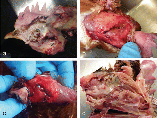
In the initial submission, one live bird with clinical signs of convulsion and head shaking, and seven dead birds were presented. The live bird weighed 1760 g and was in good body condition. At post-mortem examination, the major gross abnormality was haemorrhage and green discolouration of subcutaneous tissues immediately below the left ear canal extending posteriorly towards the neck (a).
Figure 1. 1a: Subcutaneous haemorrhage and skull fracture below the ear canal. 1b: Fracture of dorsal cranium in the first submission. 1c: Fracture of the dorsal cranium in the second submission. 1d: Extensive trauma to the cerebrum and optic lobe, deep to the fracture.

All dead birds had laceration of the skin immediately superficial to fractures of the dorsal cranium. The shape of these fractures was relatively similar; that is, 20 to 30 mm in length and 1 to 5 mm in width (b). In some cases, feathers had been forced through the broken calvarium. Subcutaneous haemorrhage was observed around the site of injury but minimal or no external haemorrhage was detected. All lesions were also associated with intracranial haemorrhage and partial disruption of cerebral parenchyma immediately deep to the fractures (data not shown).
A second batch of affected birds was submitted by the farm including a live but unconscious bird and two dead birds. The heads of the two dead birds were detached and examined using plain radiography and CT. All three birds had a fracture in the dorsal cranium (c) similar to those detected in the first submission. The sagittal section of the head of the killed bird revealed extensive destruction and haemorrhage in the cerebellar hemispheres and partial disruption to the optic lobes (d).
Digital radiography of the skulls in the dorsal and lateral projections failed to identify any fracture (data not shown). This is thought to be due to the relatively small size of the traumatic bony defects and overlap of normal skull anatomy.
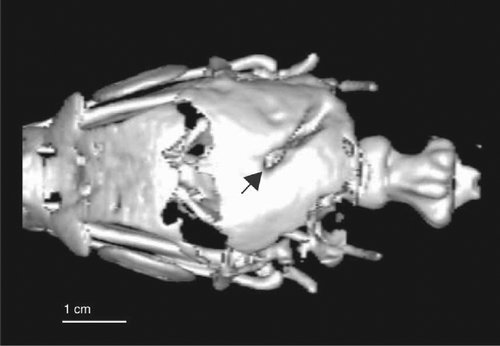
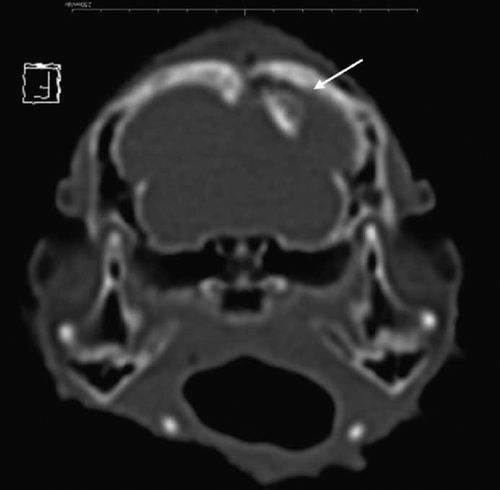
Forensic analysis of two-dimensional multiplanar reformatted images and three-dimensional (3D) volume-rendered (VR) images (see Supplementary Material, Three-dimensional CT images showing traumatic injury to a chicken skull) showed that each chicken had a focal injury to the caudo-dorsal aspect of the cranium (near the junction of the parietal and the occipital bones in mammals) with an oblique, linear defect measuring 20 mm×3 mm, passing from the right caudal to left rostral, deep to the skin wounds. Both defects were similar in size and morphology, having a signature, rounded rostral expansion (). Depressed fracture fragments were present, incorporating the thickest part of the skull vault at the cruciate eminence (). CT lesions were consistent with a single impact or stab injury through the cranium into the brain.
Figure 2. 3D VR CT image of the chicken skull viewed from the dorsal aspect, with the rostrum to the upper left of the image showing a linear defect with rounded rostral expansion matching the trough lug (arrow).

Figure 3. Short axis (or axial) multiplanar reformatted CT image of the chicken skull at the level of the temperomandibular joint, demonstrating a depressed fracture fragment entering brain (arrow).

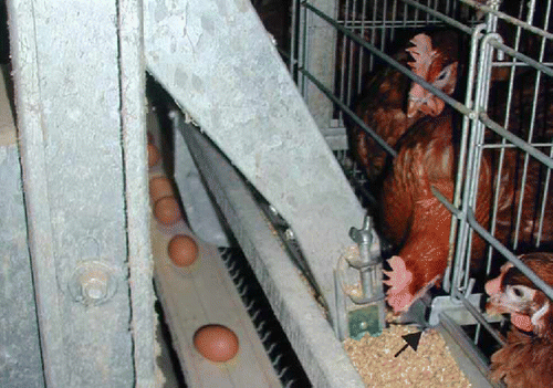
The farm was visited in the middle of the day and a moving feed hopper was identified as a probable cause of the injuries. The feed hopper was manually operated during the visit but no birds were trapped during this operation. Still, it was conjectured that the hopper forced the head of some chickens onto a lug holding the feed trough to the cages (). Initially the producer thought this was unlikely, but alerted to this possibility blood stains were observed on the suspect lugs near cages of affected birds later by the producer and farm staff. Postulating that hunger was responsible for the chicken's behaviour, the first feeding time was changed from 6:00 a.m. to 5:30 a.m., and the start of light hours was delayed until 6:00 a.m. After these modifications, no mortality associated with head trauma was recorded.
Figure 4. Moving feed hopper coming towards the camera with feed banking up on the green feed leveller (see online for colour). Birds were observed putting their heads further into the trough when less feed was present. The sheet metal lug is shown with an arrow. The producer later found blood on some of these lugs in cages where dead birds occurred.

Discussion
Post-mortem CT scanning utilizing multiplanar reformatted and 3D VR reconstructions has become accepted practice in human medico-legal death investigation (Thali et al., Citation2007). Ultra-high resolution or so-called micro-CT can, for example, be used in a specific bone to match the suspect impact object such as a knife with the bone defect (Thali et al., Citation2003). With sophisticated hardware and software, CT imaging can also be integrated into photogrammetric data to produce a merged 3D body model, and such models can be integrated into virtual scenes or objects using 3D laser surface scanning. Even more sophisticated software animation can be added to the 3D CT-generated body models and used to reconstruct the events leading to injury or death (Buck et al., Citation2007).
There is increasing interest especially in the USA, for the investigation of animal cruelty and the prosecution of individuals responsible for such activities; an area of veterinary practice known as Veterinary Forensics (Merck, Citation2007). Although plain radiographs of affected animals may be performed for the detection of fractures or penetrating foreign bodies such as bullets, there is little written about the use of more advanced cross-sectional imaging techniques such as CT scanning for the purposes of forensic analysis except for exotic animals (Thali et al., Citation2007).
Cage-associated mortality is usually easily diagnosed as the birds are found trapped where the injury occurred (Tauson, Citation1985). In this case the birds were spontaneously freed from the traumatizing equipment by the continued movement of the hopper and possible convulsive movements by the chickens. Automation of the equipment probably explains why no staff observed the event. The feed leveller in the hopper is made of a rubberized material and decapitation is very infrequent (according to the producer, less than two birds during the life of any flock in the farm). Interestingly, neither the producer nor poultry serviceman detected the external scalp lesions, as the rear of the comb covered the skin laceration and external haemorrhage was minimal (see supplementary figure). It is also not unusual for other birds to settle on the heads of deceased cage mates causing feathers to be matted, thereby further obscuring underlying skin defects.
CT findings in the skulls of both scanned birds suggested a severe impact force, penetrating injury. Only one defect was detected in each of the skull vaults, and margins were well defined with depressed fragments involving the thickest region of calvarium. In both birds the skull defects were similar in orientation and location, indicating that the impact process was stereotypic and the fractures had a signature pattern suggesting that the offending object was the same or similar in each case.
A closer examination of the feeder equipment during a farm visit suggested that if birds were trying to feed from the bottom of the trough in front of the moving feed hopper, their heads could be caught between the leveller (a flexible blade) on the front of the hopper and a metal lug of about 3 mm in diameter protruding from the cage sidewall hanging over the back of the feed trough (). Identification of this lug as the responsible impact object was assisted by the definition of wound dimensions and shape on the 3D VR CT images, and was confirmed subsequently by detection of blood on a number of these lugs in cages with affected hens (as noted by the producer)
The birds’ unusual behaviour was thought to be associated with the commencement of feeding time in relation to the lighting time. During the period when mortality was occurring, lights were switched on at 5:30 a.m. prior to the first feed at 6:00 a.m. It is probable that chickens went in search of feed in the bottom of the trough following lights on, and some subsequently had their heads trapped by the moving hopper. A simple reversal of the process (i.e. hopper operated at 5:30 a.m. and lights on at 6:00 a.m.), making sure that feed was available in the trough prior to commencement of light hours, supported this theory as it was the only intervention required to alleviate mortality.
In adjacent sheds where such injuries were not detected, the feed leveller was of a different design without flexible parts, the trough had a different shape and the feed hopper operated one extra time per day although the initial and final feeds were at the same time as in the affected shed.
cavp_a_697126_sup_27274229.jpg
Download JPEG Image (187.4 KB)cavp_a_697126_sup_27171060.avi
Download Microsoft Video (AVI) (14 MB)Acknowledgements
The authors would like to thank Dr Sandra Martig (Faculty of Veterinary Science, the University of Melbourne) for assisting with radiology.
References
- Buck , U. , Naether , S. , Braun , M. , Bolliger , S. , Friederich , H. , Jackowski , C. , Aghayev , E. , Christe , A. , Vock , P. , Dirnhofer , R. and Thali , MJ. 2007 . Application of 3D documentation and geometric reconstruction methods in traffic accident analysis: high resolution surface scanning, radiological MDCT/MRI scanning and real data based animation . Forensic Science International , 170 : 20 – 28 .
- Byard , R.W and Boardman , W. 2011 . The potential role of forensic pathologists in veterinary forensic medicine . Forensic Science, Medicine and Pathology , 7 : 231 – 232 .
- Harcke , H.T. , Levy , A.D. , Abbott , R.M. and Mallak , C.T. 2007 . Autopsy radiography: digital radiographs (DR) vs. multidetector computed tomography (MDCT) in high-velocity gunshot wound victims . American Journal of Forensic Medicine and Pathology , 28 : 13 – 19 .
- Merck , M.D. 2007 . Veterinary Forensics: Animal Cruelty Investigation , Ames , Iowa : Blackwell Publishing .
- O'Donnell , C. and Woodford , N. 2008 . Post-mortem radiology-a new sub-speciality? . Clinical Radiology , 63 : 1189 – 1194 .
- Rutty , G.N. , Morgan , B. , O'Donnell , C. , Leth , P.M. and Thali , M. 2008 . Forensic institutes across the world place CT or MRI scanners or both into their mortuaries . Journal of Trauma , 65 : 493 – 494 .
- Tauson , R. 1985 . Mortality in laying hens caused by differences in cage design . Acta Agriculturae Scandinavica , 35 : 165 – 174 .
- Tauson , R. 1998 . Health and production in improved cage designs . Poultry Science , 77 : 1820 – 1827 .
- Thali , M.J. , Taubenreuther , U. , Karolczak , M. , Braun , M. , Brueschweiler , W. , Kalender , W.A. and Dirnhofer , R. 2003 . Forensic microradiology: micro-computed tomography (Micro-CT) and analysis of patterned injuries inside of bone . Journal of Forensic Sciences , 48 : 1336 – 1342 .
- Thali , M.J. , Jackowski , C. , Oesterhelweg , L. , Ross , S.G. and Dirnhofer , R. 2007 . VIRTOPSY-the Swiss virtual autopsy approach . Legal Medicine (Tokyo, Japan) , 9 : 100 – 104 .
- Thali , M.J. , Kneubuehl , B.P. , Bolliger , S.A. , Christe , A. , Koenigsdorfer , U. , Ozdoba , C. , Spielvogel , E. and Dirnhofer , R. 2007 . Forensic veterinary radiology: ballistic-radiological 3D computer tomographic reconstruction of an illegal lynx shooting in Switzerland . Forensic Science International , 171 : 63 – 66 .