

Abstract
Interaction of a tyrosine kinase inhibitor, vandetanib (VDB), with the major transport protein in the human blood circulation, human serum albumin (HSA), was investigated using fluorescence spectroscopy, circular dichroism (CD) spectroscopy, and molecular docking analysis. The binding constant of the VDB–HSA system, as determined by fluorescence quenching titration method was found in the range, 8.92–6.89 × 10³ M−1 at three different temperatures, suggesting moderate binding affinity. Furthermore, decrease in the binding constant with increasing temperature revealed involvement of static quenching mechanism, thus affirming the formation of the VDB–HSA complex. Thermodynamic analysis of the binding reaction between VDB and HSA yielded positive ΔS (52.76 J mol−1 K−1) and negative ΔH (−6.57 kJ mol−1) values, which suggested involvement of hydrophobic interactions and hydrogen bonding in stabilizing the VDB–HSA complex. Far-UV and near-UV CD spectral results suggested alterations in both secondary and tertiary structures of HSA upon VDB-binding. Three-dimensional fluorescence spectral results also showed significant microenvironmental changes around the Trp residue of HSA consequent to the complex formation. Use of site-specific marker ligands, such as phenylbutazone (site I marker) and diazepam (site II marker) in competitive ligand displacement experiments indicated location of the VDB binding site on HSA as Sudlow’s site I (subdomain IIA), which was further established by molecular docking results. Presence of some common metal ions, such as Ca2+, Zn2+, Cu2+, Ba2+, Mg2+, and Mn2+ in the reaction mixture produced smaller but significant alterations in the binding affinity of VDB to HSA.
Graphical abstract
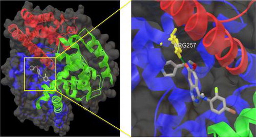
Binding orientation of the VDB in the Sudlow’s binding site I (subdomain IIA) of HSA.
Acknowledgments
The financial assistance from the University of Malaya to Md. Zahirul Kabir in the form of doctoral fellowship under the Bright Sparks Program (BSP/APP/1892/2013) is highly appreciated. We thank the Dean, Faculty of Science and the Head, Institute of Biological Sciences, University of Malaya for providing the necessary facilities.
Disclosure statement
No potential conflict of interest was reported by the authors.